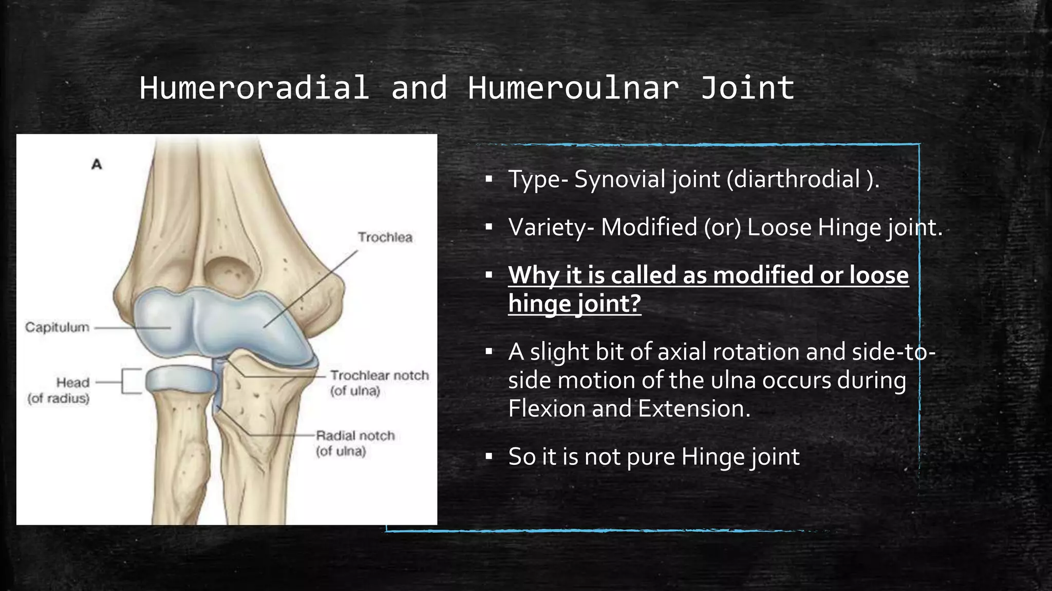
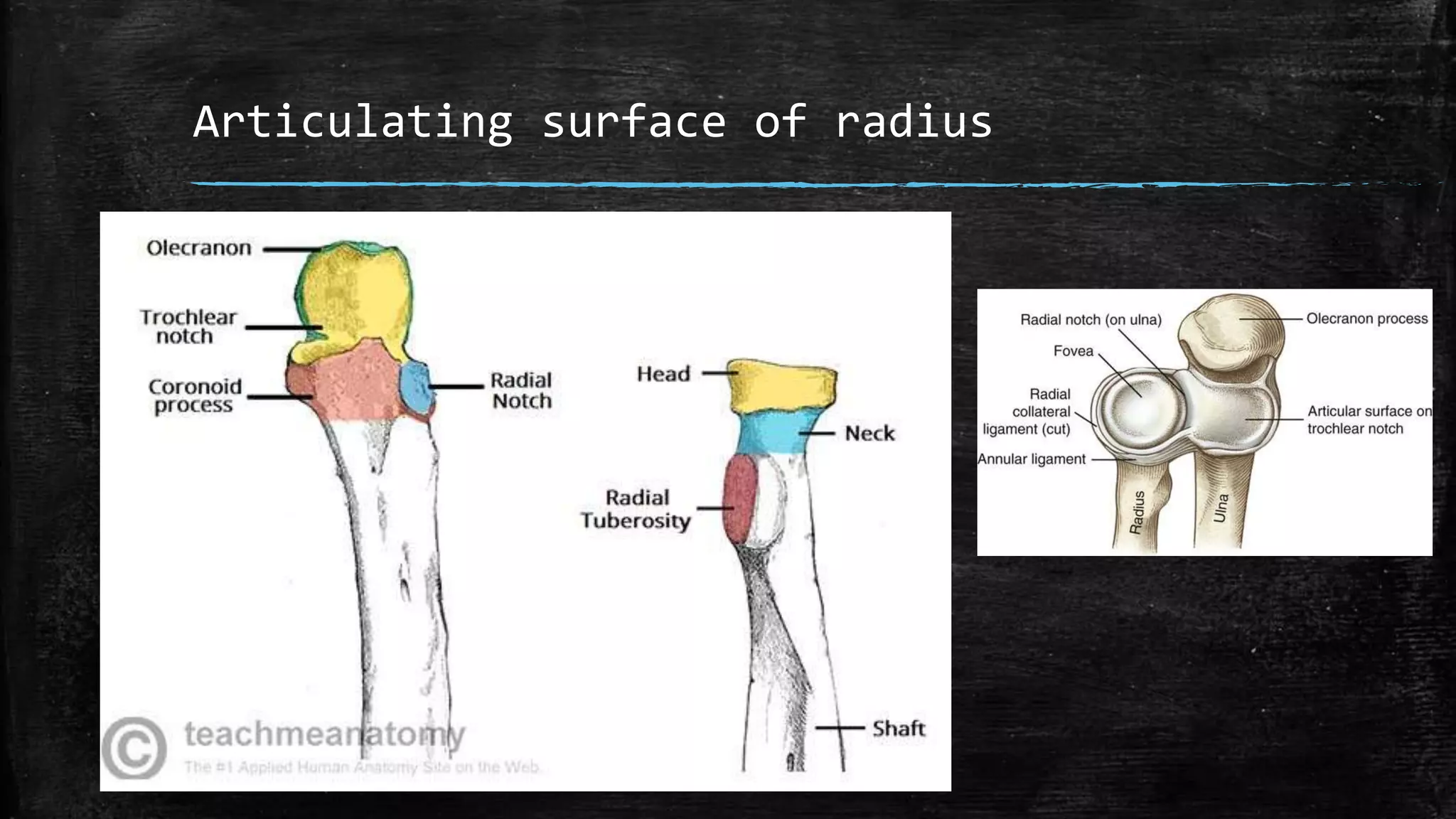
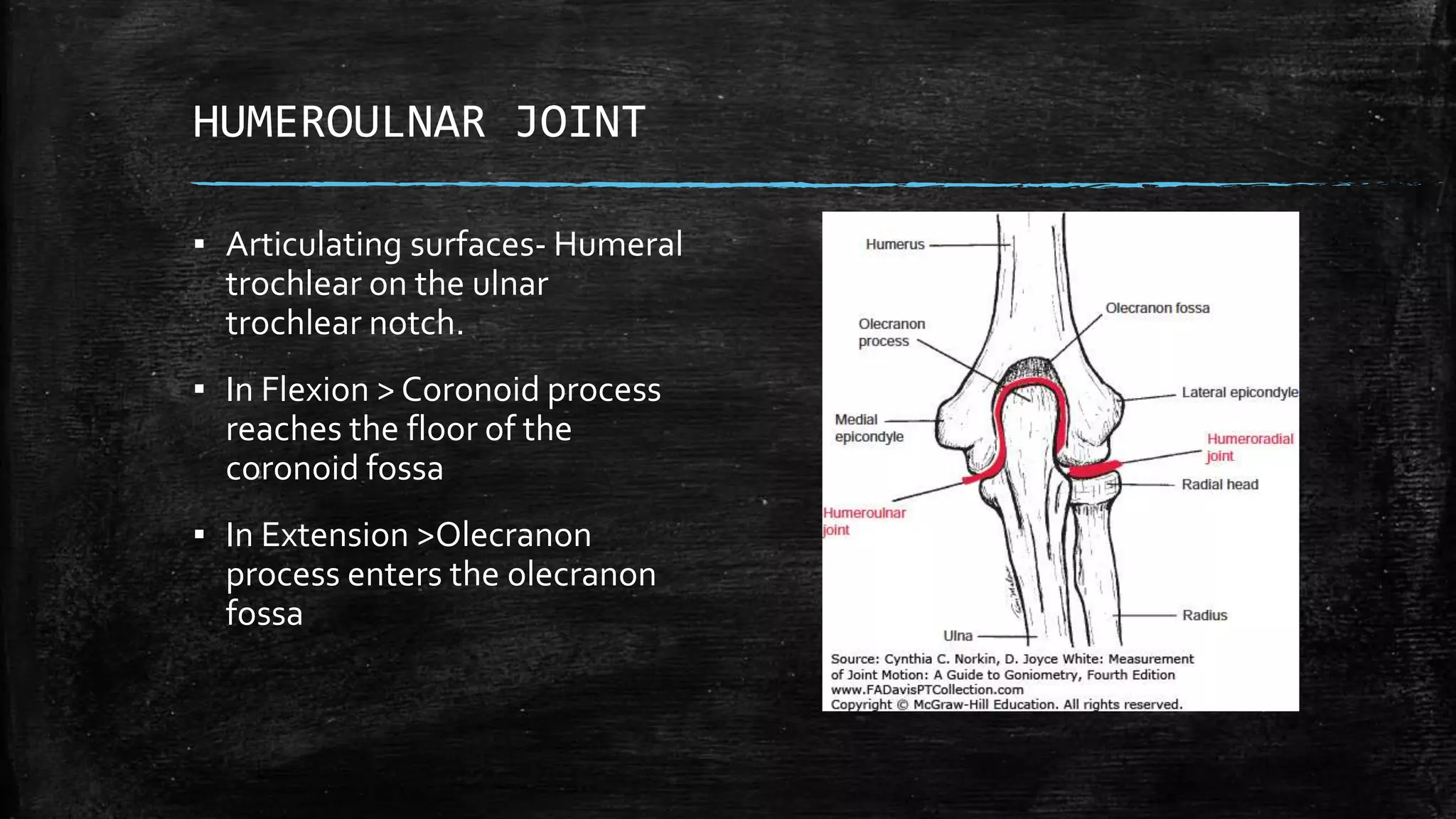
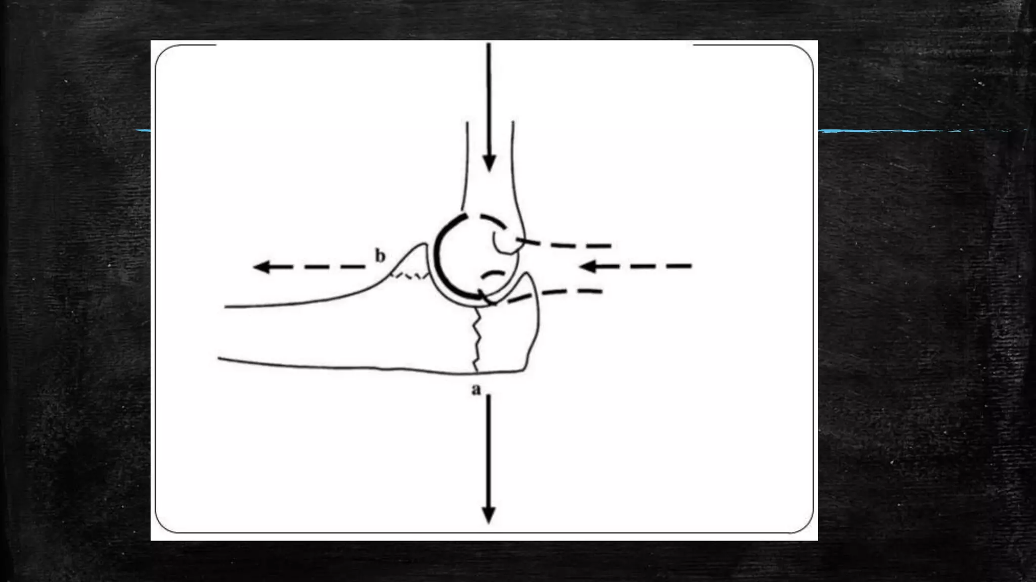
Elbow Complex
Vaibhavi Rathod
INTRODUCTION
▪ The joints and muscles of the elbow complex are designed to serve
the hand.
▪ They provide mobility for the hand in space by shortening and
lengthening the upper extremity.
▪ Rotation at the elbow complex provides additional mobility for the
hand.
▪ The elbow complex structures also provide stability for skilled (or)
forceful movements of the hand when performing activities with
tools or implements
Humeroradial and Humeroulnar Joint
▪ Type- Synovial joint (diarthrodial ).
▪ Variety- Modified (or) Loose Hinge joint.
▪ Why it is called as modified or loose
hinge joint?
▪ A slight bit of axial rotation and side-to-
side motion of the ulna occurs during
Flexion and Extension.
▪ So it is not pure Hinge joint
STRUCTURE
Articulating surface on humerus
Articulating surface of radius
Articulating surface of ulna
HUMERORADIAL JOINT
▪ Articulating surface- radial head and the
capitulum of humerus.
▪ Joint involves sliding the shallow concave
radial head over the convex surface of the
capitulum.
▪ Joint surfaces INCONGRUENT- (because of
the humeral capitulum slightly smaller than
the corresponding radial fovea).
▪ In full flexion, no contact occurs between the
articulating surfaces.
▪ In flexion, the rim of the radial head slides in
the capitulotrochlear groove.
▪ And enters the radial fossa as the end of the
Flexion range is reached.
HUMEROULNAR JOINT
▪ Articulating surfaces- Humeral
trochlear on the ulnar
trochlear notch.
▪ In Flexion > Coronoid process
reaches the floor of the
coronoid fossa
▪ In Extension >Olecranon
process enters the olecranon
fossa
LIGAMENTS
MEDIAL COLLATERAL LIGAMENT
▪ 1. Anterior medial collateral ligament Attachment: • Anterior aspect of
humerus to coronoid process. Anterior and posterior band that tightening
reciprocal manner as the elbow flexes and extends. • FUNCTION:- Restraint
of valgus stress from 20 to 120 degree of elbow flexion.
▪ 2. Posterior medial collateral ligament Attachment: • It Extends from
Posterior aspect of medial epicondyle of the humerus to ulnar coronoid and
olecranon process. • FUNCTION:- It limits elbow extension, Plays a less role
in providing valgus stability for the elbow.
▪ Transverse collateral ligament Attachment: • It extended between the
Olecranon process to ulnar coronoind process. • FUNCTION:- It Provide
little valgus stability, It May help to keep the joint surfaces in
approximation.
LATERAL COLLATERAL LIGAMENT
▪ LATERAL (radial) COLLATERAL LIGAMENT Attachment: • it extends
from inferior aspect of lateral epicondyle of humerus to merge with
the Annular ligament. • FUNCTION:- it provides reinforcement for
the Humero-radial articulation. Some protection against varus stress.
▪ LATERAL ULNAR COLLATERAL LIGAMENT Attachment: • It extend
from inferior aspect of lateral Epicondyle of humerus to edge of
Annular ligament and lateral surface of Ulna. • FUNCTION:- It acts as
a Dynamic stabilizer together with related muscles. Providing lateral
support to the Elbow joint.
MUSCLES OF ELBOW JOINT
▪ Anteriorly: 1) Pronator teres 2) Flexor
carpi radialis 3) Palmaris longus 4)
Flexor carpi ulnaris 5) Flexor
digitorum superficialis 6) Brachio
radialis 7) Brachialis 8) Biceps brachii.
▪ Posteriorly: 1) Extensor carpi radialis
longus 2) Extensor carpi radialis
brevis 3) Extensor digitorum 4)
Extensor digiti minimi 5) Extensor
carpi ulnaris 6) Supinator 7)Triceps 8)
Anconeus.
PROXIMAL AND DISTAL
RADIOULNAR JOINT
▪ TYPE:- synovial joint (Diarthrodial)
▪ VARIETY:- Pivot (trochoid) • Uniaxial-
the 2 joints acting together produce
supination and pronation of forearm
and have 1 degree of freedom of
motion (which occurs transverse pale
around a longitudinal axis)
STRUCTURE of PROXIMAL (superior) RADIO-
ULNAR JOINT
▪ Articulating surface:-
▪ Radial notch of ulna
▪ Annular ligament
▪ Head of radius
▪ Capitulum of humerus.
DISTAL(inferior) RADIO-ULNAR JOINT
▪ Articular surface:
▪ Ulnar notch of the radius
▪ Articular disc
▪ Head of ulna
ARTICULATIONS
▪ PROXIMAL RADIO-ULANR ARTICULATION:
▪ Bones taking part- Head of radius and radial
notch of the ulna.The head of radius is
attached to the radial notch of the ulna with
the help ofAnnular ligament.
▪ DISTAL RADIO-ULANR ARTICULATION:
▪ Bones taking part- Head of ulna and ulna
notch of radius.The Head of the ulna
attached to the ulna notch of radius and
Articular disc.
LIGAMENTS
▪ PROXIMAL RADIO-ULNAR JOINT: 1.
Annular ligament 2. Quadrate
ligament 3. Oblique cord.
▪ • DISTAL RADIO-ULNAR JOINT: 1.
Dorsal radio-ulnar ligament 2.
Palmar radio-ulnar ligament 3.
Interosseous membrane.
MUSCLES
▪ PRONATION: 1. Pronator teres 2. Pronator qudratus.
▪ •SUPINATION: 1. Biceps 2. supinator
Joint forces
▪ Significant compressive and shear forces at the elbow
▪ Loads across the elbow - distributed
▪ 43% across the ulnohumeral joint and 57% across the radiocapitellar joint
▪ Joint reaction forces vary with elbow position.
▪ Force transmission at the radiocapitellar joint is
▪ Greatest between 0 and 30 of flexion and is greater in pronation than in
supination.
▪ elbow - extended, the overall force on theulnohumeral joint is concentrated at
the coronoid
▪ elbow - flexed, the force moves toward the olecranon
Stability of elbow joint
▪ static and dynamic constraints
▪ 3 primary static constraints
▪ Ulnohumeral articulation, the anterior bundle of the MCL the
lateral collateral ligament (LCL) complex
▪ Secondary constraints Radiocapitellar articulation, the common
flexor tendon, the common extensor tendon, the capsule.
▪ Dynamic stabilizers - Muscles that cross the elbowjoint
Thank you

Biomechanics of Elbow complex

  • 1.
  • 2.
    INTRODUCTION ▪ The jointsand muscles of the elbow complex are designed to serve the hand. ▪ They provide mobility for the hand in space by shortening and lengthening the upper extremity. ▪ Rotation at the elbow complex provides additional mobility for the hand. ▪ The elbow complex structures also provide stability for skilled (or) forceful movements of the hand when performing activities with tools or implements
  • 3.
    Humeroradial and HumeroulnarJoint ▪ Type- Synovial joint (diarthrodial ). ▪ Variety- Modified (or) Loose Hinge joint. ▪ Why it is called as modified or loose hinge joint? ▪ A slight bit of axial rotation and side-to- side motion of the ulna occurs during Flexion and Extension. ▪ So it is not pure Hinge joint
  • 4.
  • 5.
  • 6.
  • 7.
    HUMERORADIAL JOINT ▪ Articulatingsurface- radial head and the capitulum of humerus. ▪ Joint involves sliding the shallow concave radial head over the convex surface of the capitulum. ▪ Joint surfaces INCONGRUENT- (because of the humeral capitulum slightly smaller than the corresponding radial fovea). ▪ In full flexion, no contact occurs between the articulating surfaces. ▪ In flexion, the rim of the radial head slides in the capitulotrochlear groove. ▪ And enters the radial fossa as the end of the Flexion range is reached.
  • 8.
    HUMEROULNAR JOINT ▪ Articulatingsurfaces- Humeral trochlear on the ulnar trochlear notch. ▪ In Flexion > Coronoid process reaches the floor of the coronoid fossa ▪ In Extension >Olecranon process enters the olecranon fossa
  • 9.
  • 10.
    MEDIAL COLLATERAL LIGAMENT ▪1. Anterior medial collateral ligament Attachment: • Anterior aspect of humerus to coronoid process. Anterior and posterior band that tightening reciprocal manner as the elbow flexes and extends. • FUNCTION:- Restraint of valgus stress from 20 to 120 degree of elbow flexion. ▪ 2. Posterior medial collateral ligament Attachment: • It Extends from Posterior aspect of medial epicondyle of the humerus to ulnar coronoid and olecranon process. • FUNCTION:- It limits elbow extension, Plays a less role in providing valgus stability for the elbow. ▪ Transverse collateral ligament Attachment: • It extended between the Olecranon process to ulnar coronoind process. • FUNCTION:- It Provide little valgus stability, It May help to keep the joint surfaces in approximation.
  • 11.
    LATERAL COLLATERAL LIGAMENT ▪LATERAL (radial) COLLATERAL LIGAMENT Attachment: • it extends from inferior aspect of lateral epicondyle of humerus to merge with the Annular ligament. • FUNCTION:- it provides reinforcement for the Humero-radial articulation. Some protection against varus stress. ▪ LATERAL ULNAR COLLATERAL LIGAMENT Attachment: • It extend from inferior aspect of lateral Epicondyle of humerus to edge of Annular ligament and lateral surface of Ulna. • FUNCTION:- It acts as a Dynamic stabilizer together with related muscles. Providing lateral support to the Elbow joint.
  • 12.
    MUSCLES OF ELBOWJOINT ▪ Anteriorly: 1) Pronator teres 2) Flexor carpi radialis 3) Palmaris longus 4) Flexor carpi ulnaris 5) Flexor digitorum superficialis 6) Brachio radialis 7) Brachialis 8) Biceps brachii. ▪ Posteriorly: 1) Extensor carpi radialis longus 2) Extensor carpi radialis brevis 3) Extensor digitorum 4) Extensor digiti minimi 5) Extensor carpi ulnaris 6) Supinator 7)Triceps 8) Anconeus.
  • 13.
  • 14.
    ▪ TYPE:- synovialjoint (Diarthrodial) ▪ VARIETY:- Pivot (trochoid) • Uniaxial- the 2 joints acting together produce supination and pronation of forearm and have 1 degree of freedom of motion (which occurs transverse pale around a longitudinal axis)
  • 15.
    STRUCTURE of PROXIMAL(superior) RADIO- ULNAR JOINT ▪ Articulating surface:- ▪ Radial notch of ulna ▪ Annular ligament ▪ Head of radius ▪ Capitulum of humerus.
  • 16.
    DISTAL(inferior) RADIO-ULNAR JOINT ▪Articular surface: ▪ Ulnar notch of the radius ▪ Articular disc ▪ Head of ulna
  • 17.
    ARTICULATIONS ▪ PROXIMAL RADIO-ULANRARTICULATION: ▪ Bones taking part- Head of radius and radial notch of the ulna.The head of radius is attached to the radial notch of the ulna with the help ofAnnular ligament. ▪ DISTAL RADIO-ULANR ARTICULATION: ▪ Bones taking part- Head of ulna and ulna notch of radius.The Head of the ulna attached to the ulna notch of radius and Articular disc.
  • 18.
    LIGAMENTS ▪ PROXIMAL RADIO-ULNARJOINT: 1. Annular ligament 2. Quadrate ligament 3. Oblique cord. ▪ • DISTAL RADIO-ULNAR JOINT: 1. Dorsal radio-ulnar ligament 2. Palmar radio-ulnar ligament 3. Interosseous membrane.
  • 19.
    MUSCLES ▪ PRONATION: 1.Pronator teres 2. Pronator qudratus. ▪ •SUPINATION: 1. Biceps 2. supinator
  • 20.
    Joint forces ▪ Significantcompressive and shear forces at the elbow ▪ Loads across the elbow - distributed ▪ 43% across the ulnohumeral joint and 57% across the radiocapitellar joint ▪ Joint reaction forces vary with elbow position. ▪ Force transmission at the radiocapitellar joint is ▪ Greatest between 0 and 30 of flexion and is greater in pronation than in supination. ▪ elbow - extended, the overall force on theulnohumeral joint is concentrated at the coronoid ▪ elbow - flexed, the force moves toward the olecranon
  • 22.
    Stability of elbowjoint ▪ static and dynamic constraints ▪ 3 primary static constraints ▪ Ulnohumeral articulation, the anterior bundle of the MCL the lateral collateral ligament (LCL) complex ▪ Secondary constraints Radiocapitellar articulation, the common flexor tendon, the common extensor tendon, the capsule. ▪ Dynamic stabilizers - Muscles that cross the elbowjoint
  • 23.