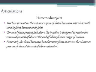
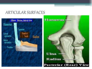
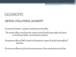
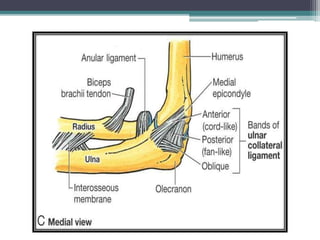
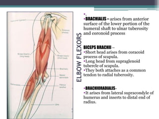
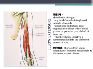
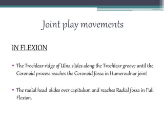
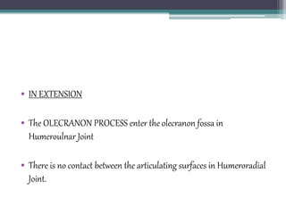
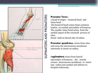
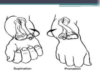
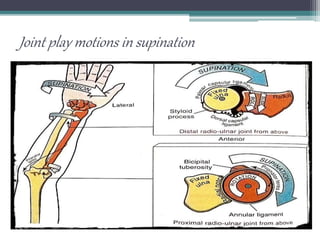
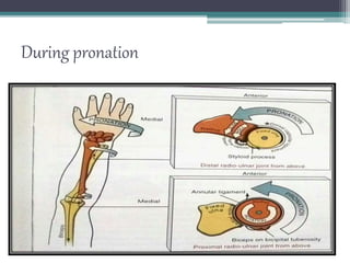
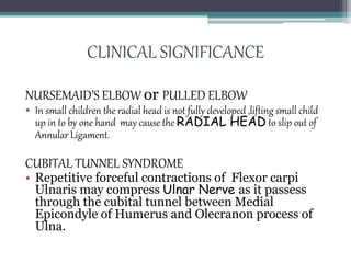
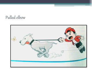
BIOMECHANICS OF
ELBOW COMPLEX
SUBMITTED TO: DR. ANKIT GAUR
SUBMITTED BY: RUKHSAR JABBAR
INTRODUCTION
• The elbow complex is composed of humero-ulnar, humero-radial,
proximal and distal radioulnar joint.
• The elbow joint is considered as a compound joint that functions as a
modified Or loose hinge joint.
• The proximal and distal radio-ulnar joints are linked and function as
one joint.
Articulations
Humero-ulnar joint
• Trochlea present on the anterior aspect of distal humerus articulates with
ulna to form humeroulnar joint.
• Coronoid fossa present just above the trochlea is designed to receive the
coronoid process of ulna at the end of elbow flexion range of motion.
• Posteriorly the distal humerus has olecranon fossa to receive the olecranon
process of ulna at the end of elbow extension.
ARTICULAR SURFACES
Humero-radial joint
• The capitulum located on the anterio-lateral surface of distal humerus
articulates with the head of radius.
• Radial fossa is present just above the capitulum is designed to receive
the head of radius in elbow flexion.
• The trochlea and capitulum is seperated by capitulotrochlear groove.
SUPERIOR RADIOULNAR JOINT
ARTICULATION
• The Radial notch on Ulna articulate with Head of Radius along with
Annular Ligament.
LIGAMENTS
• Annular Ligament-----circle the head of Radius and keeps the
Ulna together.
• Quadrate Ligament----extends from the Inferior edge of radial
notch to Neck of Radius
• Oblique cord------attached to inferior part of Radial notch on
Ulna to just below Radial Tuberosity
INFERIOR RADIOULNAR JOINT
ARTICULATION
• The Ulnar notch of Radius articulates with head of Ulna along with
Articular Disc.
LIGAMENTS
• Anterior Radio Ulnar Ligament----attached to anterior
aspect just above the Ulnar head to above Ulnar notch.
• Posterior Radio Ulnar Ligament---attached to posterior
part of Ulnar head to above Ulnar notch.
• Interosseous Membrane---binds the shaft of Radius and Ulna
together.
• An articular disc lies in between distal end of ulna called… TFCC-
Traingular fibricartilagenous complex.
DISTAL RADIO ULNAR JT
Interosseous membrane
• The radius and ulna are bound together by the interosseous
membrane of the forearm.
• The primary function of interosseous membrane are bind the radius
to ulna,serve as a stable attachment site for several extrinsic muscles of
hand and provide mechanism for transmitting force proximally
through the upper limb.
The interrosseous membrane
shunts some of the compressive
forces radius to ulna.
In this way interrosseous
membrane helps to protect the
radio-humeral jt from compressive
forces.
The fibres of interrosseous
membrane are not directed in such
a way as to resist distally applied
forces onto the radius
The distal pull from the radius
slackens the membrane rather than
tenses.
• The other structures contribute such as the oblique cord, annular
ligament and brachioradialis to accept and compensate the distractive
force.
• The contraction of brachioradialis muscle helps
in stabilising the radial head against capitulum.
LIGAMENTS
MEDIAL COLLATERAL LIGAMENT
It consists of anterior , posterior and transverse bundles.
The anterior fibres arise from the anterior part of medial epicondyle and insert
on medial part of the coronoid process of ulna.
The posterior fibres of MCL attach to the posterior aspect of medial epicondyle of
humerus
The tranverse fibres are from olecranon process to the coronoid process of ulna.
.
Lateral collateral ligamentous comlplex
It includes lateral collateral ligament,the annular ligament and lateral
ulnar collateral ligament.
The LCL extends from the inferior aspect of lateral epicondyle of
humerus to merge with annnular ligament.
Ligaments
•MCL stabilizes the joint
against lateral forces or
valgus stress.
•Mostly MCL are more
prone to injury.
•LCL stabilizes elbow
against varus forces.
Carrying angle
• The angle formed beteween the long axis of humerus and long axis of
foearm , an acute angle is formed medially when at the elbow.
• It is formed as the trochlear medial aspect extends more distally than
does the lateral aspect which results in lateral deviation of ulna to
humerus.
• It is more in females as comparitively women possess less height than
males….. Consequently the shorter forearm bones , the greater the
carrying angle.
• The medial flange of the trochlea longer in a shorter person so more
carrying angle.
Normal values
In females- 10 - 15 degress
• In males- 5-10 degress.
ELBOW
FLEXORS
.
BRACHIALIS – arises from anterior
surface of the lower portion of the
humeral shaft to ulnar tuberosity
and coronoid process
BICEPS BRACHII –
Short head arises from coracoid
process of scapula.
Long head from supraglenoid
tubercle of scapula.
They both attaches as a common
tendon to radial tuberosity.
BRACHIORADIALIS-
It arises from lateral supracondyle of
humerus and inserts to distal end of
radius.
ELBOW FLEXORS
• The brachialis muscle works in flexion of the elbow in all
positions of the forearm , with or without resistance.
• The biceps brachii is active during unresisted elbow flexion with
forearm supinated and when the forearm is midway between
supination and pronation but it tends to be more active when forearm
is pronated.
• Brachioradialis is more active in mid prone and full pronated elbow
flexion and when the speed of motion is increased or load is applied.
ELBOW
EXTENSORS
TRICEPS –
Three heads of origin-
long head-from the infraglenoid
tubercle of scapula
medial head and lateral head –
originate from either side of radial
groove on posterior part of shaft of
humerus.
the three heads insert via a
common tendon into the olecranon
process of ulna.
ANCONEUS –It arises from lateral
epicondyle of humerus and extends to
olecranon process of ulna.
EXTENSORS OF ELBOW
• TRICEPS is the powerful extensor of the elbow.
• Long head of Triceps depends on Shoulder Position.
• Medial head of Triceps is active in unresisted Elbow
extension.
• All three Heads of Triceps are active when heavy resistance is given to
Extension.
• Anconeus muscle provides stability posterolaterally to joint.
Joint play movements
IN FLEXION
• The Trochlear ridge of Ulna slides along the Trochlear groove until the
Coronoid process reaches the Coronoid fossa in Humeroulnar joint
• The radial head slides over capitulum and reaches Radial fossa in Full
Flexion.
Medial view lateral view
• IN EXTENSION
• The OLECRANON PROCESS enter the olecranon fossa in
Humeroulnar Joint
• There is no contact between the articulating surfaces in Humeroradial
Joint.
SUPINATORS
AND
PRONATORS
Pronater Teres -
2 head of origin – humeral head and
ulnar head
the humeral head comes from common
tendon on medial epicondyle of humerus.
The smaller ulnar head arises from the
medial aspect of the coronoid process of
ulna.
Insert both at lateral side of radius.
Pronator quadratus- arises from ulna
and cross the interroseous membrane
anteriorly to insert on radius.
Supinators-arises from lateral
epicondyle of humerus , the muscle
crosses interroseous membrane to insert
into radius just medial and inferior to
bicipital tuberosity.
PRONATORS AND SUPINATORS
• PRONATOR TERES----- helps in Pronation,it acts in
all position of Elbow, helps in Stabilization of
Superio Radio Ulnar Joint. Active during rapid
and resisted Pronation.
• PRONATOR QUADRATUS---- helps in Pronation in all
position of Elbow.
• SUPINATOR---------helps in Supination in all
position of Elbow
Continued’
• In Elbow extended position Pronation is limited
due to passive tension in Biceps Brachi.
• Supination is limited due to passive tension in
Interosseous Membrane.
Joint play motions in supination
During pronation
CLINICAL SIGNIFICANCE
NURSEMAID’S ELBOW or PULLED ELBOW
• In small children the radial head is not fully developed ,lifting small child
up in to by one hand may cause the RADIAL HEAD to slip out of
Annular Ligament.
CUBITAL TUNNEL SYNDROME
• Repetitive forceful contractions of Flexor carpi
Ulnaris may compress Ulnar Nerve as it passess
through the cubital tunnel between Medial
Epicondyle of Humerus and Olecranon process of
Ulna.
Pulled elbow
CONTINUED’
TENNISELBOWor LATERAL EPICONDYLITIS:
• Inflammation at origin of Extensors of Wrist at Lateral Epicondyle.
• It is caused due to repeated forceful contraction of Wrist Extensors.
• Extensor carpi radialis brevis is affected
GOLFER’S ELBOWor MEDIAL EPICONDYLITIS:
• Inflammation at origin of Flexors of Wrist at Medial Epicondyle.
• Repetitive contractions of Pronator Teres,Flexor Carpi Ulnaris .
Inflammation of common extensor
origin.
Common flexor origin inflammation.
Thank you
• IF U R NOT WILLING TO LEARN
NO ONE CAN HELP U….. BUT IF U
R DETERMINED TO LEARN NO
ONE CAN STOP U…

Elbow biomechanics

  • 1.
    BIOMECHANICS OF ELBOW COMPLEX SUBMITTEDTO: DR. ANKIT GAUR SUBMITTED BY: RUKHSAR JABBAR
  • 2.
    INTRODUCTION • The elbowcomplex is composed of humero-ulnar, humero-radial, proximal and distal radioulnar joint. • The elbow joint is considered as a compound joint that functions as a modified Or loose hinge joint. • The proximal and distal radio-ulnar joints are linked and function as one joint.
  • 3.
    Articulations Humero-ulnar joint • Trochleapresent on the anterior aspect of distal humerus articulates with ulna to form humeroulnar joint. • Coronoid fossa present just above the trochlea is designed to receive the coronoid process of ulna at the end of elbow flexion range of motion. • Posteriorly the distal humerus has olecranon fossa to receive the olecranon process of ulna at the end of elbow extension.
  • 4.
  • 5.
    Humero-radial joint • Thecapitulum located on the anterio-lateral surface of distal humerus articulates with the head of radius. • Radial fossa is present just above the capitulum is designed to receive the head of radius in elbow flexion. • The trochlea and capitulum is seperated by capitulotrochlear groove.
  • 7.
    SUPERIOR RADIOULNAR JOINT ARTICULATION •The Radial notch on Ulna articulate with Head of Radius along with Annular Ligament. LIGAMENTS • Annular Ligament-----circle the head of Radius and keeps the Ulna together. • Quadrate Ligament----extends from the Inferior edge of radial notch to Neck of Radius • Oblique cord------attached to inferior part of Radial notch on Ulna to just below Radial Tuberosity
  • 9.
    INFERIOR RADIOULNAR JOINT ARTICULATION •The Ulnar notch of Radius articulates with head of Ulna along with Articular Disc. LIGAMENTS • Anterior Radio Ulnar Ligament----attached to anterior aspect just above the Ulnar head to above Ulnar notch. • Posterior Radio Ulnar Ligament---attached to posterior part of Ulnar head to above Ulnar notch. • Interosseous Membrane---binds the shaft of Radius and Ulna together. • An articular disc lies in between distal end of ulna called… TFCC- Traingular fibricartilagenous complex.
  • 11.
  • 12.
    Interosseous membrane • Theradius and ulna are bound together by the interosseous membrane of the forearm. • The primary function of interosseous membrane are bind the radius to ulna,serve as a stable attachment site for several extrinsic muscles of hand and provide mechanism for transmitting force proximally through the upper limb.
  • 13.
    The interrosseous membrane shuntssome of the compressive forces radius to ulna. In this way interrosseous membrane helps to protect the radio-humeral jt from compressive forces.
  • 14.
    The fibres ofinterrosseous membrane are not directed in such a way as to resist distally applied forces onto the radius The distal pull from the radius slackens the membrane rather than tenses.
  • 15.
    • The otherstructures contribute such as the oblique cord, annular ligament and brachioradialis to accept and compensate the distractive force. • The contraction of brachioradialis muscle helps in stabilising the radial head against capitulum.
  • 16.
    LIGAMENTS MEDIAL COLLATERAL LIGAMENT Itconsists of anterior , posterior and transverse bundles. The anterior fibres arise from the anterior part of medial epicondyle and insert on medial part of the coronoid process of ulna. The posterior fibres of MCL attach to the posterior aspect of medial epicondyle of humerus The tranverse fibres are from olecranon process to the coronoid process of ulna. .
  • 18.
    Lateral collateral ligamentouscomlplex It includes lateral collateral ligament,the annular ligament and lateral ulnar collateral ligament. The LCL extends from the inferior aspect of lateral epicondyle of humerus to merge with annnular ligament.
  • 19.
  • 20.
    •MCL stabilizes thejoint against lateral forces or valgus stress. •Mostly MCL are more prone to injury. •LCL stabilizes elbow against varus forces.
  • 21.
    Carrying angle • Theangle formed beteween the long axis of humerus and long axis of foearm , an acute angle is formed medially when at the elbow. • It is formed as the trochlear medial aspect extends more distally than does the lateral aspect which results in lateral deviation of ulna to humerus.
  • 22.
    • It ismore in females as comparitively women possess less height than males….. Consequently the shorter forearm bones , the greater the carrying angle. • The medial flange of the trochlea longer in a shorter person so more carrying angle. Normal values In females- 10 - 15 degress • In males- 5-10 degress.
  • 24.
    ELBOW FLEXORS . BRACHIALIS – arisesfrom anterior surface of the lower portion of the humeral shaft to ulnar tuberosity and coronoid process BICEPS BRACHII – Short head arises from coracoid process of scapula. Long head from supraglenoid tubercle of scapula. They both attaches as a common tendon to radial tuberosity. BRACHIORADIALIS- It arises from lateral supracondyle of humerus and inserts to distal end of radius.
  • 25.
    ELBOW FLEXORS • Thebrachialis muscle works in flexion of the elbow in all positions of the forearm , with or without resistance. • The biceps brachii is active during unresisted elbow flexion with forearm supinated and when the forearm is midway between supination and pronation but it tends to be more active when forearm is pronated.
  • 26.
    • Brachioradialis ismore active in mid prone and full pronated elbow flexion and when the speed of motion is increased or load is applied.
  • 27.
    ELBOW EXTENSORS TRICEPS – Three headsof origin- long head-from the infraglenoid tubercle of scapula medial head and lateral head – originate from either side of radial groove on posterior part of shaft of humerus. the three heads insert via a common tendon into the olecranon process of ulna. ANCONEUS –It arises from lateral epicondyle of humerus and extends to olecranon process of ulna.
  • 28.
    EXTENSORS OF ELBOW •TRICEPS is the powerful extensor of the elbow. • Long head of Triceps depends on Shoulder Position. • Medial head of Triceps is active in unresisted Elbow extension. • All three Heads of Triceps are active when heavy resistance is given to Extension. • Anconeus muscle provides stability posterolaterally to joint.
  • 29.
    Joint play movements INFLEXION • The Trochlear ridge of Ulna slides along the Trochlear groove until the Coronoid process reaches the Coronoid fossa in Humeroulnar joint • The radial head slides over capitulum and reaches Radial fossa in Full Flexion.
  • 30.
  • 31.
    • IN EXTENSION •The OLECRANON PROCESS enter the olecranon fossa in Humeroulnar Joint • There is no contact between the articulating surfaces in Humeroradial Joint.
  • 32.
    SUPINATORS AND PRONATORS Pronater Teres - 2head of origin – humeral head and ulnar head the humeral head comes from common tendon on medial epicondyle of humerus. The smaller ulnar head arises from the medial aspect of the coronoid process of ulna. Insert both at lateral side of radius. Pronator quadratus- arises from ulna and cross the interroseous membrane anteriorly to insert on radius. Supinators-arises from lateral epicondyle of humerus , the muscle crosses interroseous membrane to insert into radius just medial and inferior to bicipital tuberosity.
  • 34.
    PRONATORS AND SUPINATORS •PRONATOR TERES----- helps in Pronation,it acts in all position of Elbow, helps in Stabilization of Superio Radio Ulnar Joint. Active during rapid and resisted Pronation. • PRONATOR QUADRATUS---- helps in Pronation in all position of Elbow. • SUPINATOR---------helps in Supination in all position of Elbow
  • 35.
    Continued’ • In Elbowextended position Pronation is limited due to passive tension in Biceps Brachi. • Supination is limited due to passive tension in Interosseous Membrane.
  • 36.
    Joint play motionsin supination
  • 37.
  • 38.
    CLINICAL SIGNIFICANCE NURSEMAID’S ELBOWor PULLED ELBOW • In small children the radial head is not fully developed ,lifting small child up in to by one hand may cause the RADIAL HEAD to slip out of Annular Ligament. CUBITAL TUNNEL SYNDROME • Repetitive forceful contractions of Flexor carpi Ulnaris may compress Ulnar Nerve as it passess through the cubital tunnel between Medial Epicondyle of Humerus and Olecranon process of Ulna.
  • 40.
  • 41.
    CONTINUED’ TENNISELBOWor LATERAL EPICONDYLITIS: •Inflammation at origin of Extensors of Wrist at Lateral Epicondyle. • It is caused due to repeated forceful contraction of Wrist Extensors. • Extensor carpi radialis brevis is affected GOLFER’S ELBOWor MEDIAL EPICONDYLITIS: • Inflammation at origin of Flexors of Wrist at Medial Epicondyle. • Repetitive contractions of Pronator Teres,Flexor Carpi Ulnaris .
  • 42.
    Inflammation of commonextensor origin.
  • 43.
    Common flexor origininflammation.
  • 44.
    Thank you • IFU R NOT WILLING TO LEARN NO ONE CAN HELP U….. BUT IF U R DETERMINED TO LEARN NO ONE CAN STOP U…