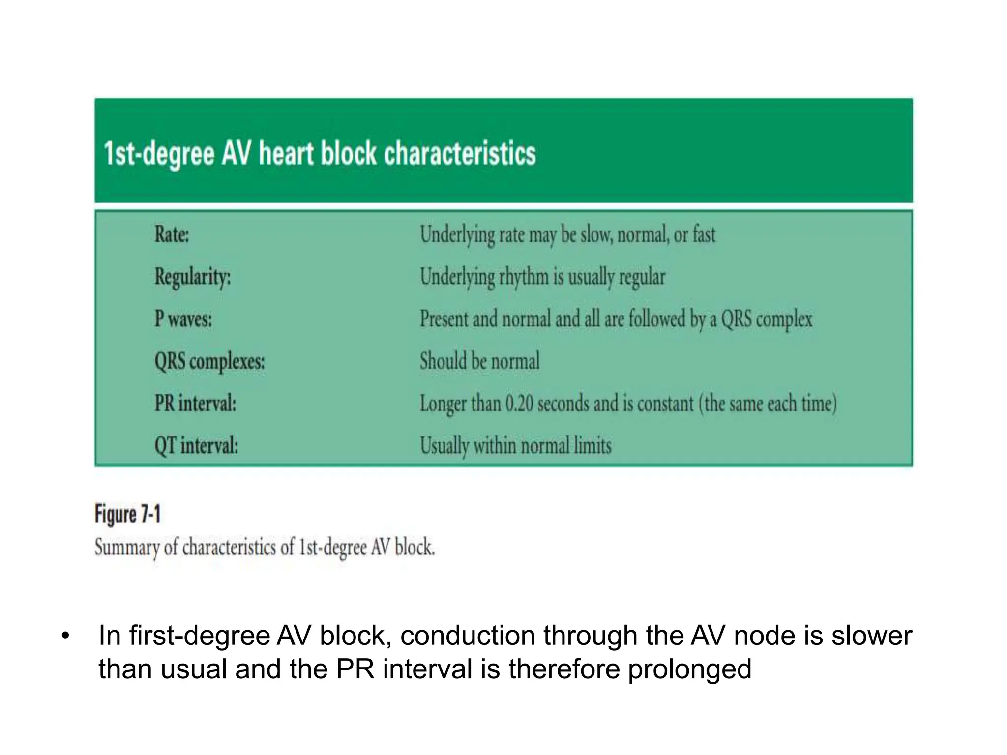
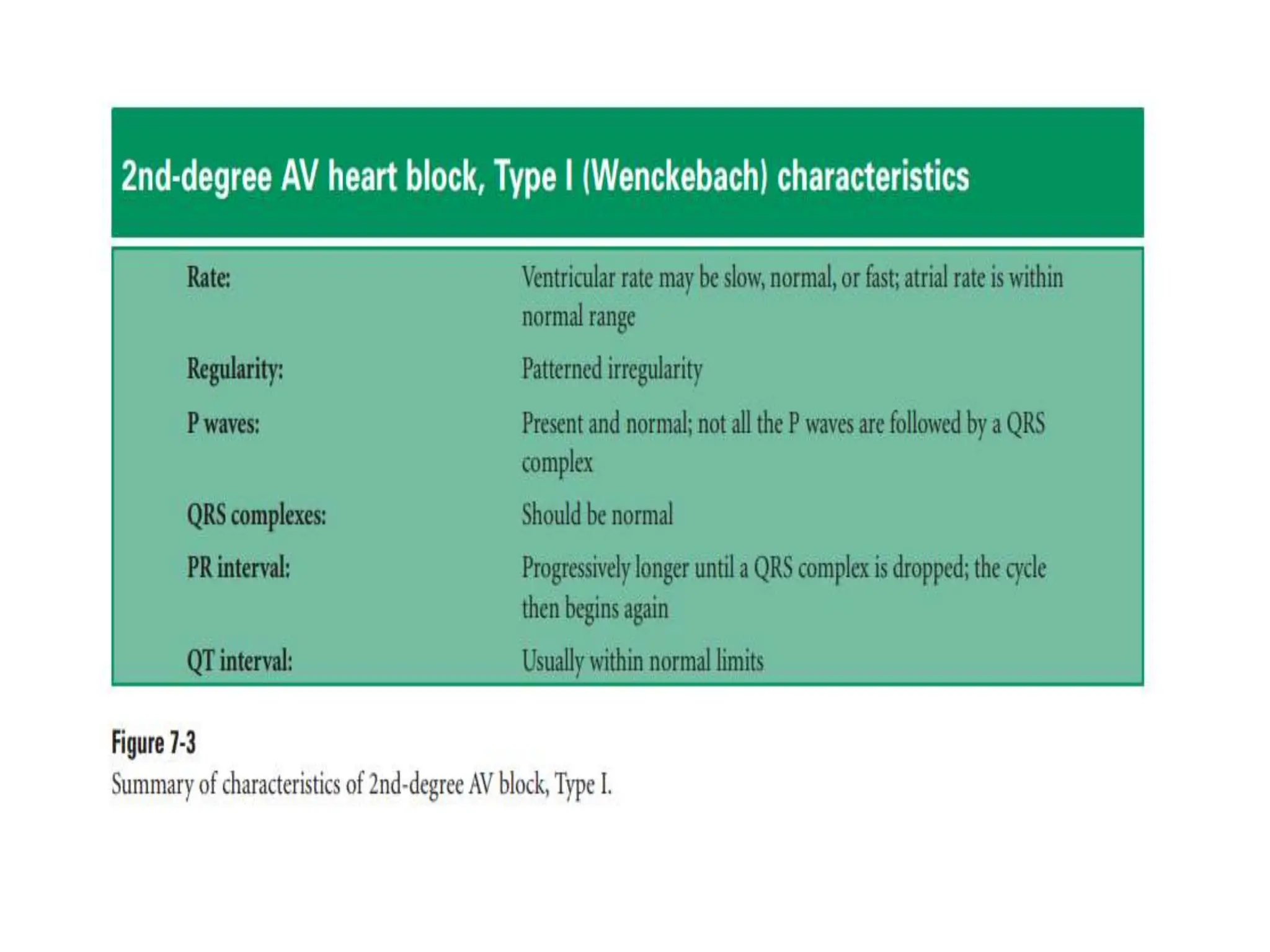
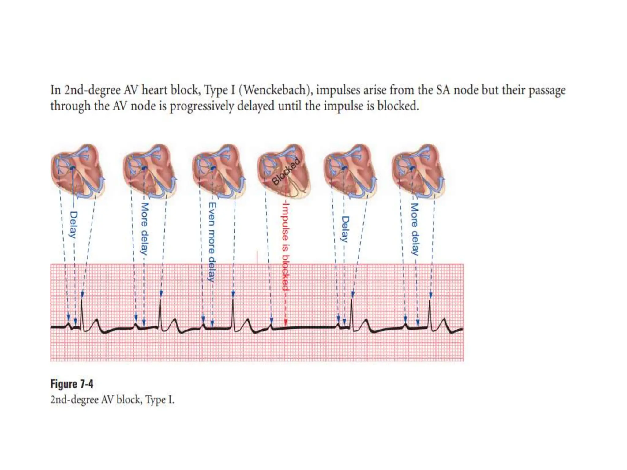
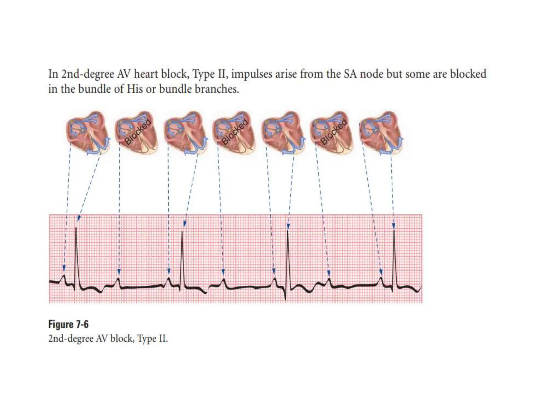
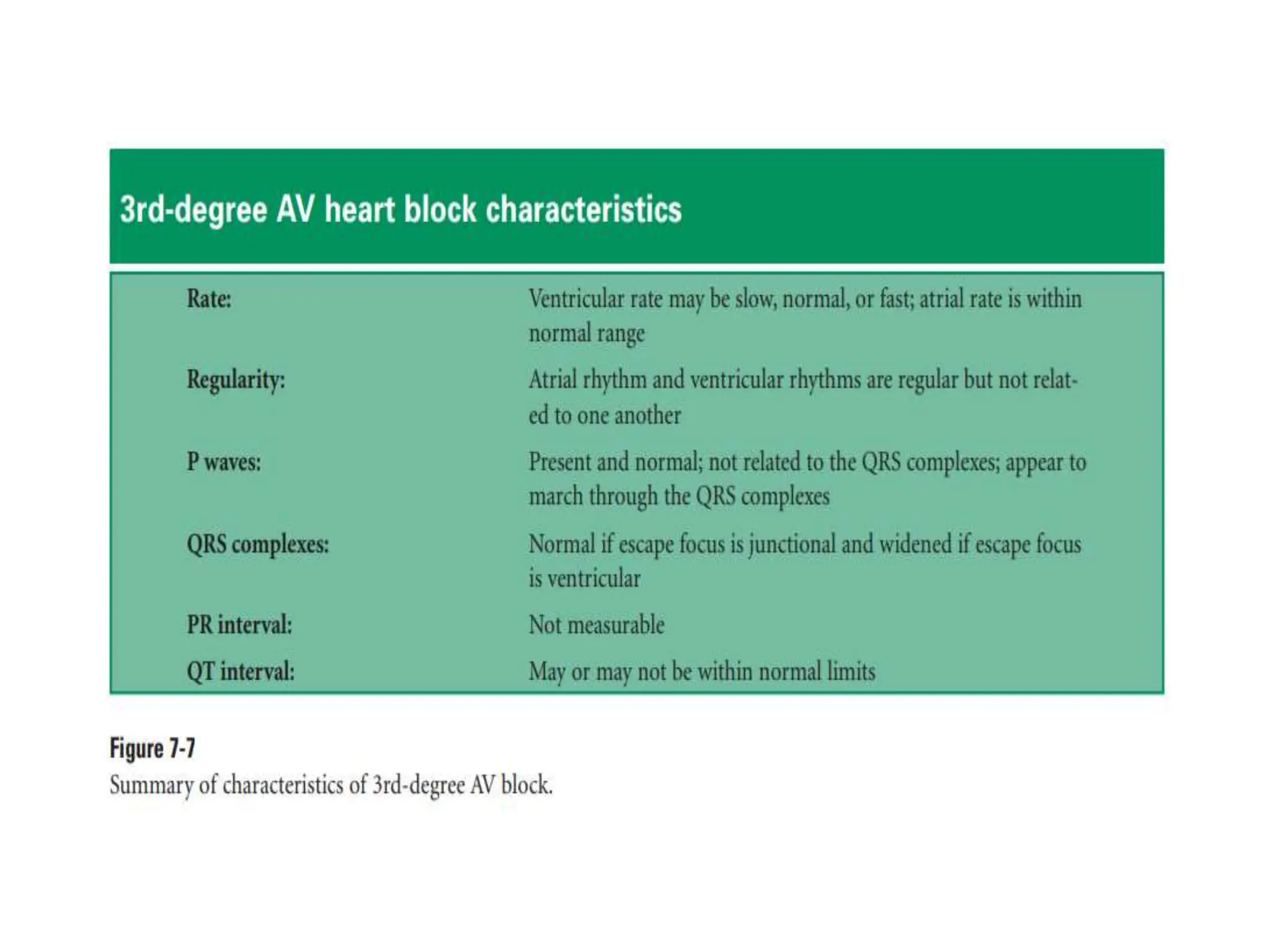
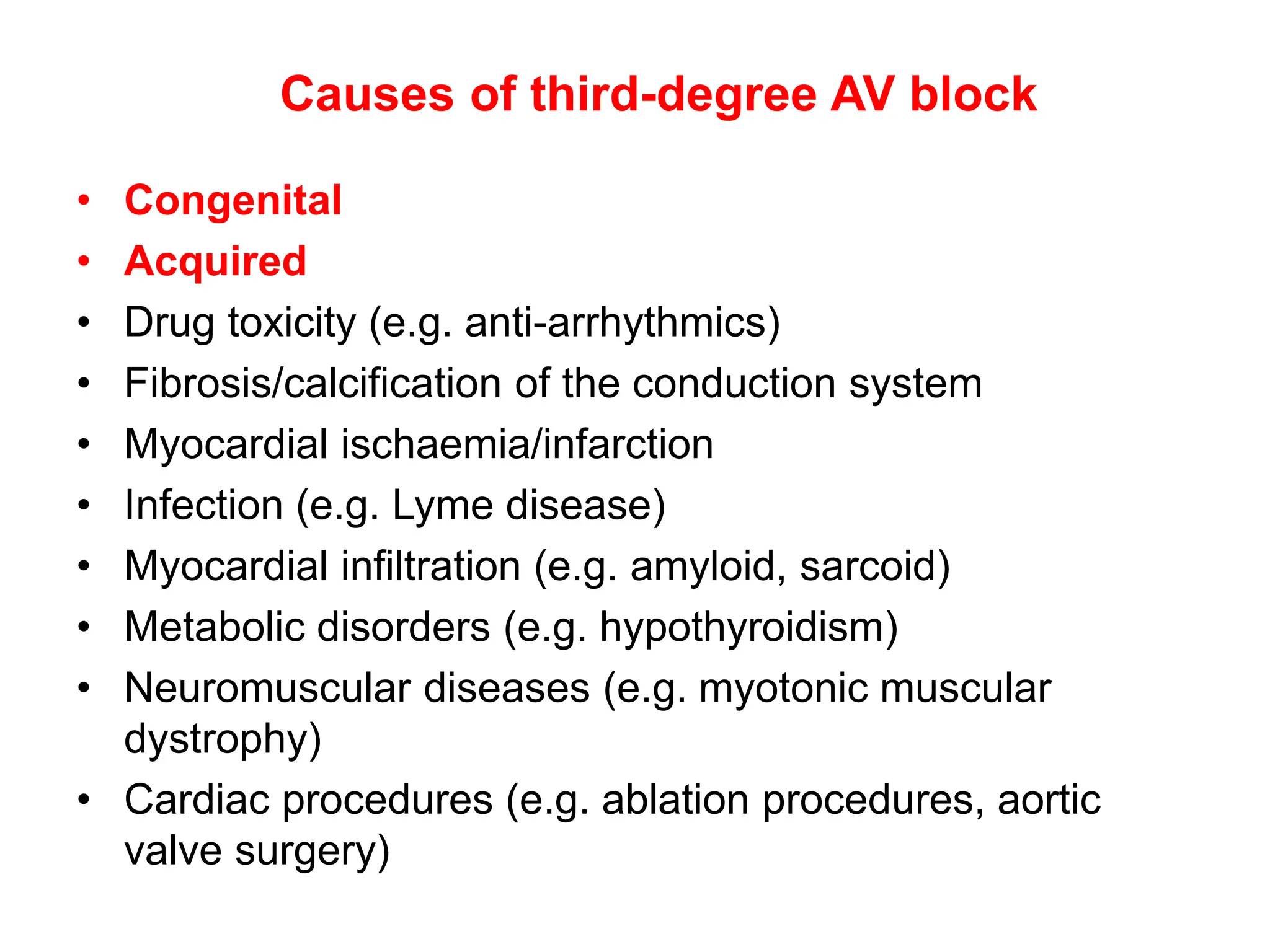
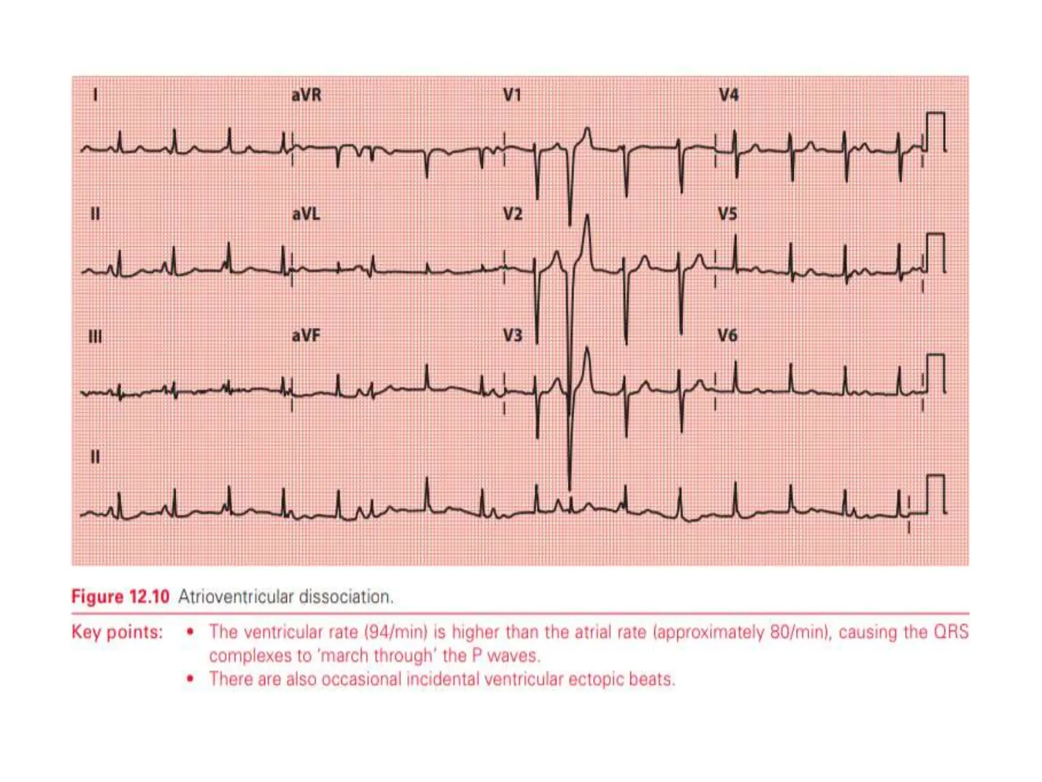
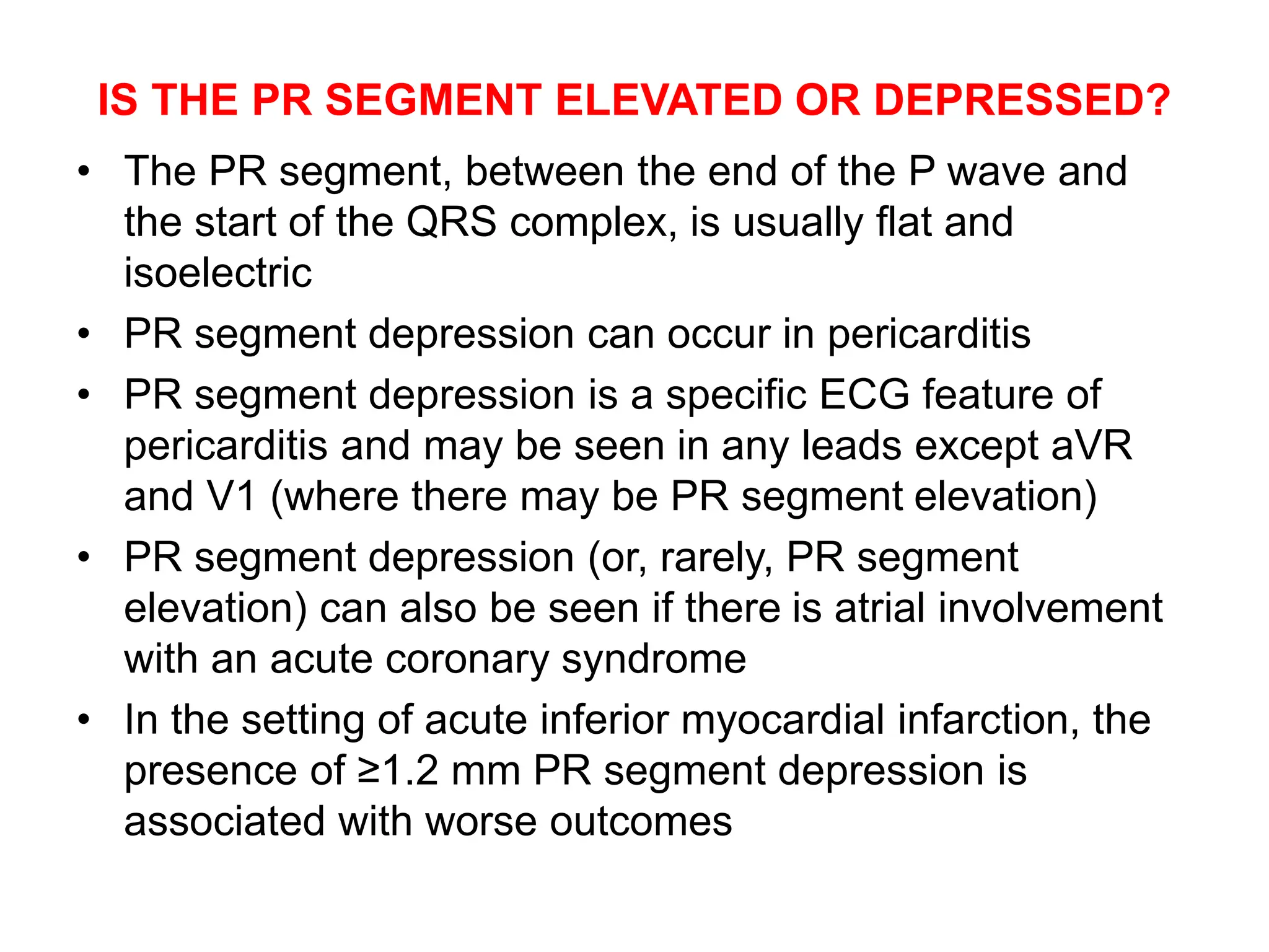
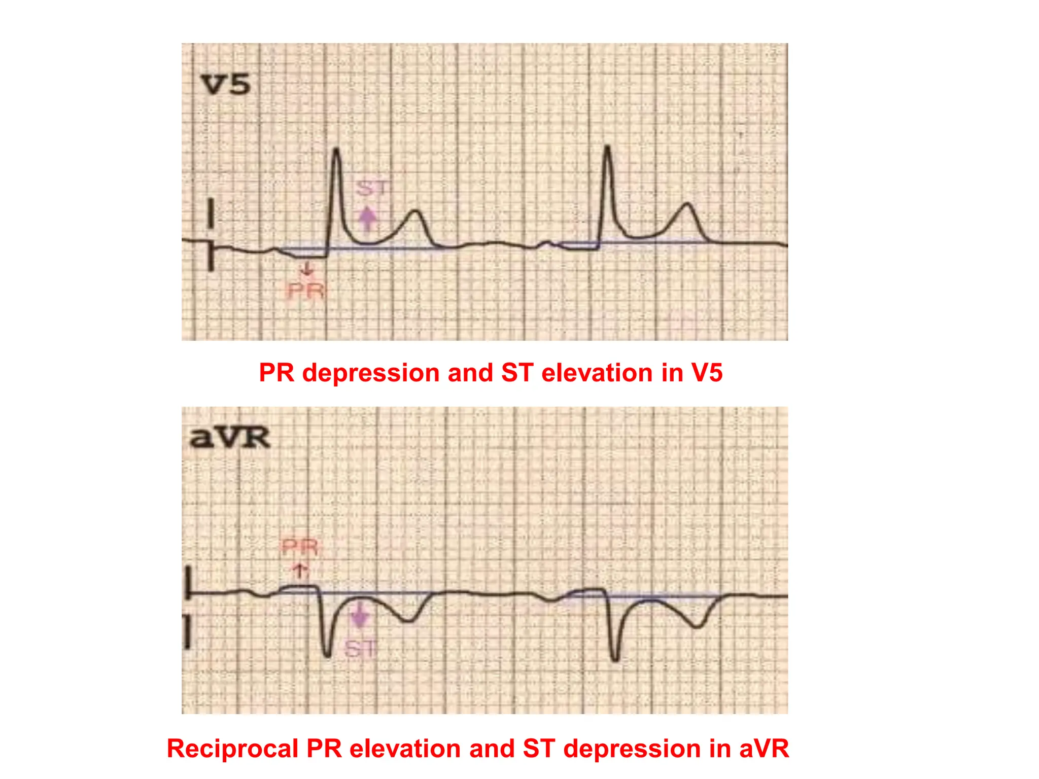
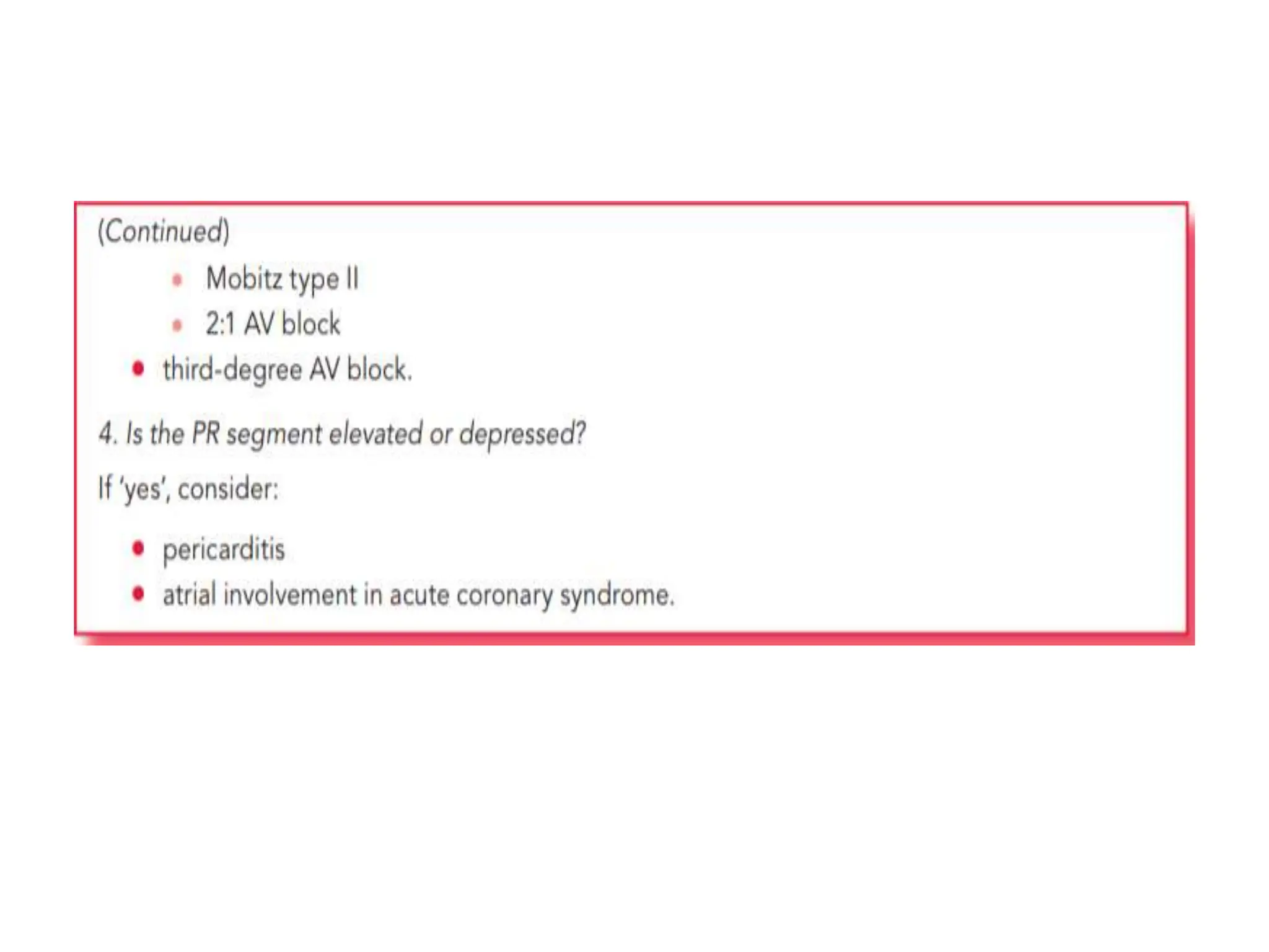
PR INTERVAL
Dr.G.VENKATA RAMANA
MBBS DNB FAMILY MEDICINE
PR INTERVAL OVERVIEW
• The time taken for the depolarization wave to pass from
its origin in the SA node, across the atria, and through
the AV node into ventricular muscle is called the PR
interval
• Normal PR interval 0.12-0.2s (3 to 5 small squares)
• PR interval > 0.2s, first degree heart block is said to be
present
• PR interval < 0.12s suggests pre-excitation (the
presence of an accessory pathway between the atria
and ventricles) or AV nodal (junctional) rhythm
PR INTERVAL
• Look for the following
• Is the PR interval less than 0.12 s long?
• Is the PR interval more than 0.2 s long?
• Does the PR interval vary or can it not be
measured?
• Is the PR segment elevated or depressed?
SHORT PR INTERVAL
• Indicates that the usual delay to conduction between the
atria and the ventricles, imposed by the AV junction, has
not occurred
• This happens if depolarization originates in the AV
junction, so that it travels up to the atria and down to the
ventricles simultaneously, or if it originates as normal in
the sinus node but bypasses the AV junction via an
additional faster conducting pathway
• Short PR interval causes
• AV nodal rhythm
• Wolff–Parkinson–White (WPW) syndrome
• Lown–Ganong–Levine (LGL) syndrome
• Accelerated AV nodal conduction
AV nodal rhythm
• If depolarization is initiated from within the AV node, the
wave of atrial depolarization will travel backwards
through the atria at the same time as setting off forwards
through the AV node towards the ventricles
• Thus, the time delay between atrial depolarization (the P
wave) and ventricular depolarization (the QRS complex)
will be reduced
• Any source of depolarization within the AV node can
therefore cause a short PR interval, including:
• AV nodal escape rhythms
• AV ectopics
• AV re-entry tachycardia
• AV nodal (junctional) rhythm
• Narrow complex, regular rhythms arising from the AV node
• P waves are either absent or abnormal (e.g. inverted) with a short
PR interval (=retrograde P waves).
• ECG: Accelerated junctional rhythm demonstrating inverted P waves
with a short PR interval (retrograde P waves)
Wolff Parkinson White syndrome
• In most people, conduction of electricity through the heart
follows just one distinct path from atria to ventricles,
namely via the AV node, bundle of His and Purkinje fibres
• Some people have an additional connection between the
atria and the ventricles– an accessory pathway – that
conducts more quickly than the AV node, so the wave of
depolarization reaches the ventricles more quickly than
usual and thus the PR interval is short
• The region of ventricle activated via the accessory pathway
slowly depolarizes, giving rise to a delta wave – the first
part of the QRS complex
• Shortly afterwards, the rest of the ventricular muscle is
depolarized rapidly with the arrival of the normally
conducted wave of depolarization via the AV node
• When a short PR interval and delta wave are seen on an
ECG, this is called a Wolff– Parkinson–White (WPW)
pattern
• In most cases, this is just an incidental finding and the
individual has no problems with their heart rhythm
• However, in some cases the presence of an accessory
pathway provides a substrate for episodes of AV re-entry
tachycardia (AVRT), in which case the patient is said to
have WPW syndrome
• Wolff-Parkinson-White syndrome
• Short PR interval (<0.12s), broad QRS and a slurred
upstroke to the QRS complex, the delta wave
Lown Ganong Levine syndrome
• The ‘classical’ view of LGL syndrome is that, like in
WPW syndrome, patients have an accessory pathway
(sometimes called the bundle of James) which bypasses
the AV node
• However, unlike the bundle of Kent in WPW syndrome,
the accessory pathway in LGL syndrome does not
activate the ventricular muscle directly
• Instead, it simply connects the atria to the bundle of His
• As a result, the AV node is bypassed (so the PR interval
is short) but there is no ventricular pre-excitation (and
therefore there is no delta wave)
• Short PR interval (in the absence of a delta wave) alone
is not sufficient to diagnose LGL syndrome
• LGL syndrome is diagnosed when patients with a short
PR interval (and no delta wave) present with re-entry
tachycardias
• Those who do not experience re-entry tachycardias are
instead considered to have a normal variant of AV
conduction known as accelerated AV nodal
conduction
• Lown-Ganong-Levine syndrome
• Very short PR interval with normal P waves and QRS complexes
and absent delta waves
Accelerated AV nodal conduction
• The presence of a short PR interval in isolation,
with no history of re-entry tachycardia, is
regarded as a normal variant of AV node
conduction (and should not be labelled as LGL
syndrome)
PROLONGED PR INTERVAL
• Prolongation of the PR interval is a common finding and
indicates that conduction through the AV node has been
delayed
• When this delay is constant for each cardiac cycle, and
each P wave is followed by a QRS complex, it is referred
to as first-degree AV block
• In first-degree AV block, conduction through the AV node is slower
than usual and the PR interval is therefore prolonged
• First-degree AV block is a common feature of vagally
induced bradycardia, as an increase in vagal tone
decreases AV nodal conduction
• It may also be a feature of:
• Ischaemic heart disease
• Hyperkalemia or hypokalemia
• Acute rheumatic myocarditis
• Lyme disease
• Drugs
• Beta blockers
• Rate-modifying calcium channel blockers
• Digoxin
DOES THE PR INTERVAL VARY OR CAN IT NOT BE
MEASURED?
• Normally, the PR interval is constant
• In some conditions, however, the interval between P waves
and QRS complexes changes, giving rise to a variable PR
interval
• Sometimes a P wave is not followed by a QRS complex at all
and so the PR interval cannot be measured
• If the PR interval gradually lengthens with each beat, until one
P wave fails to produce a QRS complex, the patient has
Mobitz type I AV block
• If the PR interval is fixed and normal, but occasionally a P
wave fails to produce a QRS complex, the patient has Mobitz
type II AV block
• If alternate P waves are not followed by QRS complexes, the
patient has 2:1 AV block
• If there is no relationship between P waves and QRS
complexes, the patient has third-degree (complete) AV block
• When Mobitz type I AV block occurs at the level of the AV
node it is generally regarded as ‘benign’,and a
permanent pacemaker is not required unless the
frequency of ‘dropped’ ventricular beats causes a
symptomatic bradycardia
• When the block is infranodal (as identified by
electrophysiological testing) there is a stronger indication
for pacing,even if patients are asymptomatic
• Mobitz type I AV block may require pacing prior to
surgery
• Mobitz type II AV block
• Characteristic features are:
• Most P waves are followed by a QRS complex
• The PR interval is normal and constant
• Occasionally, a P wave is not followed by a QRS
complex.
• Mobitz type II AV block is thought to result from abnormal
conduction below the AV node (infranodal) and is
considered more serious than Mobitz type I as it can
progress without warning to third-degree (complete)
heart block
• Referral to a cardiologist is therefore recommended, as
a pacemaker may be required
2:1 AV block
Special form of second-degree heart block in which alternate P
waves are not followed by QRS complexes
Usually requires pacing
• Third-degree AV block
• In third-degree AV block (‘complete heart block’), there is
complete interruption of conduction between atria and
ventricles, so that the two are working independently
• QRS complexes usually arise as the result of a
ventricular escape rhythm in which the QRS complexes
are usually broad
• However, if the level of AV block is located in or just
below the AV node, a junctional escape rhythm may
arise with narrow QRS complexes
• Third-degree AV block usually requires pacing
• Characteristic features of complete heart block are:
• P wave rate is faster than ventricular QRS complexes
• P waves bear no relationship to the ventricular QRS
complexes
• If block occurs in the AV node, QRS complexes are
usually narrow due to a subsidiary pacemaker arising in
the bundle of His
• If block occurs below the AV node, QRS complexes are
usually broad due to a subsidiary pacemaker arising in
the left or right bundle branches
Causes of third-degree AV block
• Congenital
• Acquired
• Drug toxicity (e.g. anti-arrhythmics)
• Fibrosis/calcification of the conduction system
• Myocardial ischaemia/infarction
• Infection (e.g. Lyme disease)
• Myocardial infiltration (e.g. amyloid, sarcoid)
• Metabolic disorders (e.g. hypothyroidism)
• Neuromuscular diseases (e.g. myotonic muscular
dystrophy)
• Cardiac procedures (e.g. ablation procedures, aortic
valve surgery)
ATRIOVENTRICULAR DISSOCIATION
• Atrioventricular dissociation occurs when the ventricular
(QRS) rate is higher than the atrial (P wave) rate
• The opposite is found in third degree AV block
• Atrioventricular dissociation usually occurs in the context
of an escape rhythm(from the AV junction or ventricles)
during sinus bradycardia, or an acceleration in a
subsidiary focus in the AV junction or ventricles which
then overtakes the sinoatrial node, which continues firing
independently
IS THE PR SEGMENT ELEVATED OR DEPRESSED?
• The PR segment, between the end of the P wave and
the start of the QRS complex, is usually flat and
isoelectric
• PR segment depression can occur in pericarditis
• PR segment depression is a specific ECG feature of
pericarditis and may be seen in any leads except aVR
and V1 (where there may be PR segment elevation)
• PR segment depression (or, rarely, PR segment
elevation) can also be seen if there is atrial involvement
with an acute coronary syndrome
• In the setting of acute inferior myocardial infarction, the
presence of ≥1.2 mm PR segment depression is
associated with worse outcomes
PR depression and ST elevation in V5
Reciprocal PR elevation and ST depression in aVR
PR INTERVAL,CAUSES OF SHORT AND LONG PR INTERVAL

PR INTERVAL,CAUSES OF SHORT AND LONG PR INTERVAL

  • 1.
  • 2.
    PR INTERVAL OVERVIEW •The time taken for the depolarization wave to pass from its origin in the SA node, across the atria, and through the AV node into ventricular muscle is called the PR interval • Normal PR interval 0.12-0.2s (3 to 5 small squares) • PR interval > 0.2s, first degree heart block is said to be present • PR interval < 0.12s suggests pre-excitation (the presence of an accessory pathway between the atria and ventricles) or AV nodal (junctional) rhythm
  • 3.
    PR INTERVAL • Lookfor the following • Is the PR interval less than 0.12 s long? • Is the PR interval more than 0.2 s long? • Does the PR interval vary or can it not be measured? • Is the PR segment elevated or depressed?
  • 4.
    SHORT PR INTERVAL •Indicates that the usual delay to conduction between the atria and the ventricles, imposed by the AV junction, has not occurred • This happens if depolarization originates in the AV junction, so that it travels up to the atria and down to the ventricles simultaneously, or if it originates as normal in the sinus node but bypasses the AV junction via an additional faster conducting pathway • Short PR interval causes • AV nodal rhythm • Wolff–Parkinson–White (WPW) syndrome • Lown–Ganong–Levine (LGL) syndrome • Accelerated AV nodal conduction
  • 5.
    AV nodal rhythm •If depolarization is initiated from within the AV node, the wave of atrial depolarization will travel backwards through the atria at the same time as setting off forwards through the AV node towards the ventricles • Thus, the time delay between atrial depolarization (the P wave) and ventricular depolarization (the QRS complex) will be reduced • Any source of depolarization within the AV node can therefore cause a short PR interval, including: • AV nodal escape rhythms • AV ectopics • AV re-entry tachycardia
  • 7.
    • AV nodal(junctional) rhythm • Narrow complex, regular rhythms arising from the AV node • P waves are either absent or abnormal (e.g. inverted) with a short PR interval (=retrograde P waves). • ECG: Accelerated junctional rhythm demonstrating inverted P waves with a short PR interval (retrograde P waves)
  • 8.
    Wolff Parkinson Whitesyndrome • In most people, conduction of electricity through the heart follows just one distinct path from atria to ventricles, namely via the AV node, bundle of His and Purkinje fibres • Some people have an additional connection between the atria and the ventricles– an accessory pathway – that conducts more quickly than the AV node, so the wave of depolarization reaches the ventricles more quickly than usual and thus the PR interval is short • The region of ventricle activated via the accessory pathway slowly depolarizes, giving rise to a delta wave – the first part of the QRS complex • Shortly afterwards, the rest of the ventricular muscle is depolarized rapidly with the arrival of the normally conducted wave of depolarization via the AV node
  • 9.
    • When ashort PR interval and delta wave are seen on an ECG, this is called a Wolff– Parkinson–White (WPW) pattern • In most cases, this is just an incidental finding and the individual has no problems with their heart rhythm • However, in some cases the presence of an accessory pathway provides a substrate for episodes of AV re-entry tachycardia (AVRT), in which case the patient is said to have WPW syndrome
  • 11.
    • Wolff-Parkinson-White syndrome •Short PR interval (<0.12s), broad QRS and a slurred upstroke to the QRS complex, the delta wave
  • 12.
    Lown Ganong Levinesyndrome • The ‘classical’ view of LGL syndrome is that, like in WPW syndrome, patients have an accessory pathway (sometimes called the bundle of James) which bypasses the AV node • However, unlike the bundle of Kent in WPW syndrome, the accessory pathway in LGL syndrome does not activate the ventricular muscle directly • Instead, it simply connects the atria to the bundle of His • As a result, the AV node is bypassed (so the PR interval is short) but there is no ventricular pre-excitation (and therefore there is no delta wave)
  • 13.
    • Short PRinterval (in the absence of a delta wave) alone is not sufficient to diagnose LGL syndrome • LGL syndrome is diagnosed when patients with a short PR interval (and no delta wave) present with re-entry tachycardias • Those who do not experience re-entry tachycardias are instead considered to have a normal variant of AV conduction known as accelerated AV nodal conduction
  • 14.
    • Lown-Ganong-Levine syndrome •Very short PR interval with normal P waves and QRS complexes and absent delta waves
  • 15.
    Accelerated AV nodalconduction • The presence of a short PR interval in isolation, with no history of re-entry tachycardia, is regarded as a normal variant of AV node conduction (and should not be labelled as LGL syndrome)
  • 17.
    PROLONGED PR INTERVAL •Prolongation of the PR interval is a common finding and indicates that conduction through the AV node has been delayed • When this delay is constant for each cardiac cycle, and each P wave is followed by a QRS complex, it is referred to as first-degree AV block
  • 18.
    • In first-degreeAV block, conduction through the AV node is slower than usual and the PR interval is therefore prolonged
  • 20.
    • First-degree AVblock is a common feature of vagally induced bradycardia, as an increase in vagal tone decreases AV nodal conduction • It may also be a feature of: • Ischaemic heart disease • Hyperkalemia or hypokalemia • Acute rheumatic myocarditis • Lyme disease • Drugs • Beta blockers • Rate-modifying calcium channel blockers • Digoxin
  • 21.
    DOES THE PRINTERVAL VARY OR CAN IT NOT BE MEASURED? • Normally, the PR interval is constant • In some conditions, however, the interval between P waves and QRS complexes changes, giving rise to a variable PR interval • Sometimes a P wave is not followed by a QRS complex at all and so the PR interval cannot be measured • If the PR interval gradually lengthens with each beat, until one P wave fails to produce a QRS complex, the patient has Mobitz type I AV block • If the PR interval is fixed and normal, but occasionally a P wave fails to produce a QRS complex, the patient has Mobitz type II AV block • If alternate P waves are not followed by QRS complexes, the patient has 2:1 AV block • If there is no relationship between P waves and QRS complexes, the patient has third-degree (complete) AV block
  • 23.
    • When Mobitztype I AV block occurs at the level of the AV node it is generally regarded as ‘benign’,and a permanent pacemaker is not required unless the frequency of ‘dropped’ ventricular beats causes a symptomatic bradycardia • When the block is infranodal (as identified by electrophysiological testing) there is a stronger indication for pacing,even if patients are asymptomatic • Mobitz type I AV block may require pacing prior to surgery
  • 26.
    • Mobitz typeII AV block • Characteristic features are: • Most P waves are followed by a QRS complex • The PR interval is normal and constant • Occasionally, a P wave is not followed by a QRS complex. • Mobitz type II AV block is thought to result from abnormal conduction below the AV node (infranodal) and is considered more serious than Mobitz type I as it can progress without warning to third-degree (complete) heart block • Referral to a cardiologist is therefore recommended, as a pacemaker may be required
  • 29.
    2:1 AV block Specialform of second-degree heart block in which alternate P waves are not followed by QRS complexes Usually requires pacing
  • 30.
    • Third-degree AVblock • In third-degree AV block (‘complete heart block’), there is complete interruption of conduction between atria and ventricles, so that the two are working independently • QRS complexes usually arise as the result of a ventricular escape rhythm in which the QRS complexes are usually broad • However, if the level of AV block is located in or just below the AV node, a junctional escape rhythm may arise with narrow QRS complexes • Third-degree AV block usually requires pacing
  • 31.
    • Characteristic featuresof complete heart block are: • P wave rate is faster than ventricular QRS complexes • P waves bear no relationship to the ventricular QRS complexes • If block occurs in the AV node, QRS complexes are usually narrow due to a subsidiary pacemaker arising in the bundle of His • If block occurs below the AV node, QRS complexes are usually broad due to a subsidiary pacemaker arising in the left or right bundle branches
  • 35.
    Causes of third-degreeAV block • Congenital • Acquired • Drug toxicity (e.g. anti-arrhythmics) • Fibrosis/calcification of the conduction system • Myocardial ischaemia/infarction • Infection (e.g. Lyme disease) • Myocardial infiltration (e.g. amyloid, sarcoid) • Metabolic disorders (e.g. hypothyroidism) • Neuromuscular diseases (e.g. myotonic muscular dystrophy) • Cardiac procedures (e.g. ablation procedures, aortic valve surgery)
  • 36.
    ATRIOVENTRICULAR DISSOCIATION • Atrioventriculardissociation occurs when the ventricular (QRS) rate is higher than the atrial (P wave) rate • The opposite is found in third degree AV block • Atrioventricular dissociation usually occurs in the context of an escape rhythm(from the AV junction or ventricles) during sinus bradycardia, or an acceleration in a subsidiary focus in the AV junction or ventricles which then overtakes the sinoatrial node, which continues firing independently
  • 38.
    IS THE PRSEGMENT ELEVATED OR DEPRESSED? • The PR segment, between the end of the P wave and the start of the QRS complex, is usually flat and isoelectric • PR segment depression can occur in pericarditis • PR segment depression is a specific ECG feature of pericarditis and may be seen in any leads except aVR and V1 (where there may be PR segment elevation) • PR segment depression (or, rarely, PR segment elevation) can also be seen if there is atrial involvement with an acute coronary syndrome • In the setting of acute inferior myocardial infarction, the presence of ≥1.2 mm PR segment depression is associated with worse outcomes
  • 39.
    PR depression andST elevation in V5 Reciprocal PR elevation and ST depression in aVR