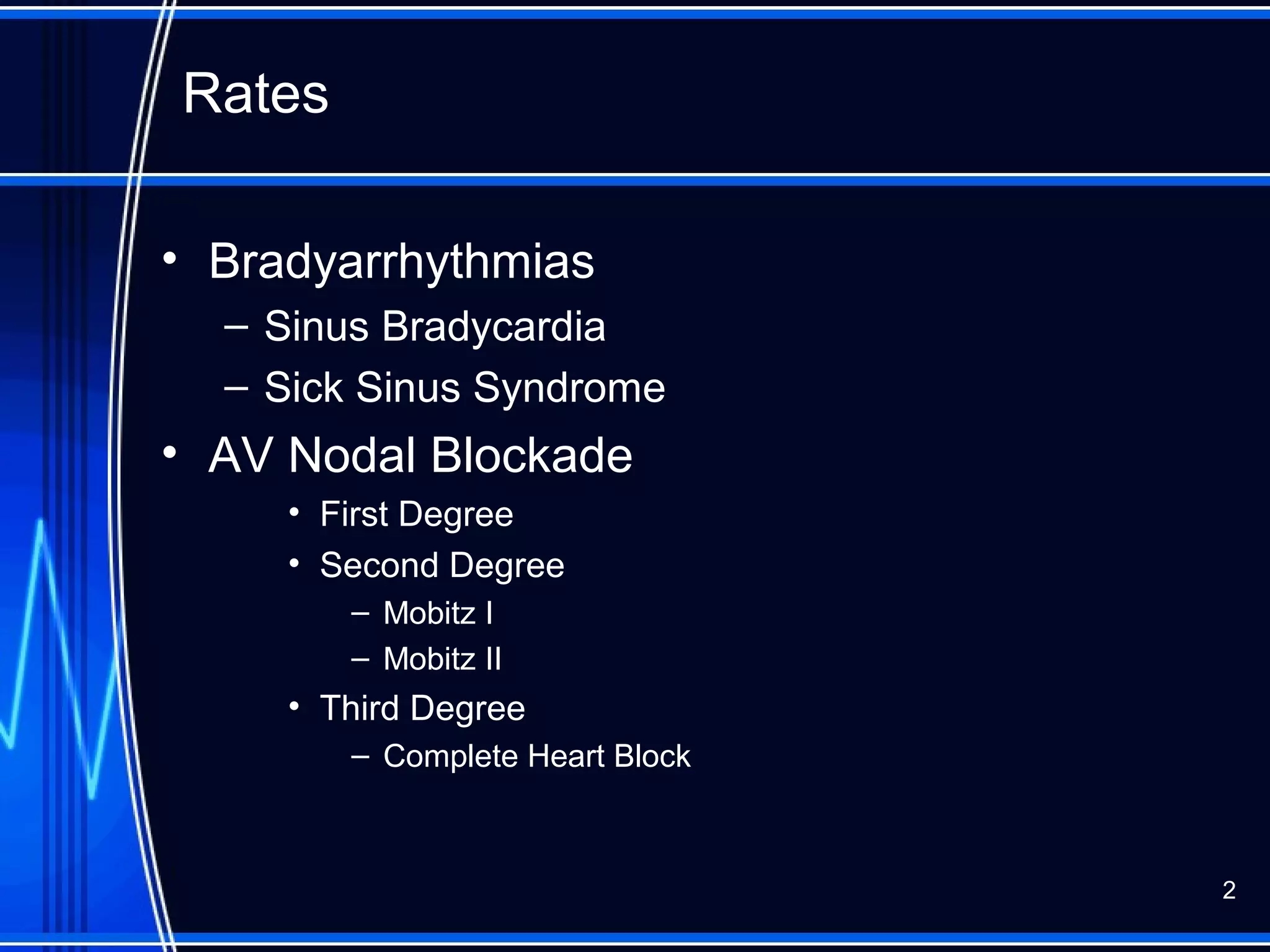
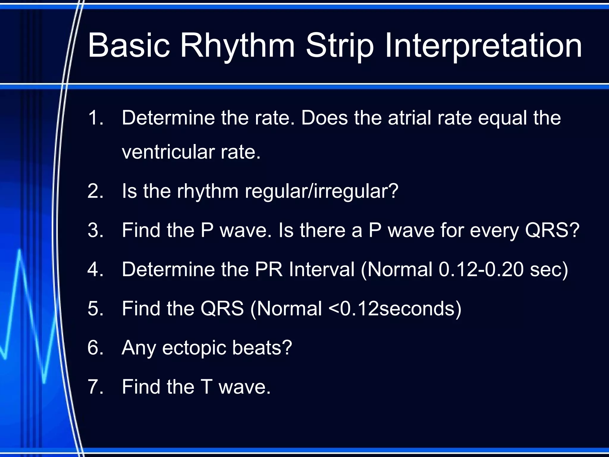
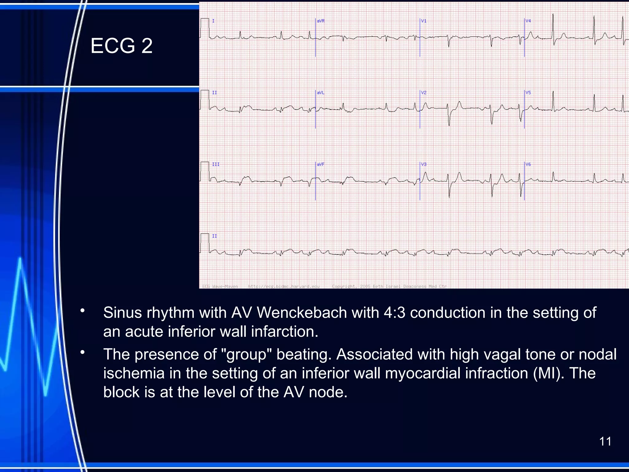
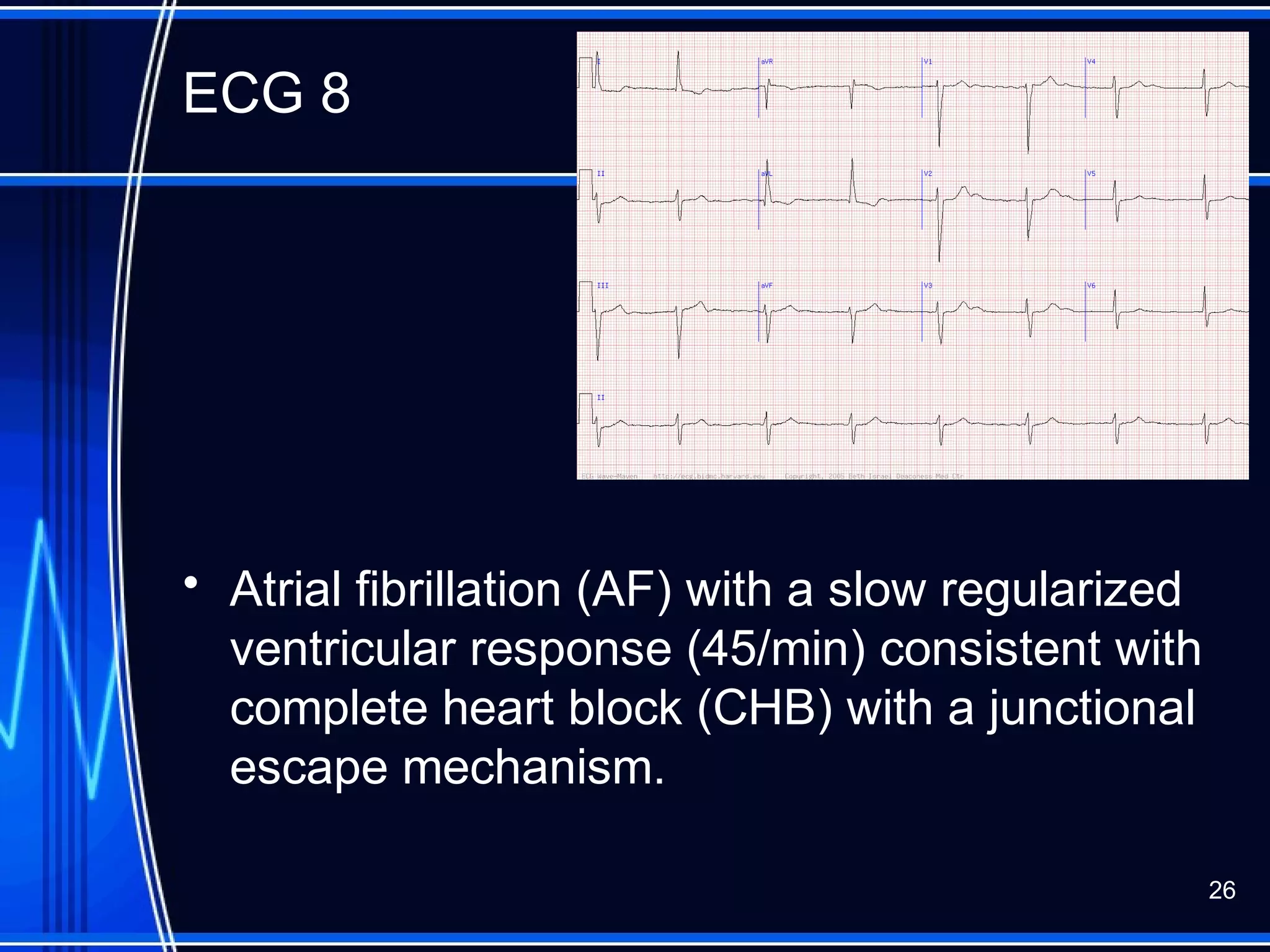
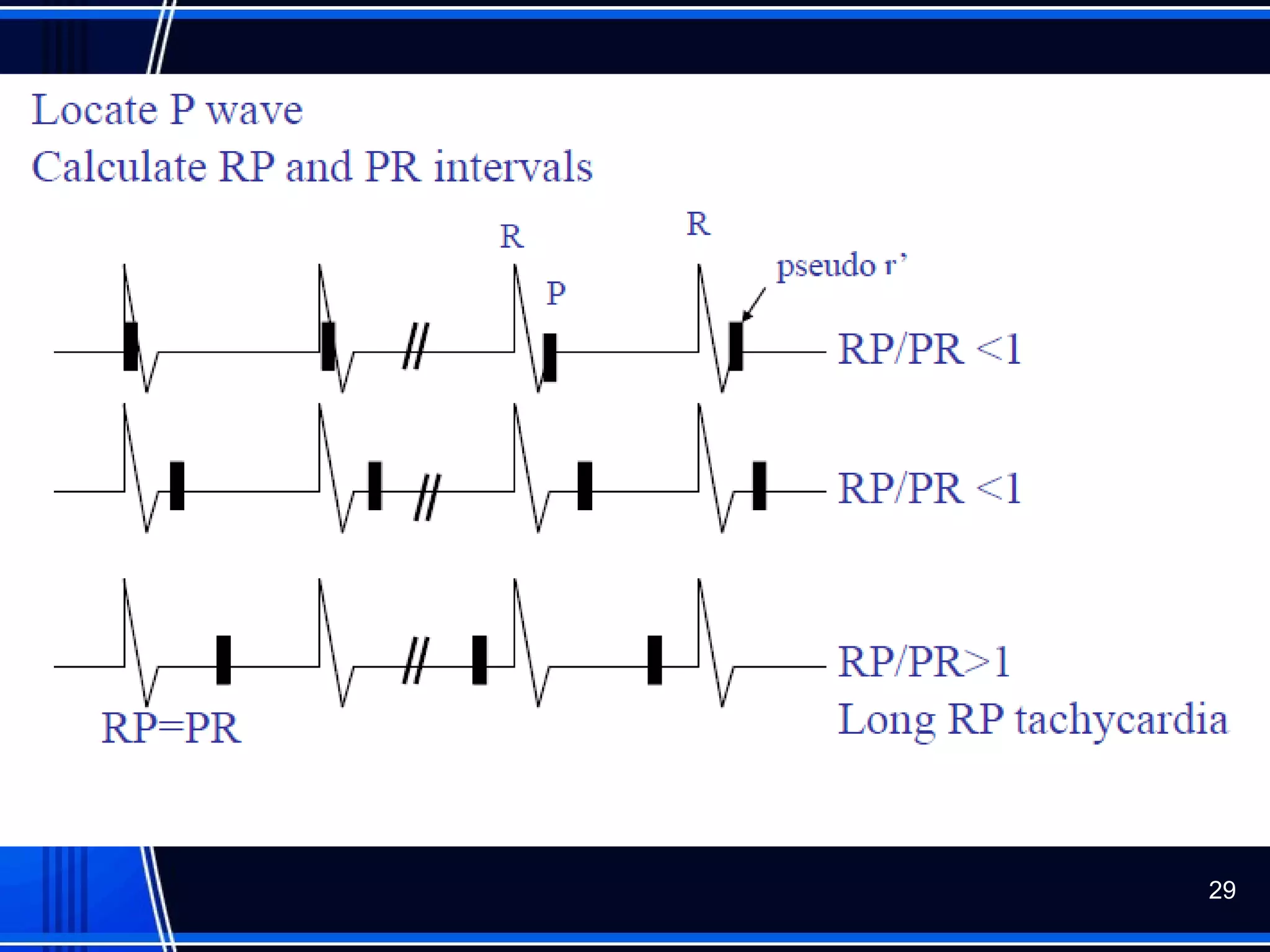
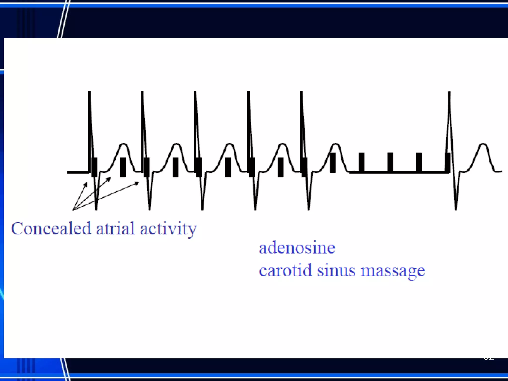
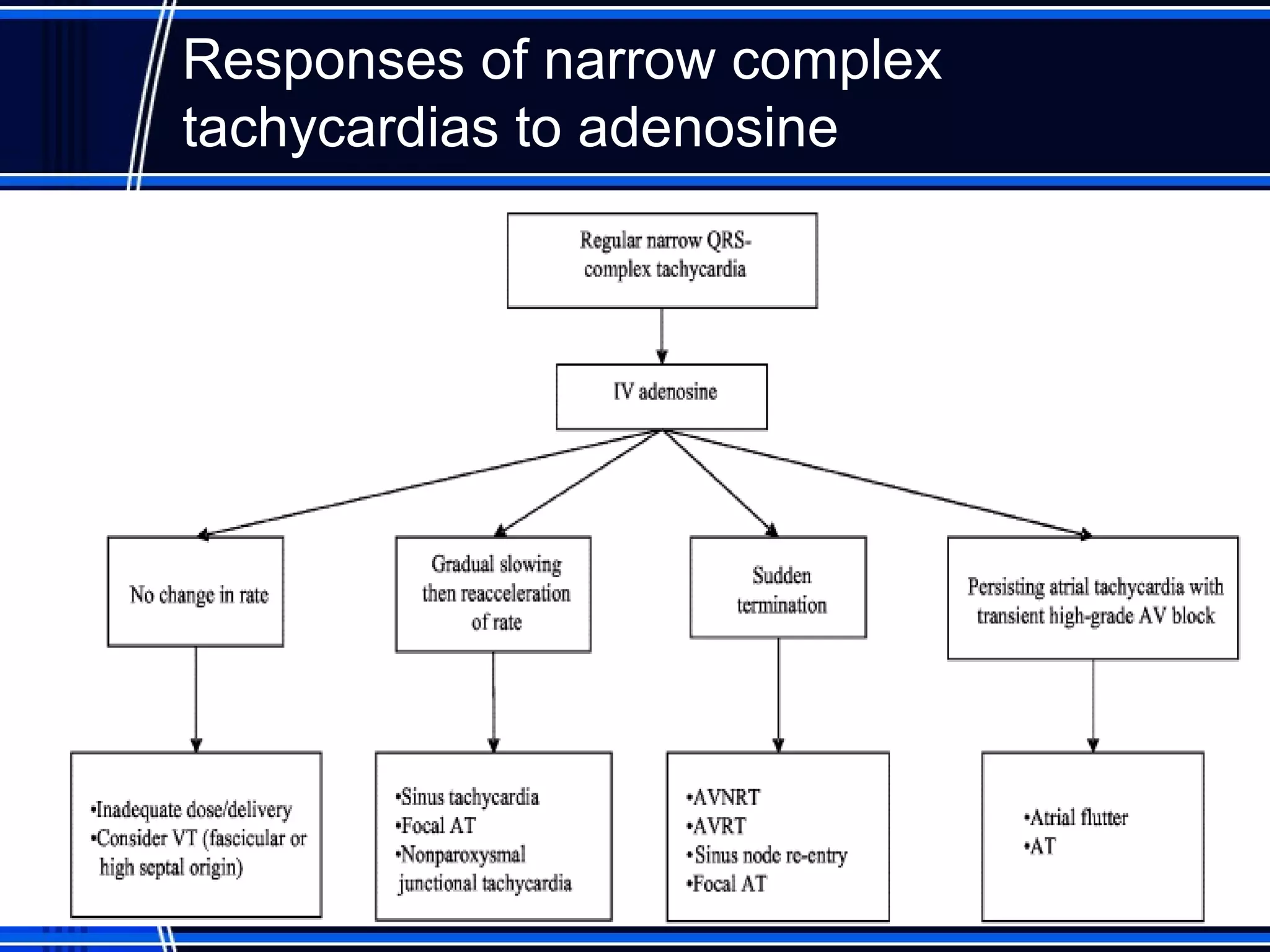
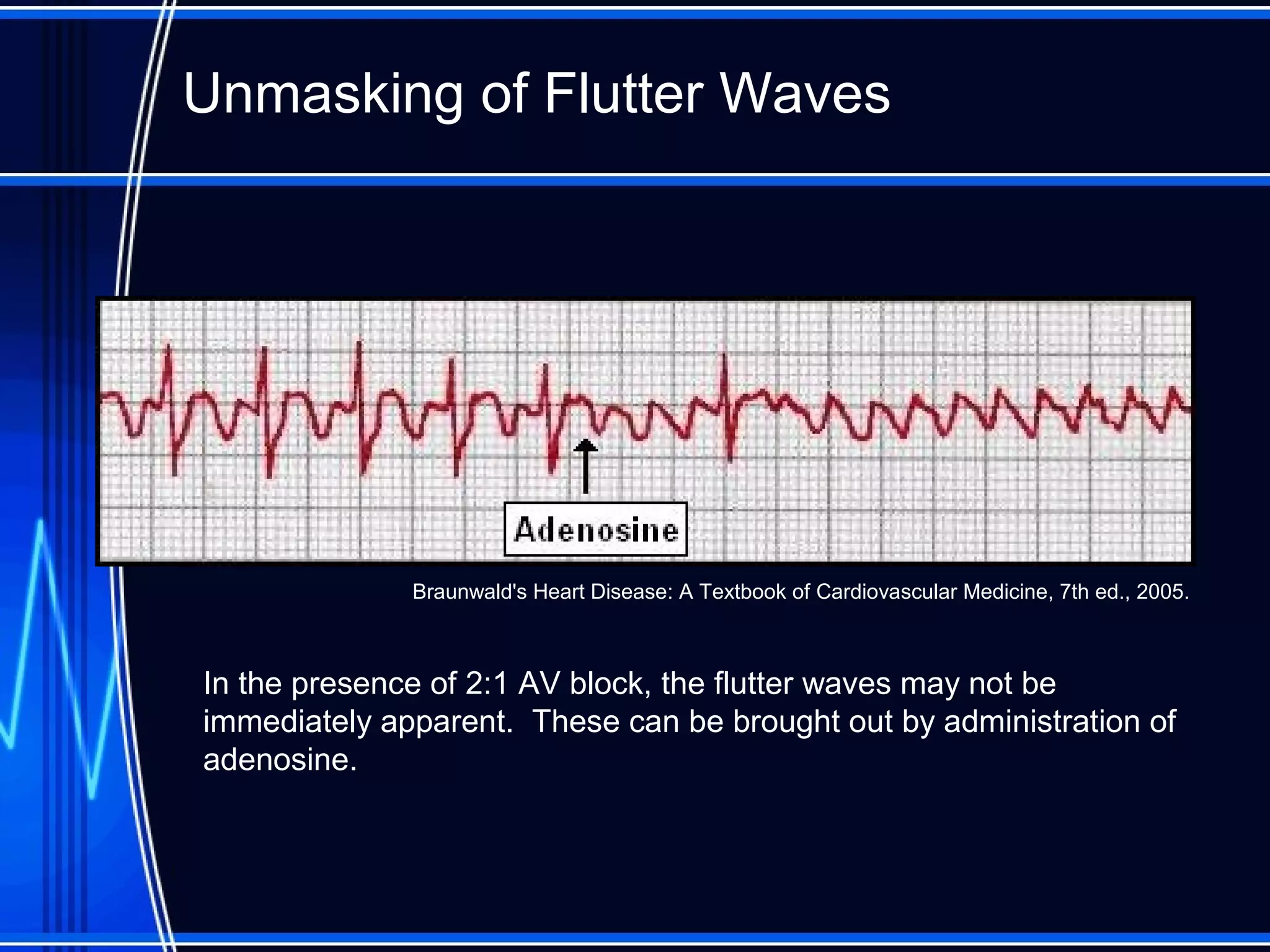
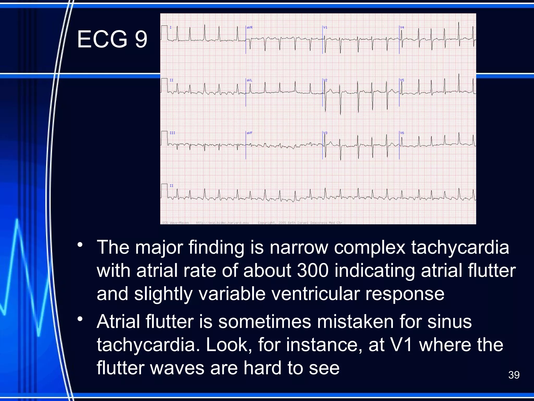
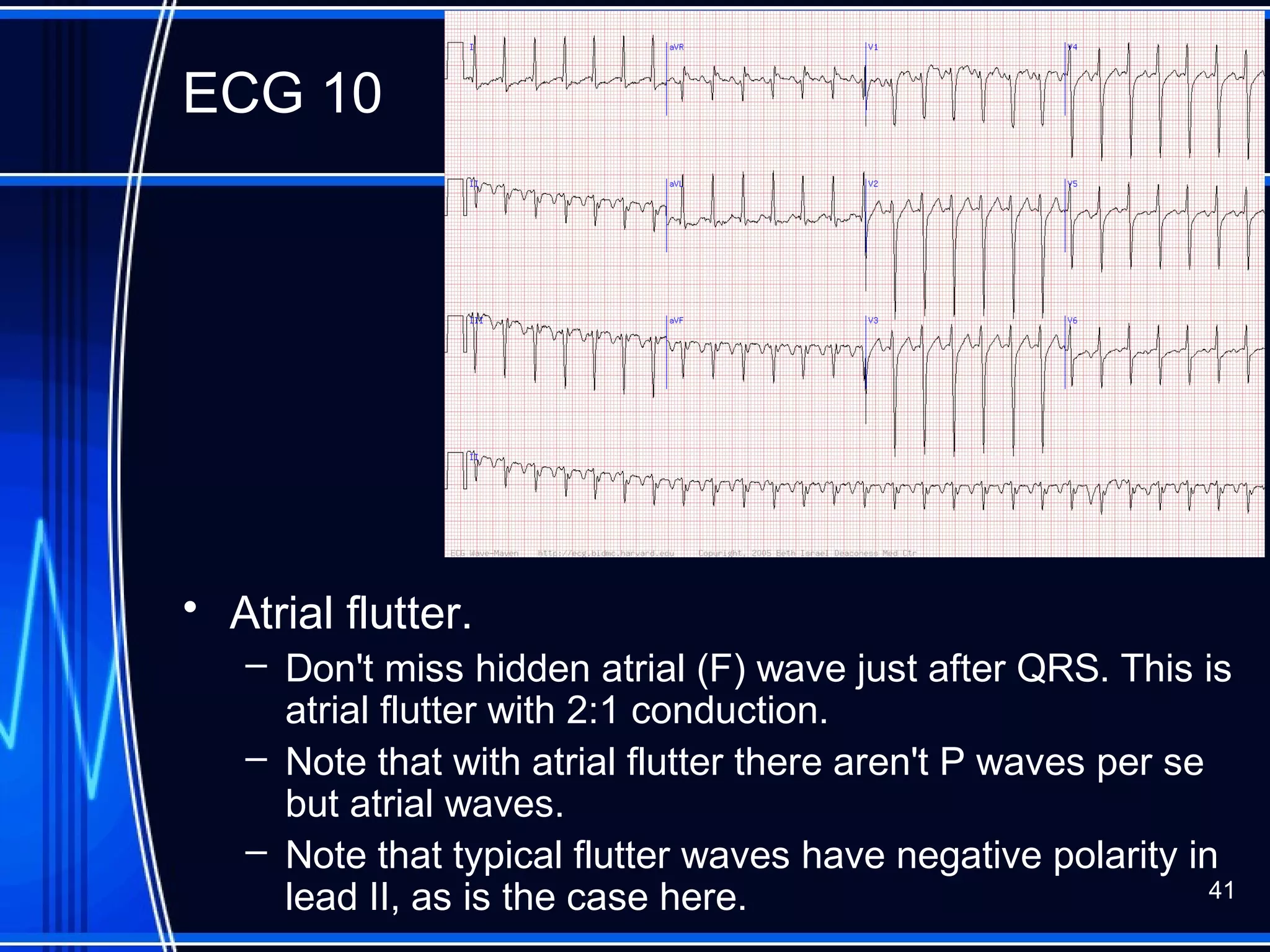
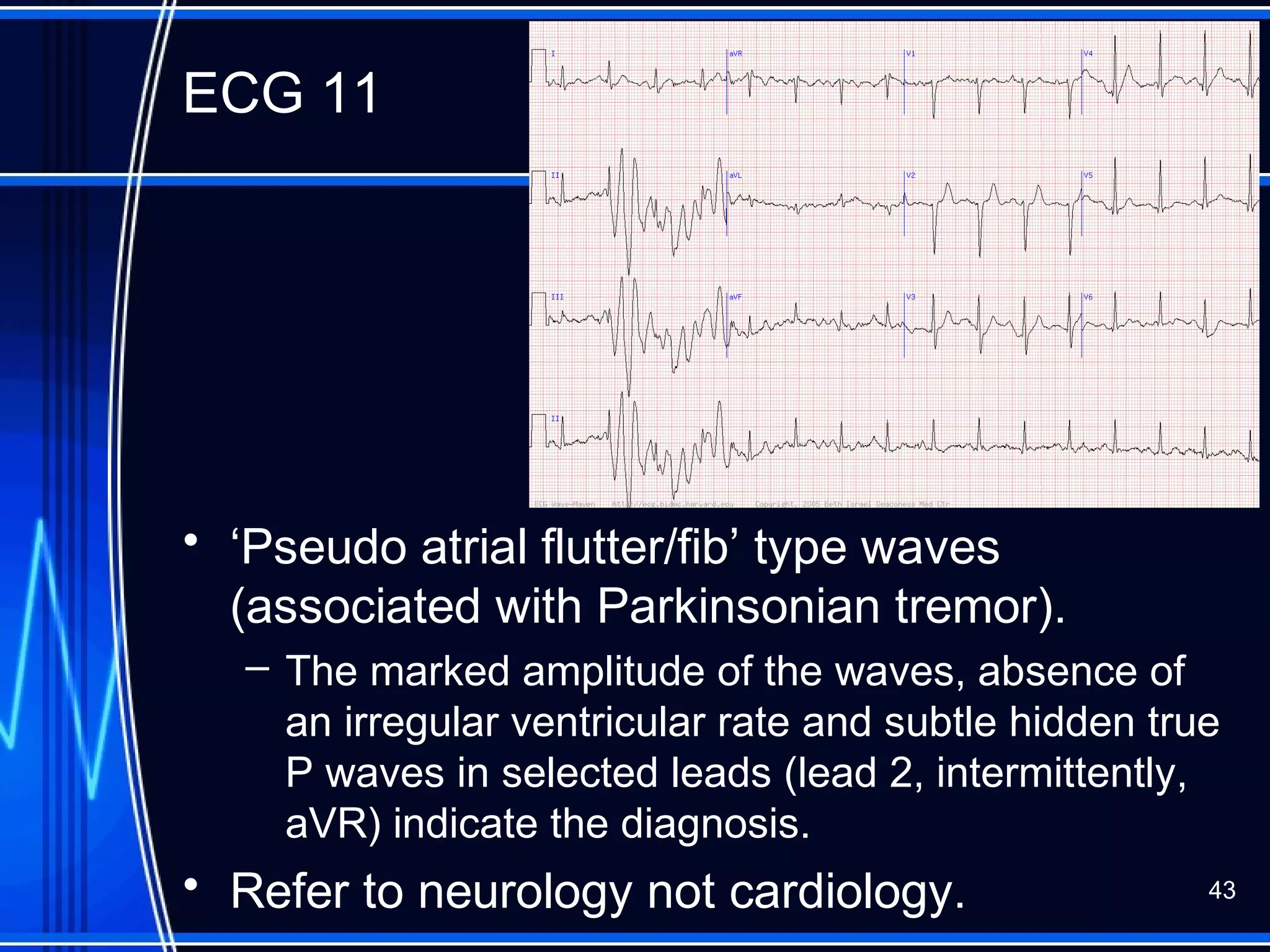
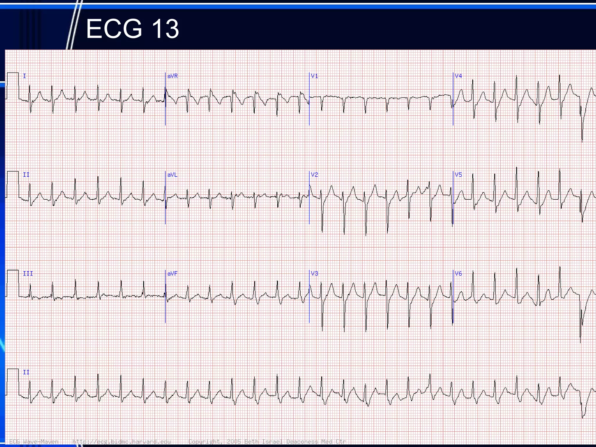
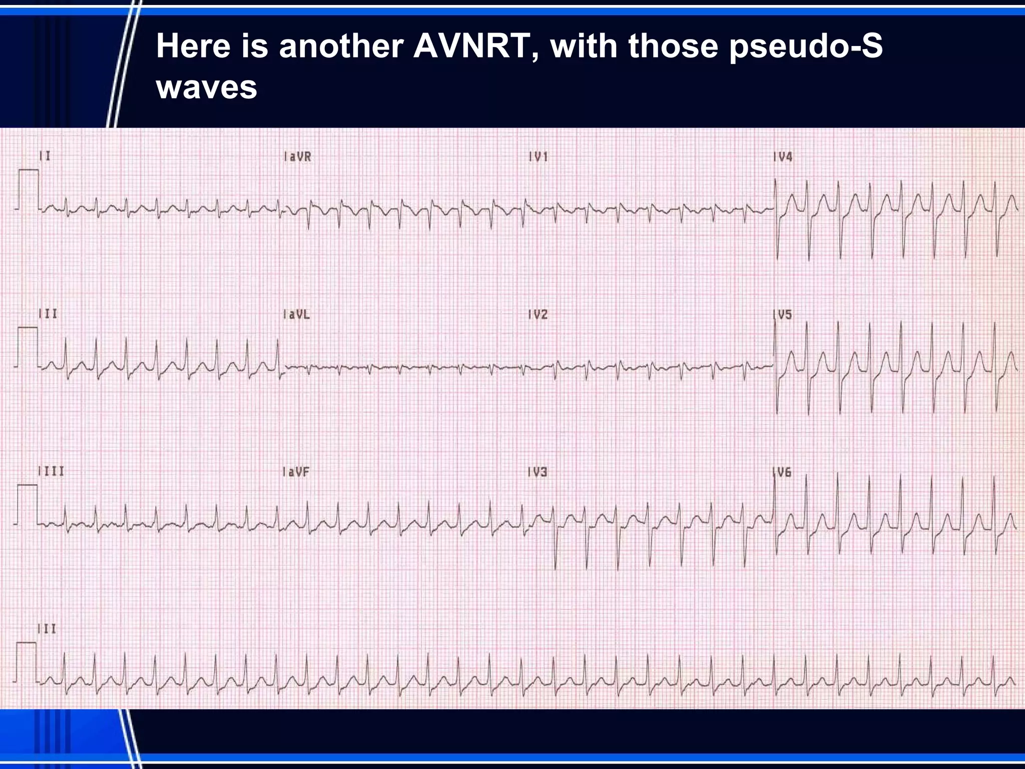
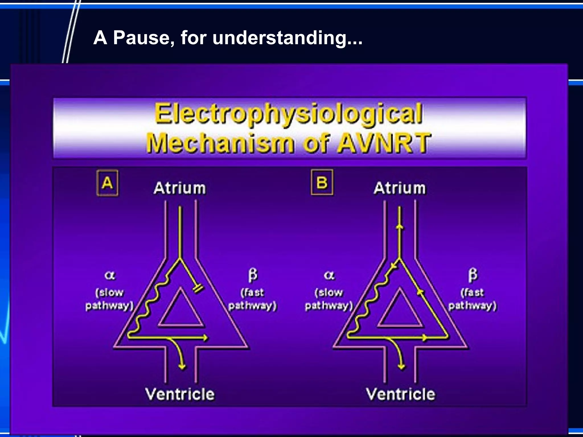
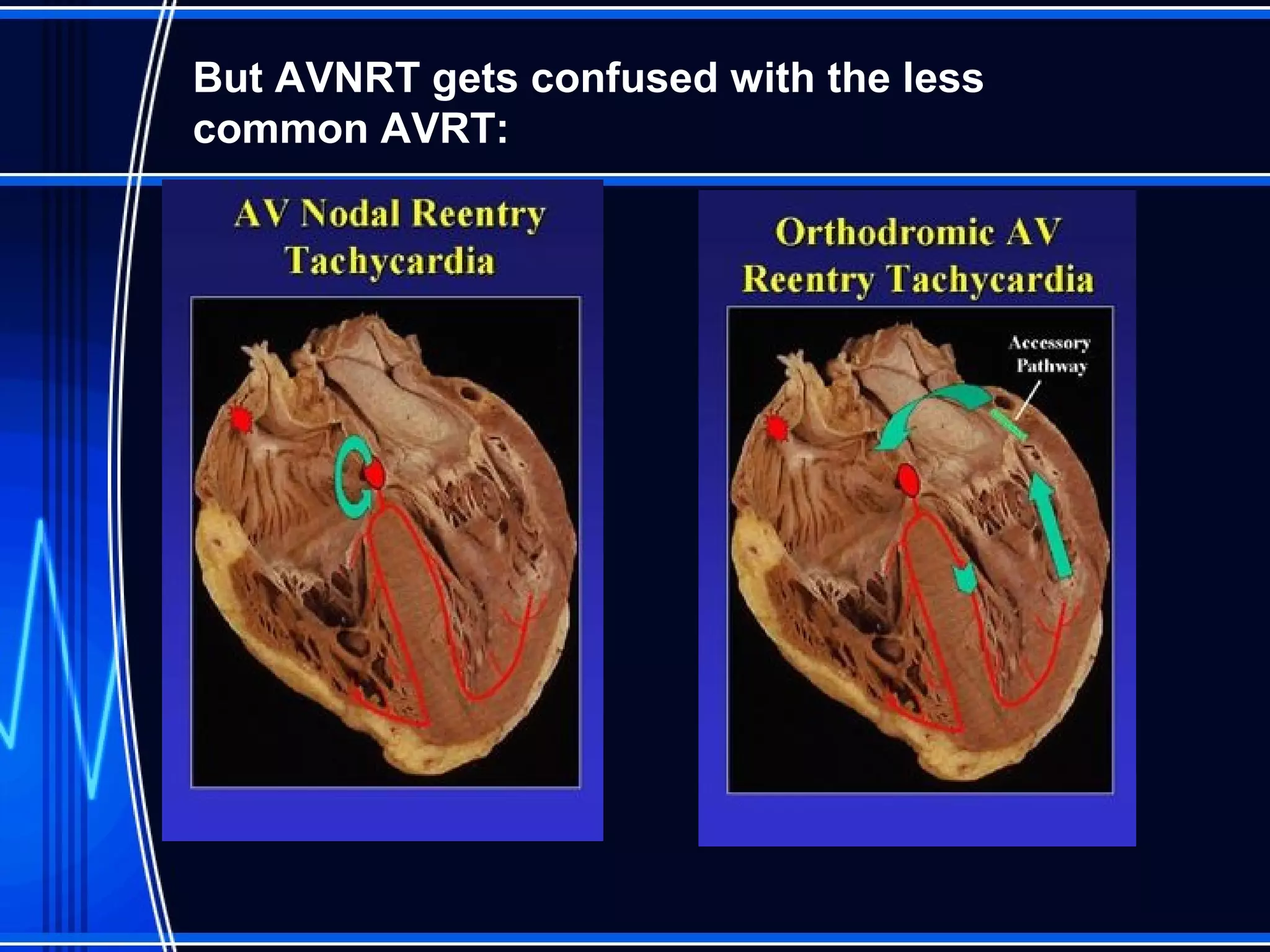
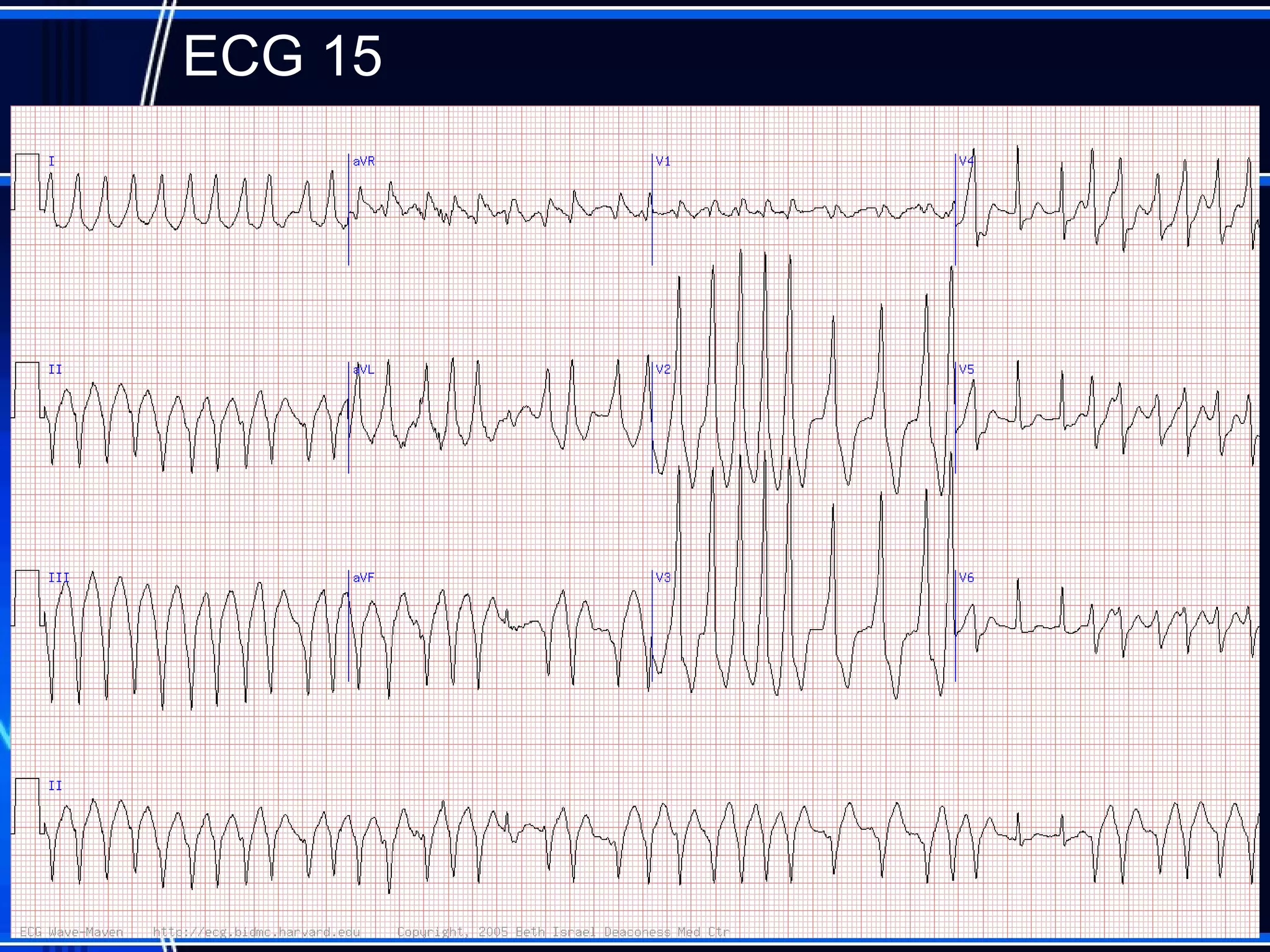
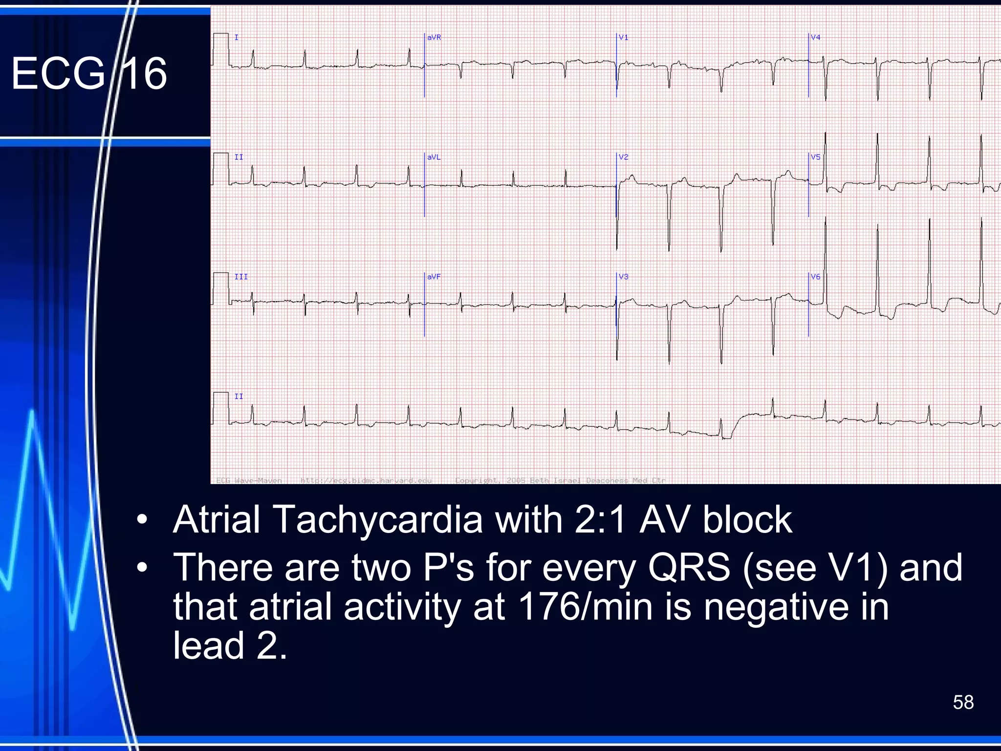
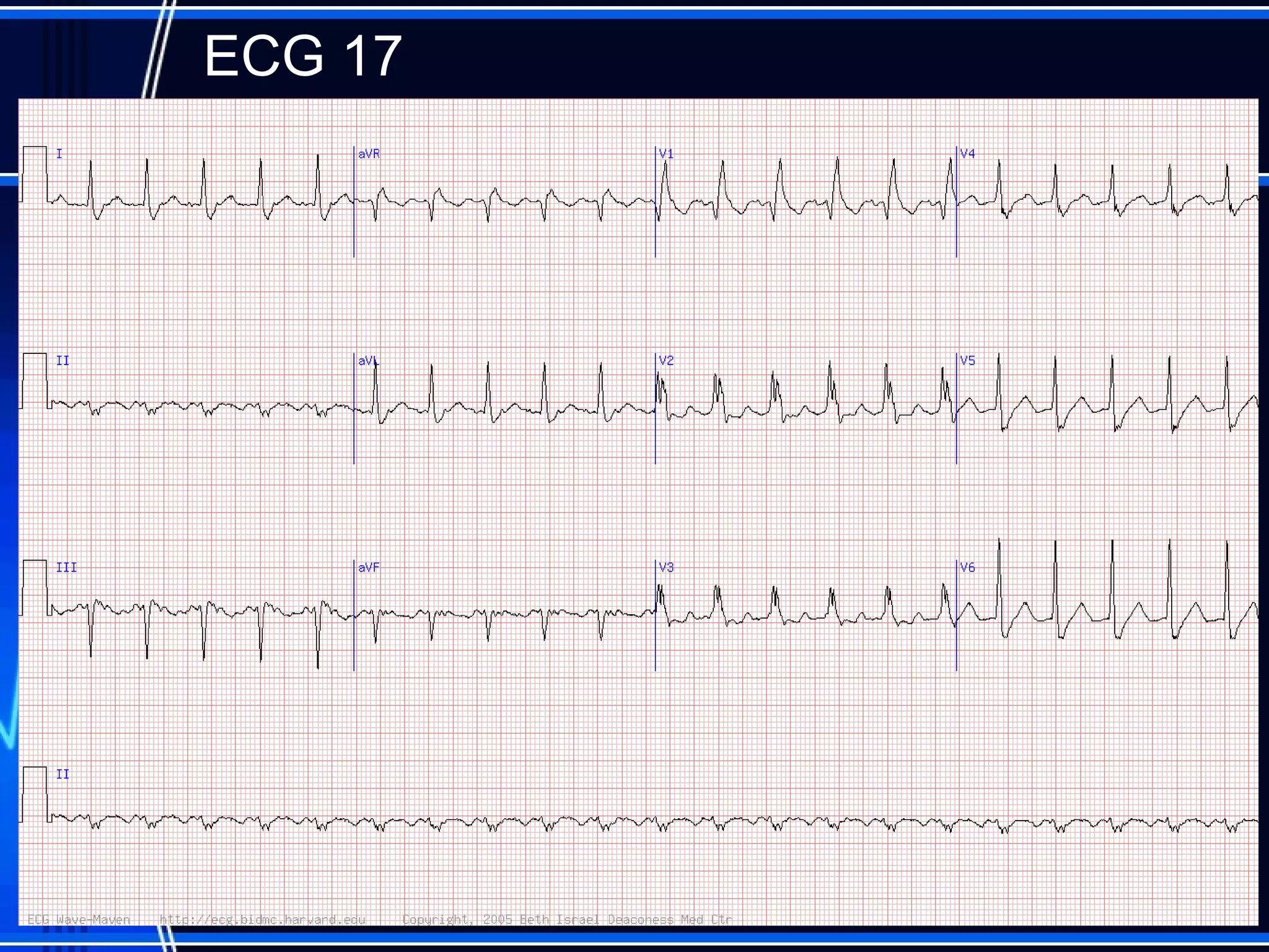
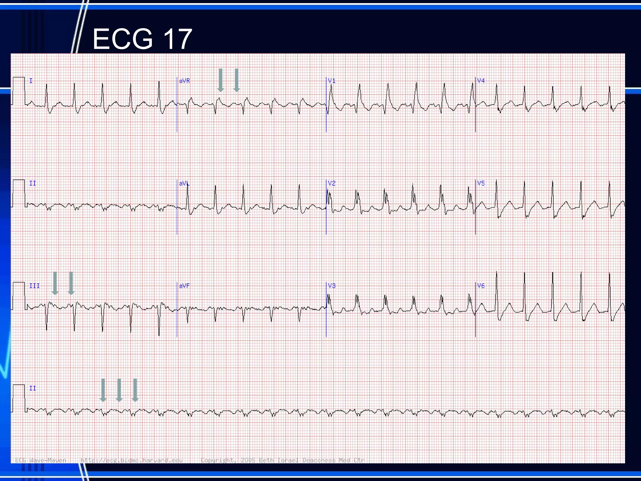
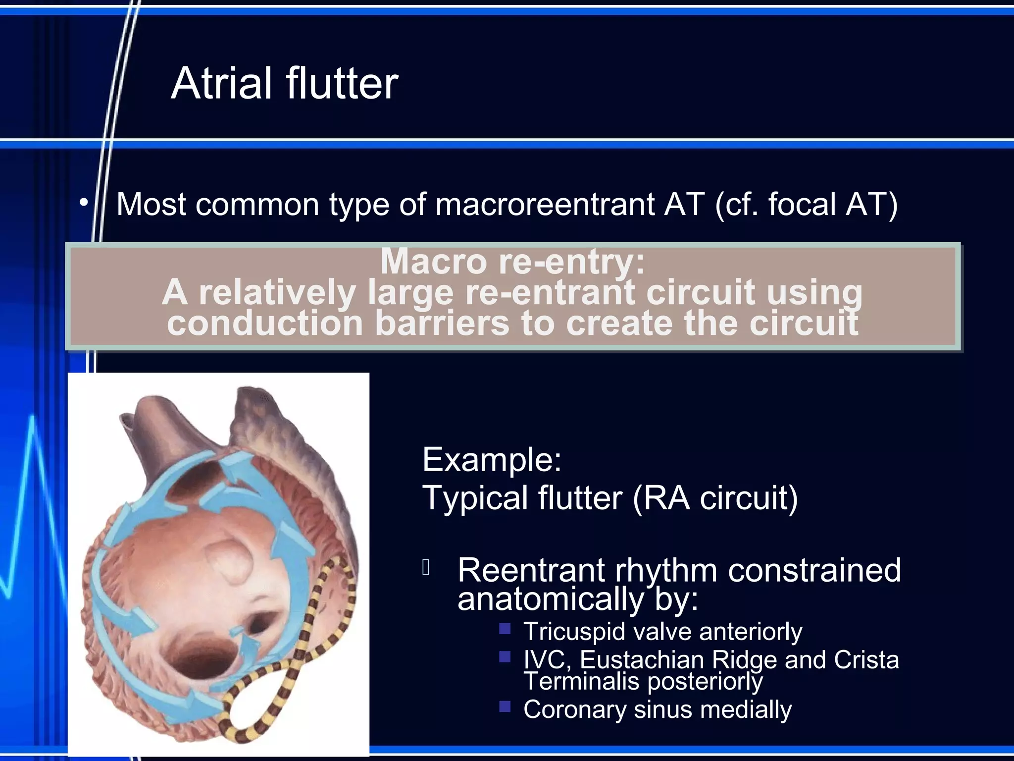
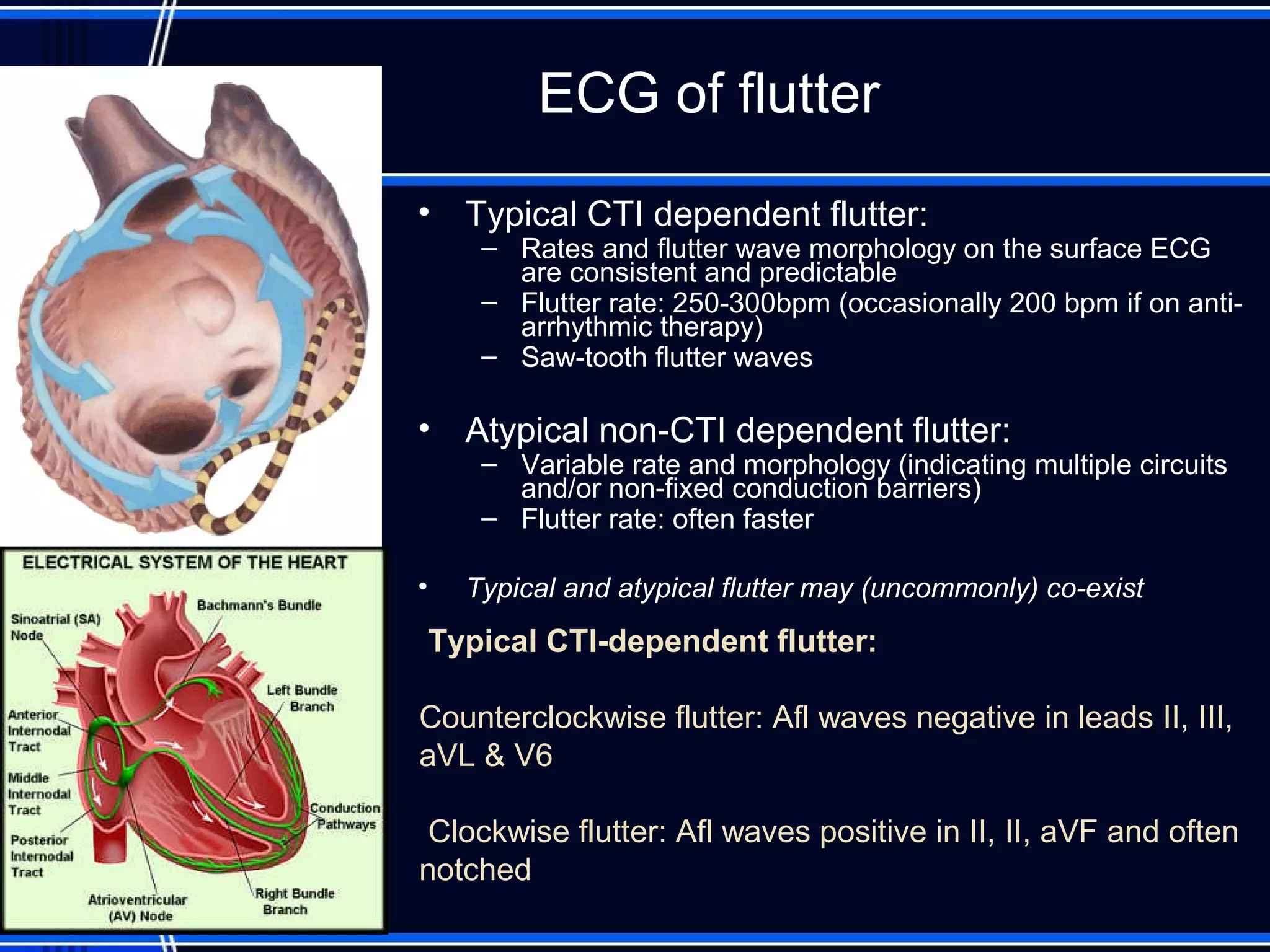
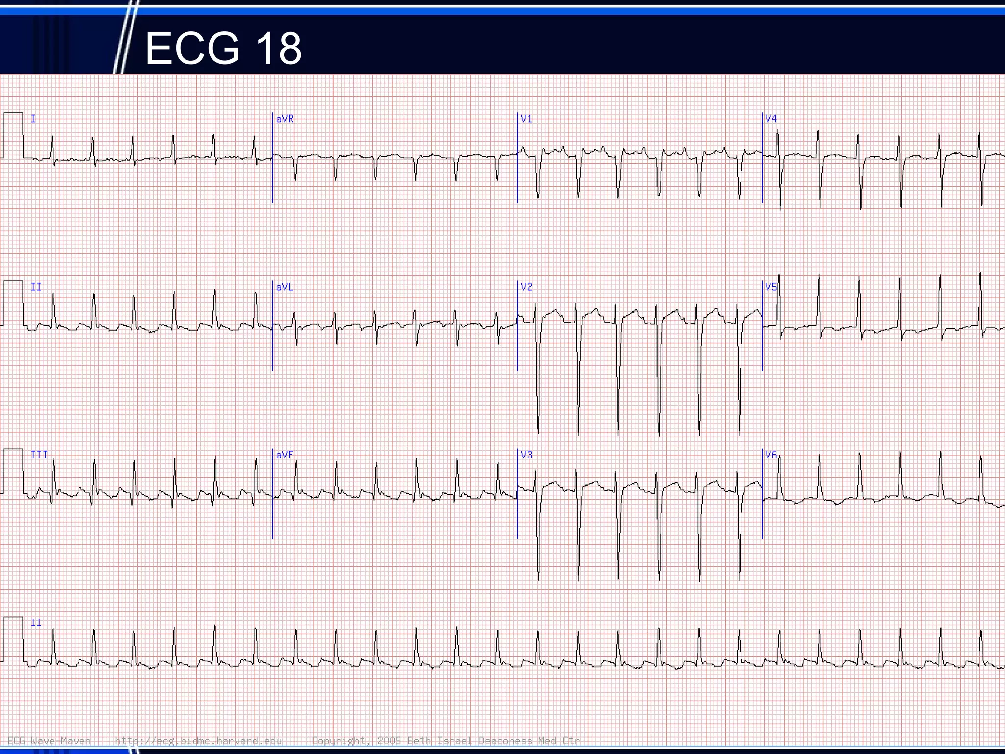
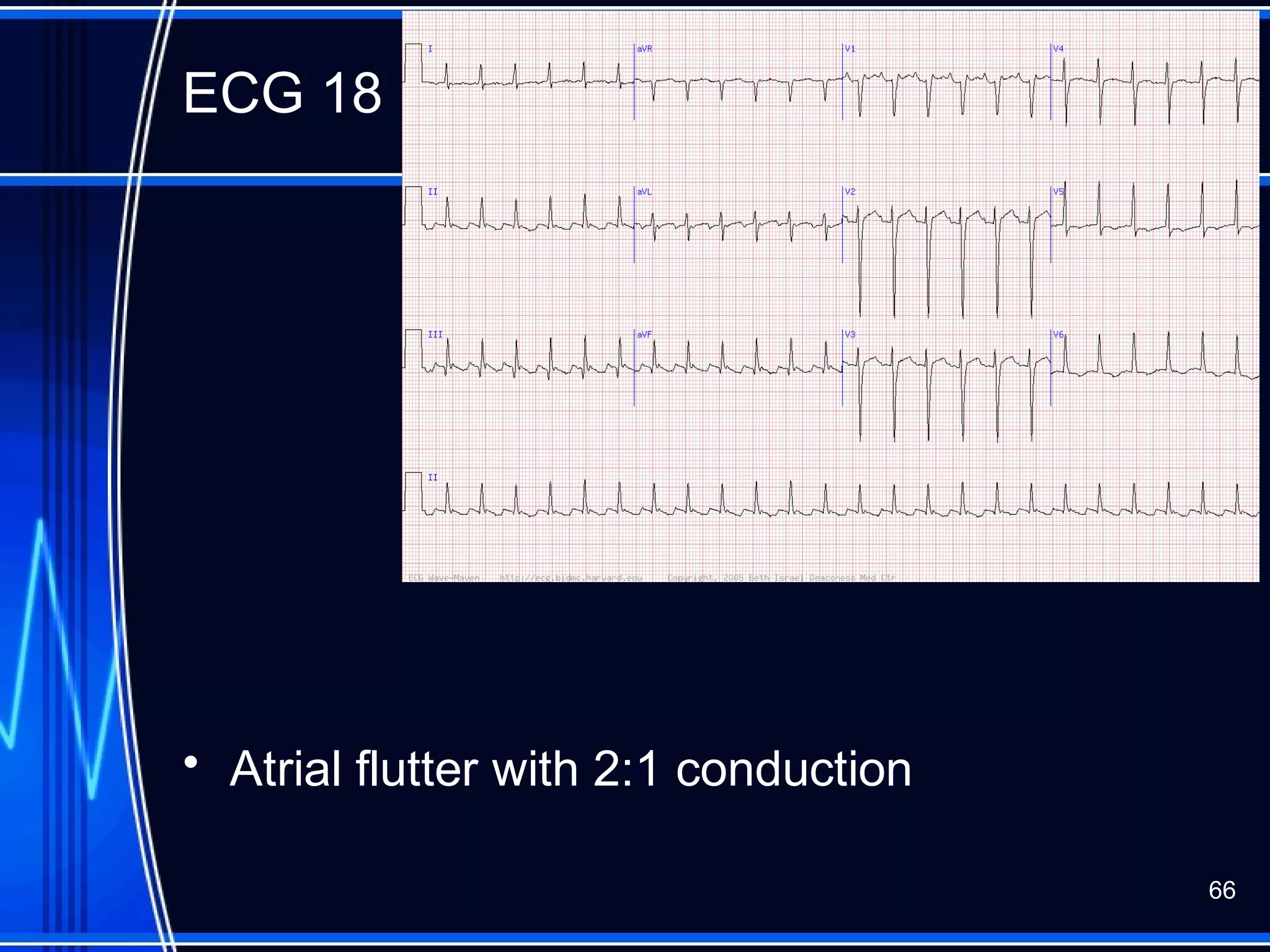
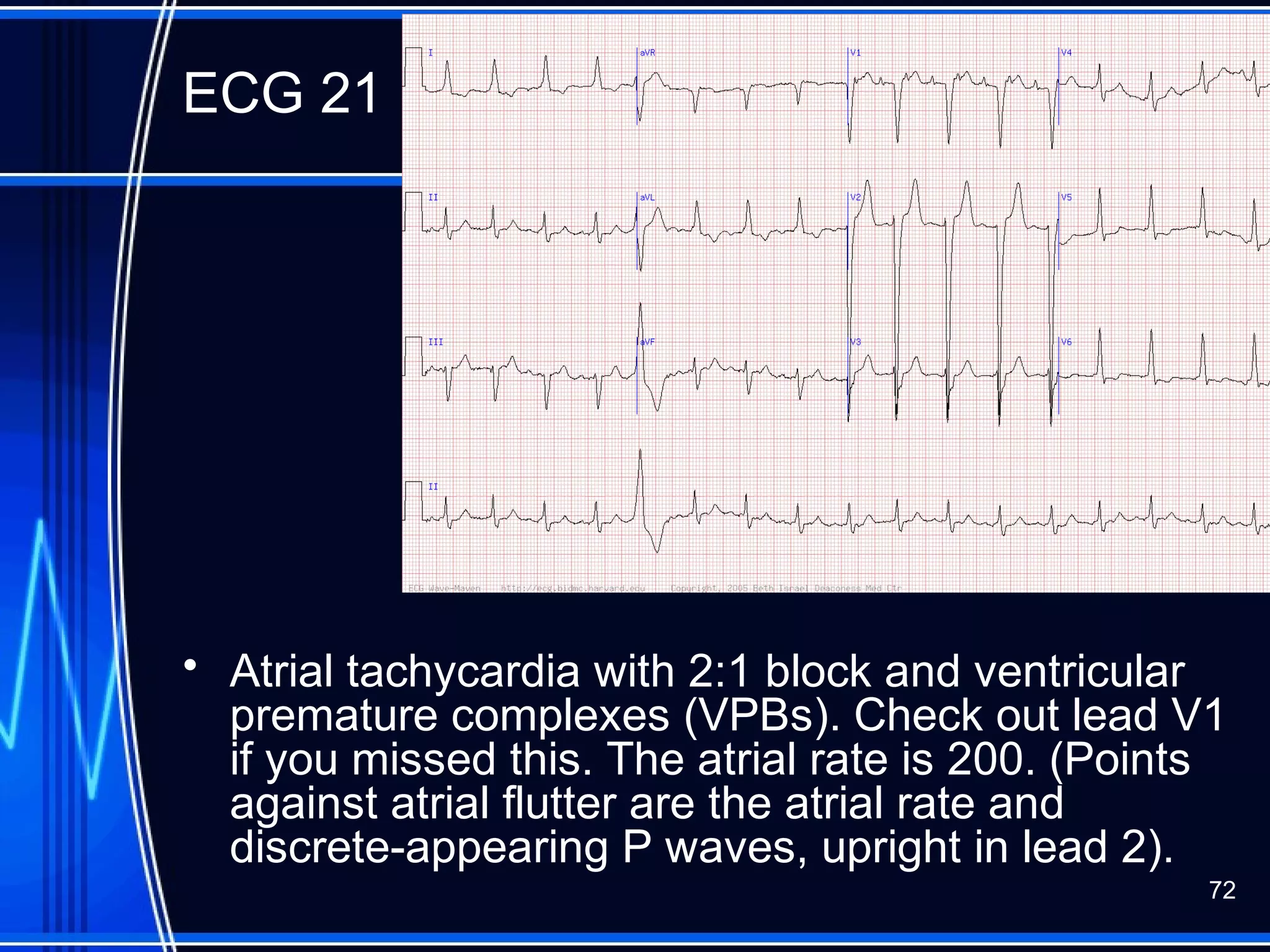
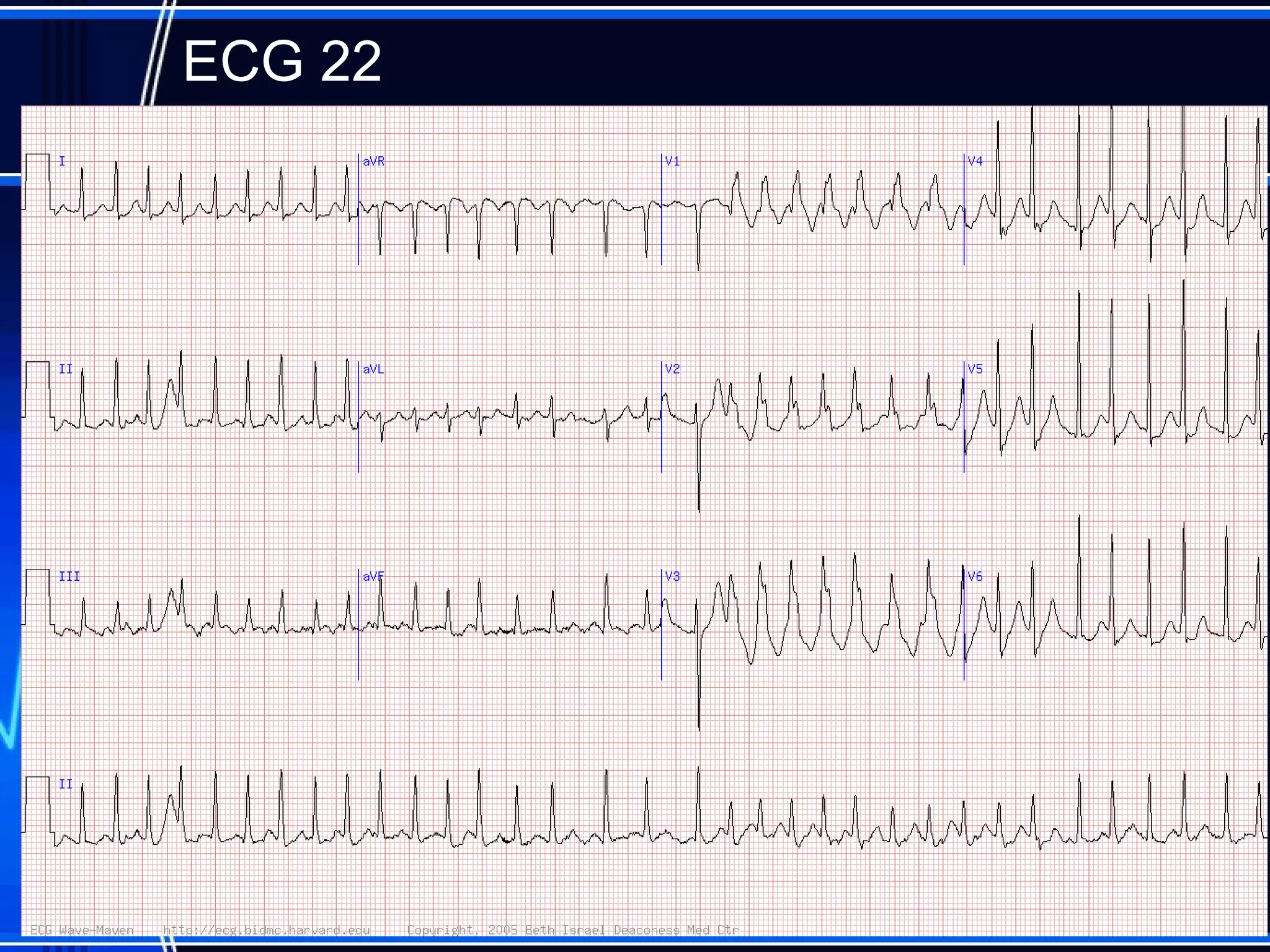
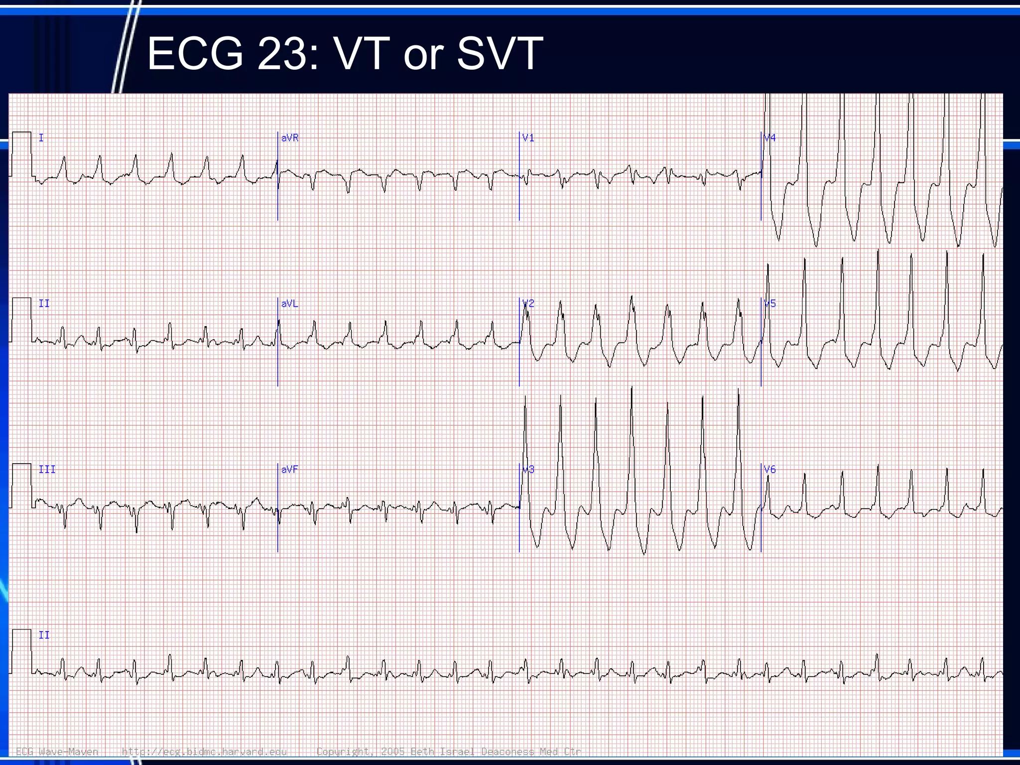
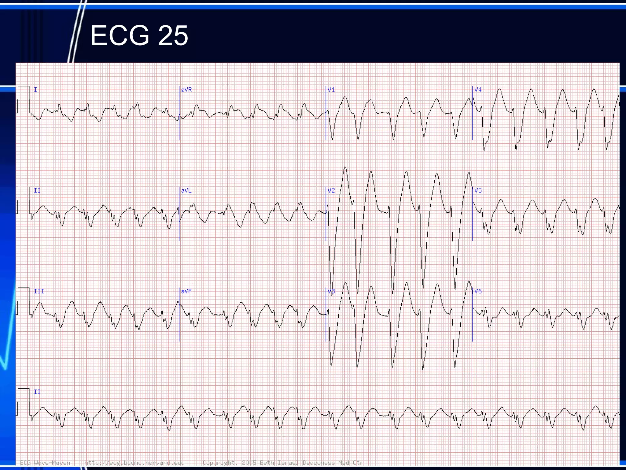
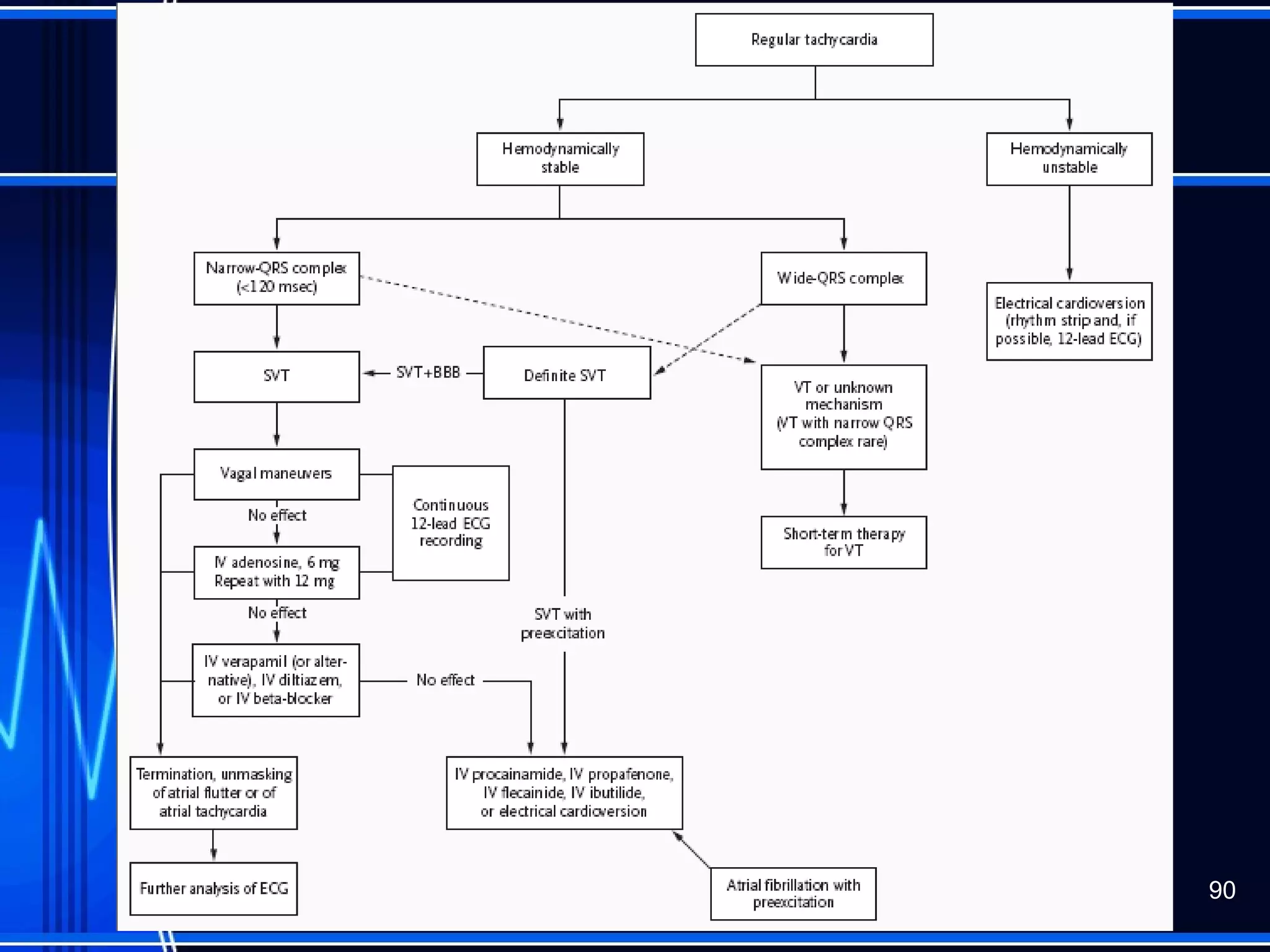
This document provides an overview of approaches to arrhythmias from an electrophysiological perspective. It begins by classifying bradyarrhythmias and tachyarrhythmias based on location and rate. Common supraventricular arrhythmias like AV nodal reentrant tachycardia, atrial flutter, and atrial fibrillation are described. Ventricular arrhythmias including nonsustained and sustained VT are also covered. The document reviews ECG interpretation and provides examples of various arrhythmias, discussing distinguishing characteristics and treatment approaches. Case examples include sinus bradycardia with heart block, Wenckebach block, junctional escape rhythms, atrial flutter, AVNRT, preexcitation
4. Location
• Supraventricular
• Originate from foci above or within the atrioventricular
node
• Main players in outpatient setting
• All the favorites
– AV nodal reentrant tachycardia and AV reentrant
tachycardias (SVT)
– Atrial flutter
– Atrial fibrillation
4
5. Location
• Ventricular Arrhythmias
– Non-sustained ventricular tachycardia
– Sustained ventricular tachycardia
• Stable
– Know the neighborhood
– Do no harm
• Unstable
– ACLS
– Ventricular fibrillation
• Never a stable rhythm
• Immediate ACLS
5
6. Basic Rhythm Strip Interpretation
1. Determine the rate. Does the atrial rate equal the
ventricular rate.
2. Is the rhythm regular/irregular?
3. Find the P wave. Is there a P wave for every QRS?
4. Determine the PR Interval (Normal 0.12-0.20 sec)
5. Find the QRS (Normal <0.12seconds)
6. Any ectopic beats?
7. Find the T wave.
9. ECG 1: Sinus rhythm with 2:1 AV heart
block.
• It is not possible to reliably identify the point of block
(nodal vs. infranodal).
– In general, with 2:1 block, involvement of the AV node is
favored by a narrow QRS complex and a prolonged PR
interval, or by the presence of intermittent AV Wenckebach.
– Block (infranodal) in the His-Purkinje system would be
favored by a concomitant bundle branch block and/or with a
PR interval of 160 ms or less.
• Useful bedside diagnostic test : increase the sinus rate (mild
exercise). A resumption of 1:1 conduction favors AV node block
while worsening of block strongly favors infranodal disease.
• Pacemaker placement is indicated for symptomatic
2:1 block without reversible cause (e.g., drug effect)
and generally for asymptomatic 2:1 block due to
infranodal disease.
• Intracardiac His bundle electrogram would definitively
identify the site of block.
11. ECG 2
• Sinus rhythm with AV Wenckebach with 4:3 conduction in the setting of
an acute inferior wall infarction.
• The presence of "group" beating. Associated with high vagal tone or nodal
ischemia in the setting of an inferior wall myocardial infraction (MI). The
block is at the level of the AV node.
11
13. ECG 3
• Sinus rhythm with complete heart block and
an A-V junctional type escape rhythm.
• The P-P interval surrounding an individual
QRS complex is narrower (shorter) than the
P-P interval between two QRS complexes.
Sinus rate variation with complete heart block
is called ventriculophasic sinus arrhythmia.
• The escape rhythm is proximal in the
conduction system and a pacemaker may not
be required at this time.
13
16. ECG 4
• Acute infero-lateral and probably posterior myocardial
infarction (MI).
• Second degree AV block (Type 1) with 2:1 block
initially and then 3:2 AV
• Since this is an inferior MI, a pacemaker is not
usually indicated: block here is in the AV node
usually due to ischemia and increased vagal tone.
• In contrast, new right bundle with left axis with acute
anterior-septal MI would be indication for prophylactic
pacemaker. AV block in that setting is due to Type II
mechanism associated with severe involvement of
His-Purkinje system and carries ominous prognosis
with high risk of complete heart block with slow (or
no) escape rhythm
16
19. ECG 5: ‘Meet me halfway’ rule
• Sinus tachycardia with 2:1 block (hidden P
waves in T waves, e.g. leads V2 and V3) and
left bundle branch block (LBBB).
– This most likely is type 2 block given its
occurrence with sinus tachycardia and presence
of LBBB.
• “Meet me halfway" rule: always look halfway
in between obvious P waves for hidden ones.
19
21. ECG 6: 80 year old woman with
recurrent syncope and pre-syncope
22. ECG 6
• Sick Sinus Syndrome
– Needs pacemaker
• ? Atrial based single chamber, or
• ? Dual chamber, or
• ? Ventricular based single chamber
– Is there AV conduction disease as well ?
22
34. Unmasking of Flutter Waves
In the presence of 2:1 AV block, the flutter waves may not be
immediately apparent. These can be brought out by administration of
adenosine.
Braunwald's Heart Disease: A Textbook of Cardiovascular Medicine, 7th ed., 2005.
36. Differential diagnosis of wide
complex tachycardia
• Regular
– SVT with bundle branch block aberrancy
• Pre-existing
• Rate-related
– Antidromic AVRT
– Atrial flutter / tachycardia with aberrancy or conduction over
acc.pathway (bystander)
– VT
• Irregular
– AF
– Atrial flutter / atrial tachycardia with variable conduction and
• Bundle branch block
• Antegrade conduction via accessory pathway
37. Wide complex tachycardia
• Stable vital signs during WCT are not
useful for distinguishing SVT from VT
• In the event of previous MI or LV
dysfunction VT is by far more likely
• If in doubt , avoid iv verapamil / diltiazem -
can be catastrophic in VT
• Response to adenosine is not proof that
the WCT was SVT
39. ECG 9
• The major finding is narrow complex tachycardia
with atrial rate of about 300 indicating atrial flutter
and slightly variable ventricular response
• Atrial flutter is sometimes mistaken for sinus
tachycardia. Look, for instance, at V1 where the
flutter waves are hard to see 39
41. ECG 10
• Atrial flutter.
– Don't miss hidden atrial (F) wave just after QRS. This is
atrial flutter with 2:1 conduction.
– Note that with atrial flutter there aren't P waves per se
but atrial waves.
– Note that typical flutter waves have negative polarity in
lead II, as is the case here. 41
43. ECG 11
• ‘Pseudo atrial flutter/fib’ type waves
(associated with Parkinsonian tremor).
– The marked amplitude of the waves, absence of
an irregular ventricular rate and subtle hidden true
P waves in selected leads (lead 2, intermittently,
aVR) indicate the diagnosis.
• Refer to neurology not cardiology. 43
45. ECG 12
• This is sinus rhythm.
• Pseudo-flutter waves due to artifact (probably
Parkinsonian tremor). Some leads show clear
sinus P wave, e.g. V2, V3. So, remember,
look at all the leads.
45
47. ECG 13
• AV nodal reentrant tachycardia (AVNRT).
• P waves can be located at the end of the
QRS in lead II and aVF, producing a "pseudo
S" wave.
47
55. ECG 15
• Atrial fibrillation with the Wolff-Parkinson-White
(WPW) syndrome, with conduction down the bypass
tract.
• The differential diagnosis includes 1) ventricular
tachycardia, 2) supraventricular tachycardia with
aberrancy, and 3) WPW with conduction down the
bypass tract.
• The major clues include the "irregularly irregular"
rhythm and the extremely rapid rate.
• A correct diagnosis is very important because drugs
that slow AV conduction (verapamil, beta blockers,
digoxin, adenosine) are contraindicated. These drugs
can facilitate preferential conduction down the bypass
tract and the atrial fibrillation can degenerate to
ventricular fibrillation.
55
58. ECG 16
• Atrial Tachycardia with 2:1 AV block
• There are two P's for every QRS (see V1) and
that atrial activity at 176/min is negative in
lead 2.
58
60. ECG 17
• The rhythm is NOT sinus but atrial flutter with
subtle flutter waves (aVR, II, V1).
• Note the negative polarity of atrial waves in
lead II, characteristic of typical atrial flutter.
• Atrial flutter with 2:1 conduction is among
most commonly missed major rhythms.
60
62. Atrial flutter
• Most common type of macroreentrant AT (cf. focal AT)
Example:
Typical flutter (RA circuit)
Reentrant rhythm constrained
anatomically by:
Tricuspid valve anteriorly
IVC, Eustachian Ridge and Crista
Terminalis posteriorly
Coronary sinus medially
Macro re-entry:
A relatively large re-entrant circuit using
conduction barriers to create the circuit
Macro re-entry:
A relatively large re-entrant circuit using
conduction barriers to create the circuit
63. ECG of flutter
• Typical CTI dependent flutter:
– Rates and flutter wave morphology on the surface ECG
are consistent and predictable
– Flutter rate: 250-300bpm (occasionally 200 bpm if on anti-
arrhythmic therapy)
– Saw-tooth flutter waves
• Atypical non-CTI dependent flutter:
– Variable rate and morphology (indicating multiple circuits
and/or non-fixed conduction barriers)
– Flutter rate: often faster
• Typical and atypical flutter may (uncommonly) co-exist
Typical CTI-dependent flutter:
Counterclockwise flutter: Afl waves negative in leads II, III,
aVL & V6
Clockwise flutter: Afl waves positive in II, II, aVF and often
notched
68. ECG 19
• There is a wide-complex tachycardia. The QRS
shows a classic left bundle branch block (LBBB)
pattern. If you look carefully, you will see atrial
activity in the limb leads, with negative polarity in
lead II, at rate of 320/min. Hence, atrial flutter
with 2:1 conduction and LBBB. 68
70. ECG 20
• Narrow complex tachyardia at 180/min with probable
subtle retrograde P waves just after the QRS
• Therefore, likely diagnosis is AV nodal reentrant
tachycardia (AVNRT); although, concealed bypass
tract tachycardia or atrial tachycardia not excluded.
• NOT atrial flutter (no flutter waves and ventricular
rate is too fast for flutter with 2:1 conduction). 70
72. ECG 21
• Atrial tachycardia with 2:1 block and ventricular
premature complexes (VPBs). Check out lead V1
if you missed this. The atrial rate is 200. (Points
against atrial flutter are the atrial rate and
discrete-appearing P waves, upright in lead 2).
72
74. ECG 22
• Atrial fibrillation with an rapid ventricular
response (overall average about 204 beats per
minute).
• There is intermittent right bundle branch block
(RBBB) aberrancy
74
77. Differential criteria considering the QRS morphology in
leads V1 and V6 in case of right bundle branch block
(RBBB) or left bundle branch block (LBBB) aspect.
78. But here’s an important point:
if there is hemodynamic collapse, shoot
first and ask questions later:
Often the best 'drug' is in fact electricity, as
sustained VT often deteriorates into VF if left
unmanaged.
79. Many of these are very difficult.
The algorithms are hard to commit to
memory.
For family medicine practitioners who see
this stuff infrequently, this can be an
impossible situation,
80. Always assume the patient has the worst
treatable condition you can do something
about, and proceed from there.
In the case of wide-complex tachycardias,
to say: “This is probably just SVT with
aberrated conduction” and send the
patient home, is to court disaster.
81. You do not have to know everything
to be a great doctor.
You just have to think.
83. ECG 23
• AV dissociation is noted, confirming
the diagnosis of ventricular
tachycardia (VT)
83
84. • VA dissociation on ECG proves VT but is
clearly discernible in only 30% - evidence of
VA dissociation on physical examination may
be useful
– Irregular cannon A waves in the JVP
– Variability in loudness of S1
• Fusion complexes / capture beats
86. The tracing has a classic RBBB
configuration
And is triphasic both in V1 and V6
87. ECG 24
• There is a classic right bundle branch
block (RBBB) morphology (rsR'
in V1)
making VT unlikely. No definite atrial
activity is seen (P waves or flutter
waves). The very regular rate at 150/min
excludes AF. Therefore, the rhythm is
most consistent with paroxysmal
supraventricular tachycardia (PSVT).
87
89. ECG 25
• Atrial fibrillation with a rapid ventricular response
(irregular rate at about 120 beats/min). There is an
extremely wide QRS of left bundle branch block
morphology; the QRS duration of over 200ms is
suggestive of drug effect or hyperkalemia 89