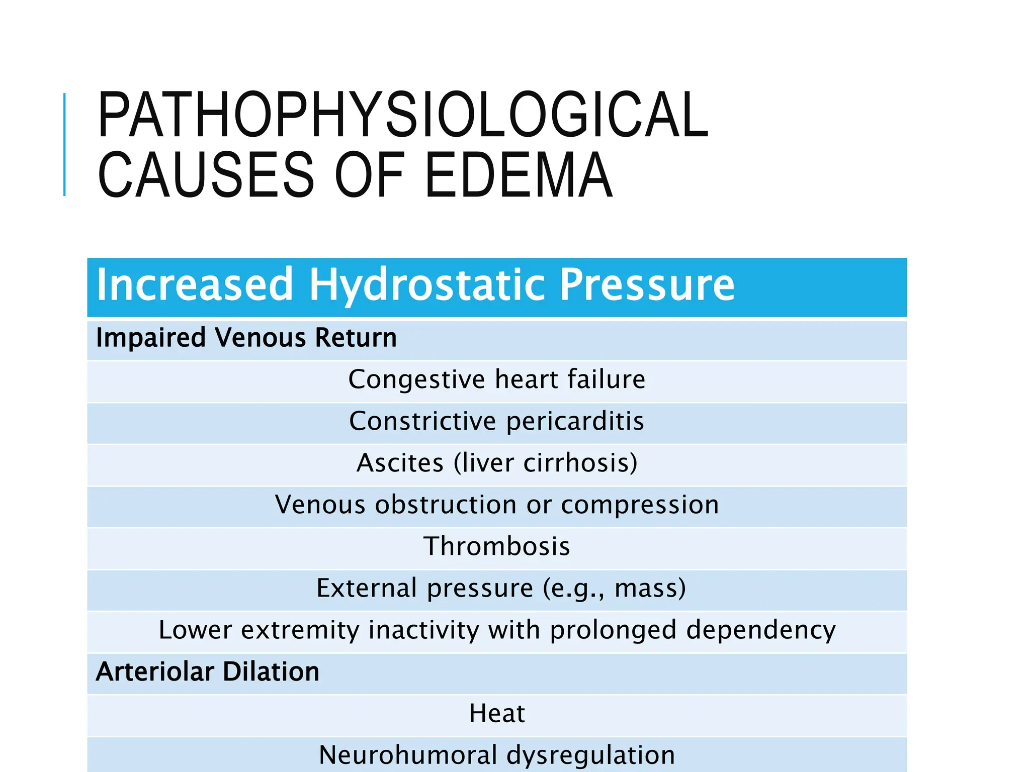
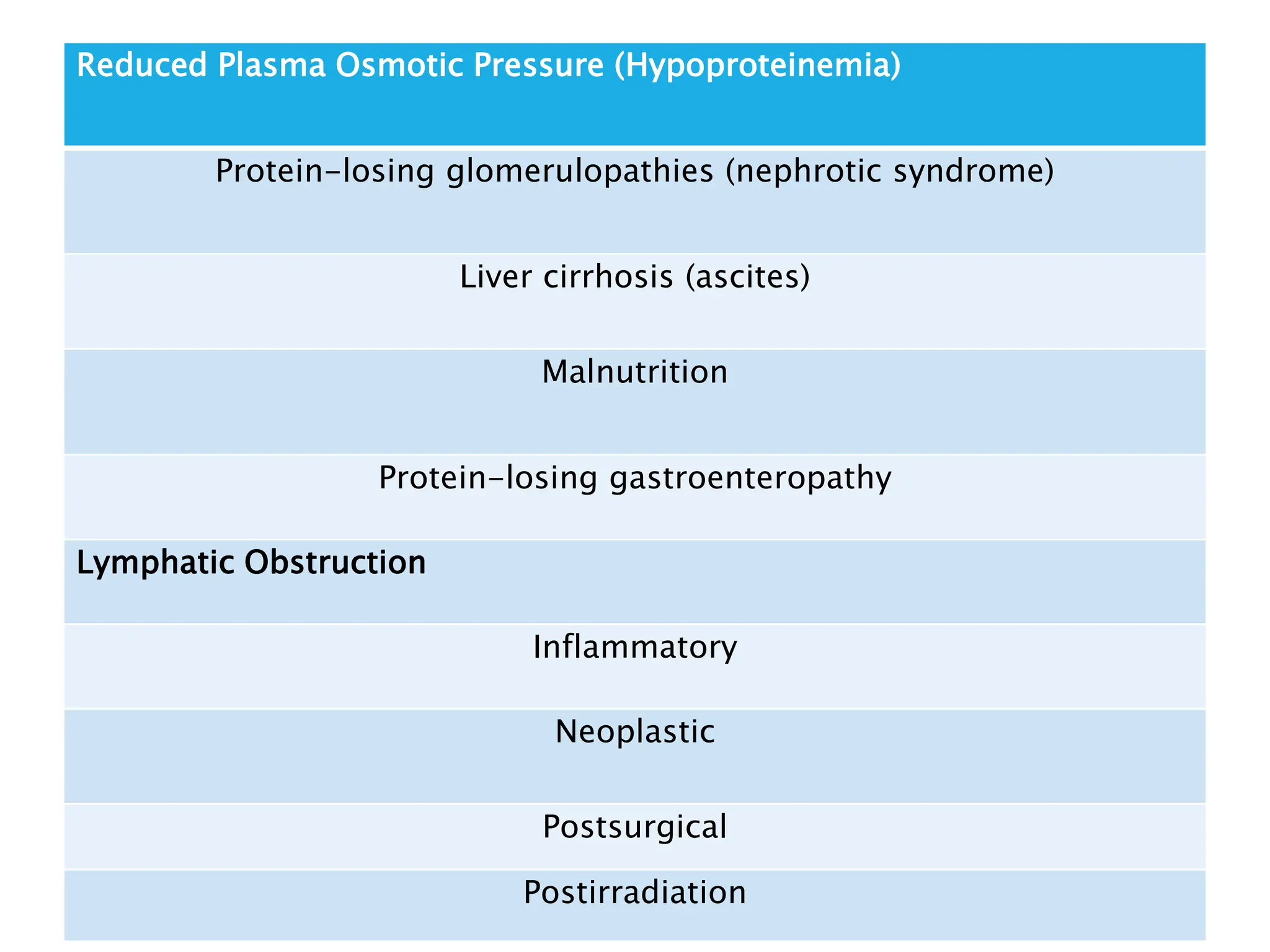
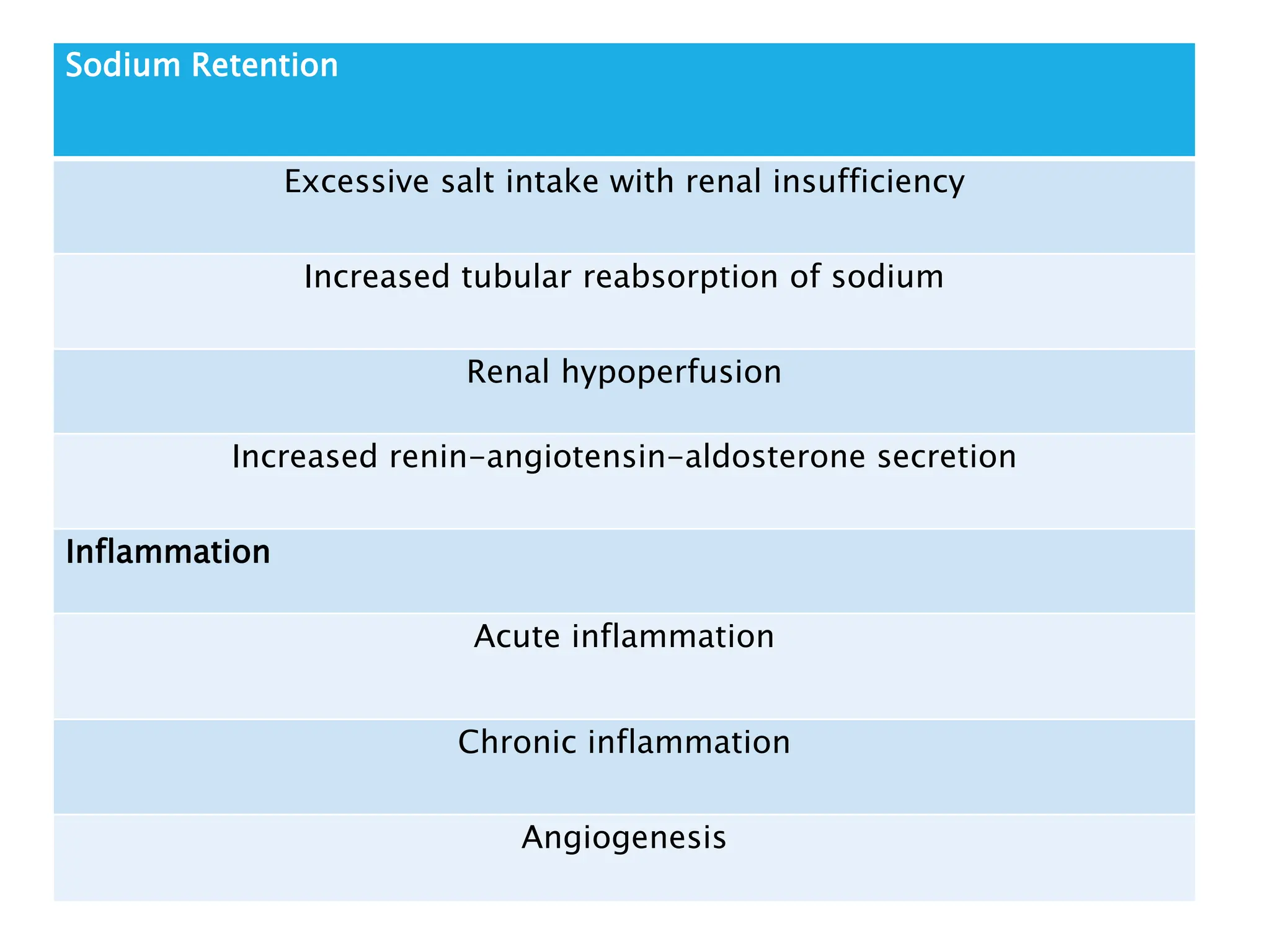
HEMODYNAMIC
DISORDERS
CONTENTS
Edema
Pathophysiological causes of edema
Morphology and clinical correlation
of edema
Hemorrhage
Manifestations and clinical
significance of hemorrhage
EDEMA
Edema is an accumulation of
interstitial fluid within tissues.
Extravascular fluid can also collect in
body cavities such as the pleural
cavity (hydrothorax), the pericardial
cavity (hydropericardium), or the
peritoneal cavity (hydroperitoneum,
or ascites).
Anasarca is severe, generalized
edema marked by profound swelling
of subcutaneous tissues and
accumulation of fluid in body cavities.
Fluid movement abetween the
vascular and interstitial spaces is
governed mainly by two opposing
forces—the vascular hydrostatic
pressure and the colloid osmotic
pressure produced by plasma
Normally, the outflow of fluid
produced by hydrostatic pressure at
the arteriolar end of the
microcirculation is neatly balanced by
inflow due to the slightly elevated
osmotic pressure at the venular end;
hence there is only a small net
outflow of fluid into the interstitial
space, which is drained by lymphatic
vessels.
Either increased hydrostatic pressure
or diminished colloid osmotic
pressure causes increased movement
of water into the interstitium.
The edema fluid that accumulates
owing to increased hydrostatic
pressure or reduced intravascular
colloid typically is a protein-poor
transudate; it has a specific gravity
less than 1.012.
By contrast, because of increased
vascular permeability, inflammatory
edema fluid is a protein-rich exudate
with a specific gravity usually greater
than 1.020.
PATHOPHYSIOLOGICAL
CAUSES OF EDEMA
Increased Hydrostatic Pressure
Impaired Venous Return
Congestive heart failure
Constrictive pericarditis
Ascites (liver cirrhosis)
Venous obstruction or compression
Thrombosis
External pressure (e.g., mass)
Lower extremity inactivity with prolonged dependency
Arteriolar Dilation
Heat
Neurohumoral dysregulation
Reduced Plasma Osmotic Pressure (Hypoproteinemia)
Protein-losing glomerulopathies (nephrotic syndrome)
Liver cirrhosis (ascites)
Malnutrition
Protein-losing gastroenteropathy
Lymphatic Obstruction
Inflammatory
Neoplastic
Postsurgical
Postirradiation
Sodium Retention
Excessive salt intake with renal insufficiency
Increased tubular reabsorption of sodium
Renal hypoperfusion
Increased renin-angiotensin-aldosterone secretion
Inflammation
Acute inflammation
Chronic inflammation
Angiogenesis
MORPHOLOGY
Edema is easily recognized on gross
inspection; microscopic examination
shows clearing and separation of the
extracellular matrix elements.
Subcutaneous edema can be diffuse
but usually accumulates preferentially
in parts of the body positioned the
greatest distance below the heart
where hydrostatic pressures are
highest.
Thus, edema typically is most
pronounced in the legs with standing,
a relationship termed dependent
edema.
Finger pressure over edematous
subcutaneous tissue displaces the
interstitial fluid, leaving a finger-
shaped depression; this appearance is
called pitting edema.
Edema due to renal dysfunction or
nephrotic syndrome often manifests
first in loose connective tissues (e.g.,
the eyelids, causing periorbital
edema).
With pulmonary edema, the lungs
often are two to three times their
normal waeight, and sectioning
reveals frothy, sometimes blood-
tinged fluid consisting of a mixture of
Brain edema can be localized (e.g.,
due to abscess or tumor) or
generalized, depending on the nature
and extent of the pathologic process
or injury.
CLINICAL CORRELATION
Subcutaneous edema, signals
potential underlying cardiac or renal
disease; however, when significant, it
also can impair wound healing or the
clearance of infections.
Pulmonary edema, frequently is seen
in the setting of left ventricular failure
but also may occur in renal failure,
acute respiratory distress syndrome,
and inflammatory and infectious
Brain edema is life-threatening; if the
swelling is severe, the brain can
herniate (extrude) through the
foramen magnum. With increased
intracranial pressure, the brain stem
vascular supply can be compressed.
Either condition can cause death by
injuring the medullary centers
HEMORRHAGE
Hemorrhage, defined as the
extravasation of blood from vessels,
occurs in a variety of settings.
The risk of hemorrhage (often after a
seemingly insignificant injury) is
increased in a wide variety of clinical
disorders collectively called
hemorrhagic diatheses.
Trauma, atherosclerosis, or
inflammatory or neoplastic erosion of
a vessel wall also may lead to
hemorrhage, which may be extensive
if the affected vessel is a large vein or
artery.
MANIFESTATIONS OF
HEMORRHAGE
Hemorrhage may be external or
accumulate within a tissue as a
hematoma, which ranges in
significance from trivial (e.g., a
bruise) to fatal.
Large bleeds into body cavities are
given various names according to
location—hemothorax,
hemopericardium, hemoperitoneum,
or hemarthrosis (in joints).
PETECHIAE
Petechiae are minute (1 to 2 mm in
diameter) hemorrhages into skin,
mucous membranes, or serosal
surfaces, ; causes include low platelet
counts (thrombocytopenia), defective
platelet function.
PURPURA AND ECCHYMOSES
Purpura are slightly larger (3 to 5
mm) hemorrhages, causes include
trauma, vascular inflammation
(vasculitis), and increased vascular
fragility.
Ecchymoses are larger (1 to 2 cm)
subcutaneous hematomas
(colloquially called bruises).
CLINICAL SIGNIFICANCE OF
HEMORRHAGE
Depends on the volume of blood lost
and the rate of bleeding.
The site of hemorrhage also is
important.
Chronic or recurrent external blood
loss (e.g., due to peptic ulcer)
frequently culminates in iron
deficiency anemia
Hemodynamic Disorders in detail with there mechanisms

Hemodynamic Disorders in detail with there mechanisms

  • 2.
  • 3.
    CONTENTS Edema Pathophysiological causes ofedema Morphology and clinical correlation of edema Hemorrhage Manifestations and clinical significance of hemorrhage
  • 4.
    EDEMA Edema is anaccumulation of interstitial fluid within tissues. Extravascular fluid can also collect in body cavities such as the pleural cavity (hydrothorax), the pericardial cavity (hydropericardium), or the peritoneal cavity (hydroperitoneum, or ascites).
  • 5.
    Anasarca is severe,generalized edema marked by profound swelling of subcutaneous tissues and accumulation of fluid in body cavities. Fluid movement abetween the vascular and interstitial spaces is governed mainly by two opposing forces—the vascular hydrostatic pressure and the colloid osmotic pressure produced by plasma
  • 6.
    Normally, the outflowof fluid produced by hydrostatic pressure at the arteriolar end of the microcirculation is neatly balanced by inflow due to the slightly elevated osmotic pressure at the venular end; hence there is only a small net outflow of fluid into the interstitial space, which is drained by lymphatic vessels.
  • 7.
    Either increased hydrostaticpressure or diminished colloid osmotic pressure causes increased movement of water into the interstitium. The edema fluid that accumulates owing to increased hydrostatic pressure or reduced intravascular colloid typically is a protein-poor transudate; it has a specific gravity less than 1.012.
  • 8.
    By contrast, becauseof increased vascular permeability, inflammatory edema fluid is a protein-rich exudate with a specific gravity usually greater than 1.020.
  • 9.
    PATHOPHYSIOLOGICAL CAUSES OF EDEMA IncreasedHydrostatic Pressure Impaired Venous Return Congestive heart failure Constrictive pericarditis Ascites (liver cirrhosis) Venous obstruction or compression Thrombosis External pressure (e.g., mass) Lower extremity inactivity with prolonged dependency Arteriolar Dilation Heat Neurohumoral dysregulation
  • 10.
    Reduced Plasma OsmoticPressure (Hypoproteinemia) Protein-losing glomerulopathies (nephrotic syndrome) Liver cirrhosis (ascites) Malnutrition Protein-losing gastroenteropathy Lymphatic Obstruction Inflammatory Neoplastic Postsurgical Postirradiation
  • 11.
    Sodium Retention Excessive saltintake with renal insufficiency Increased tubular reabsorption of sodium Renal hypoperfusion Increased renin-angiotensin-aldosterone secretion Inflammation Acute inflammation Chronic inflammation Angiogenesis
  • 12.
    MORPHOLOGY Edema is easilyrecognized on gross inspection; microscopic examination shows clearing and separation of the extracellular matrix elements. Subcutaneous edema can be diffuse but usually accumulates preferentially in parts of the body positioned the greatest distance below the heart where hydrostatic pressures are highest.
  • 13.
    Thus, edema typicallyis most pronounced in the legs with standing, a relationship termed dependent edema. Finger pressure over edematous subcutaneous tissue displaces the interstitial fluid, leaving a finger- shaped depression; this appearance is called pitting edema.
  • 14.
    Edema due torenal dysfunction or nephrotic syndrome often manifests first in loose connective tissues (e.g., the eyelids, causing periorbital edema). With pulmonary edema, the lungs often are two to three times their normal waeight, and sectioning reveals frothy, sometimes blood- tinged fluid consisting of a mixture of
  • 15.
    Brain edema canbe localized (e.g., due to abscess or tumor) or generalized, depending on the nature and extent of the pathologic process or injury.
  • 16.
    CLINICAL CORRELATION Subcutaneous edema,signals potential underlying cardiac or renal disease; however, when significant, it also can impair wound healing or the clearance of infections. Pulmonary edema, frequently is seen in the setting of left ventricular failure but also may occur in renal failure, acute respiratory distress syndrome, and inflammatory and infectious
  • 17.
    Brain edema islife-threatening; if the swelling is severe, the brain can herniate (extrude) through the foramen magnum. With increased intracranial pressure, the brain stem vascular supply can be compressed. Either condition can cause death by injuring the medullary centers
  • 18.
    HEMORRHAGE Hemorrhage, defined asthe extravasation of blood from vessels, occurs in a variety of settings. The risk of hemorrhage (often after a seemingly insignificant injury) is increased in a wide variety of clinical disorders collectively called hemorrhagic diatheses.
  • 19.
    Trauma, atherosclerosis, or inflammatoryor neoplastic erosion of a vessel wall also may lead to hemorrhage, which may be extensive if the affected vessel is a large vein or artery.
  • 20.
    MANIFESTATIONS OF HEMORRHAGE Hemorrhage maybe external or accumulate within a tissue as a hematoma, which ranges in significance from trivial (e.g., a bruise) to fatal. Large bleeds into body cavities are given various names according to location—hemothorax, hemopericardium, hemoperitoneum, or hemarthrosis (in joints).
  • 21.
    PETECHIAE Petechiae are minute(1 to 2 mm in diameter) hemorrhages into skin, mucous membranes, or serosal surfaces, ; causes include low platelet counts (thrombocytopenia), defective platelet function.
  • 22.
    PURPURA AND ECCHYMOSES Purpuraare slightly larger (3 to 5 mm) hemorrhages, causes include trauma, vascular inflammation (vasculitis), and increased vascular fragility. Ecchymoses are larger (1 to 2 cm) subcutaneous hematomas (colloquially called bruises).
  • 23.
    CLINICAL SIGNIFICANCE OF HEMORRHAGE Dependson the volume of blood lost and the rate of bleeding. The site of hemorrhage also is important. Chronic or recurrent external blood loss (e.g., due to peptic ulcer) frequently culminates in iron deficiency anemia