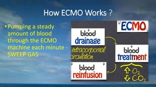
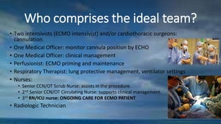
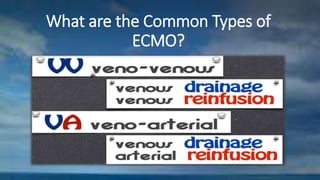
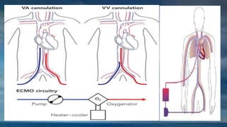
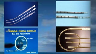
Extracorporeal
Membrane Oxygenation
Presented by Group B
Intensive Care Unit
SQUH,OMAN
What is an ECMO?
• ECMO is the use of extracorporeal
circulation and gas exchange to
provide temporary life support in
patients with reversible pulmonary
or cardiac failure.
GOALS
•Ensure the body has enough oxygen.
•ECMO does not heal the heart or lungs but gives
them time to rest and recover.
•The ventilator settings can be decreased to
levels that will not damage the lungs.
•Able to decrease other drugs that support the
heart.
How ECMO Works ?
•Pumping a steady
amount of blood
through the ECMO
machine each minute -
SWEEP GAS
ECMO Indications
• Neonatal
• Congenital Diaphragmatic Hernia
• Meconium aspiration
• ARDS
• Pneumonia and sepsis
• Pulmonary HTN
• VV ECMO in Peds and Adults
• Pneumonia and sepsis
• ARDS
• Aspiration Pneumonia
• Drowning & Trauma
• Respiratory Failure
• VA ECMO Pedia &
Adults
• Cardiomyopathy
• Sepsis
• Perioperative Cardiac Surgery
• Poisoning
• Post Cardiac Arrest
ECMO Contraindications
• Age: <34 weeks gestational age, > 75 years old
• Severe chronic organ failures, e.g. cirrhosis, ESRD,
hepatic failure
• <2000gms for paediatrics
• Coagulopathy that is ongoing/uncorrectable
• Severe neurologic insult or asphyxia
Who comprises the ideal team?
• Two intensivists (ECMO intensivist) and/or cardiothoracic surgeons:
cannulation
• One Medical Officer: monitor cannula position by ECHO
• One Medical Officer: clinical management
• Perfusionist: ECMO priming and maintenance
• Respiratory Therapist: lung protective management, ventilator settings
• Nurses:
• Senior CCN/OT Scrub Nurse: assists in the procedure.
• 2nd Senior CCN/OT Circulating Nurse: supports clinical management.
• 3rd RN/ICU nurse: ONGOING CARE FOR ECMO PATIENT
• Radiologic Technician
What are the Common Types of
ECMO?
• VA ECMO is mainly used in
people with heart problems or
blood pressure problems. A
catheter takes blood from the
superior vena cava, runs it
through the ECMO machine,
and then replaces the blood,
under pressure, to arterial
circulation, usually via the
aorta. This method supports
both the heart and the lungs.
• VV ECMO is used for patients
who need mainly lung support.
In this case, the blood is taken
from a major vein and returned
to another major vein after
oxygenation
CENTRAL VA ECMO: cannula into
the ascending aorta or subclavian
artery
Nursing Considerations:
•
Nursing Actions
•Maintain strict infection control. No alcohol swabs or
chlorhexidine wash. Only USE BETADINE
•Restrict access to essential personnel.
•Remove unnecessary invasive lines.
•Ensure that all required invasive access are present,
eg. NGT, core temp probe.
•Secure ET tube to maintain access during procedure
maintaining the sterile field.
• Ensure crash trolley in close proximity
• Insert fecal management system, ensure fecal
softeners as prescribed.
• Prepare and position patient. Place appropriate
mattress on bed.
• Clip hair on the proposed site with electric razor.
• Move the bed so the ECHO machine, ECMO trolley
and sterile field can be positioned
Nursing Actions
• Collaborate with the members of the ECMO management
team about the necessary equipment and materials to be
prepared.
• Ensure liberal supply of gauze.
• Be aware that it is the responsibility of the ECMO intensivist
to dress and secure ECMO cannula.
• Ensure that baseline ECMO observations and pump settings
are monitored, liaised with the perfusionist.
Cannulation
• Ensure contact numbers of the on call perfusionist
and ECMO intensivist is updated and noted.
• Ensure four tubing clamps are readily available.
• Liaised with respiratory therapist and ECMO
intensivist about the rescue ventilation orders for
emergencies.
Shift Checks
• Power
• Flow Mode (liters per minute)
• Mode
• Alarms
• Hand crank
• Emergency Checks
• Battery Check
• Circuit Assessment
• Cannula Assessment
• ECMO checks
Nursing Assessments
• Routine Assessments:
• HR, SaO2, SBP, MAP
• Hourly Assessments:
• Neuro-vascular observation
• Urine output
• Core temperature
• EtC02
• Ventilator observations
• Regular Assessments:
• CVP
• Neurological assessments
• Sedation level
Pressure area care and positioning:
•Restriction in mobility; ensure appropriate
mattress. Once a day pressure care, preferably
in day shift.
•Log rolled. One RN ensures circuit and cannula
safety and directs turning.
Mouth, eye and catheter care
•Use swabs; no toothbrushes.
•No shaving with wet razor; use clippers.
•Don’t do routine tracheal and mouth
suctioning.
•Do not dislodge clots covering wound and
insertion sites.
•No venipuncture.
Dressing the cannula
• Only if there is significant exudates or if not intact or
secured.
• Do not remove lower adhesion tape and stitching. Do
dressing of cannula site first before, the replacing
adhesion tapes.
• Required two nurses for dressing. Dressing changes
preferable in day shift.
• Pull the dressing off towards the insertion site.
• If there is ongoing oozing, calcium/sorbalgon alginate
dressing can be applied.
Blood Works and Diagnostics
• Ensure current crossmatch PRBCs are available.
• Daily U/E, Mg and LFT.
• CBC BD and as prn. PLATELET COUNT
• Daily blood cultures during spike of fever or ideally beginning at 5th
day of therapy.
• Pre and post oxygenator ABG c/o perfusionist.
• ACT every 2 hours x 24 hours.
• APTT every 6 hours, target 55-75 s or as specified by intensivist.
Weaning
• Be knowledgeable of the signs for weaning:
• Improving oxygenation
• Reduced CO2 retention
• Improving chest xray
• Blood flow unchanged in ECMO
• Stable ABG > 6 hours without oxygenator support
• Note clotting and platelet levels before decannulation procedure.
• Discontinue heparin infusion 2-4 hours or as ordered prior to
decannulation.
Decannulation
• Ensure two medical staff are involved in the removal of the cannulas, while
a third medical staff clinically manage the patient.
• Coordinate with perfusionist and respiratory technician about the plans.
• Ensure that direct pressure is applied on the insertion site for at least 20
minutes and the ECMO intensivist will remain with the patient until
hemostasis achieved.
• Coordinate with intensivist about the need for sedation and pain
medication before the procedure.
• Carry out successive Doppler assessment of the decannulated limbs after
catheter removal.
Preventing and Managing Complications
• Patient Related:
• Bleeding
• Hemolysis
• Recirculation
• Infection
• Circuit Related:
• Clot formation
• Plasma Leak
• Oxygenator Failure
• Heat Exchanger
Failure
• Tubing, Connector
Cracks and Blood
Leakage
• Emergencies:
• Pump Failure
• Decannulation
• Air Embolism
• Cardiac Arrest
Bleeding
• Hourly cannula site assessment.
• Monitor clotting time, Hb, platelets.
• Ensure access sites are stabilized. Do not dislodge clots
directly from wounds or insertion sites.
• Maintain enteral feeding if tolerated; ulcer prophylaxis.
• Report blood loss.
• Ensure current crossmatched PRBC.
• Give blood products as ordered.
Hemolysis
• Monitor the lab results( CBC, U/E and urine)
• Hourly assessment for movement(kinking) of the access cannula
• Hourly assessment of the temperature of the heat exchanger
Recirculation
• Monitor the SPO2 and ABG values
• Colour of RETURNED BLOOD
• Ensure catheter security during patient movement.
Infection
• Prevent high risk of nosocomial infection.
• Closely watched for signs of infection
Circuit Related Complications
• Clot formation
Maintain anti-coagulation as prescribed
• Plasma Leak,ALARMS
• Oxygenator Failure
• Heat Exchanged Failure
• Tubing Cracks or blood leakage
ECMO Emergencies
• Pump failure- power check
• Decannulation- reinsertion
• Air embolism- trendlenberg position
• Cardiac Arrest- VA ECMO-
No Chest compression
VV ECMO-chest compression
Ecmo nurse presentation

Ecmo nurse presentation

  • 1.
    Extracorporeal Membrane Oxygenation Presented byGroup B Intensive Care Unit SQUH,OMAN
  • 2.
    What is anECMO? • ECMO is the use of extracorporeal circulation and gas exchange to provide temporary life support in patients with reversible pulmonary or cardiac failure.
  • 3.
    GOALS •Ensure the bodyhas enough oxygen. •ECMO does not heal the heart or lungs but gives them time to rest and recover. •The ventilator settings can be decreased to levels that will not damage the lungs. •Able to decrease other drugs that support the heart.
  • 4.
    How ECMO Works? •Pumping a steady amount of blood through the ECMO machine each minute - SWEEP GAS
  • 5.
    ECMO Indications • Neonatal •Congenital Diaphragmatic Hernia • Meconium aspiration • ARDS • Pneumonia and sepsis • Pulmonary HTN • VV ECMO in Peds and Adults • Pneumonia and sepsis • ARDS • Aspiration Pneumonia • Drowning & Trauma • Respiratory Failure • VA ECMO Pedia & Adults • Cardiomyopathy • Sepsis • Perioperative Cardiac Surgery • Poisoning • Post Cardiac Arrest
  • 6.
    ECMO Contraindications • Age:<34 weeks gestational age, > 75 years old • Severe chronic organ failures, e.g. cirrhosis, ESRD, hepatic failure • <2000gms for paediatrics • Coagulopathy that is ongoing/uncorrectable • Severe neurologic insult or asphyxia
  • 7.
    Who comprises theideal team? • Two intensivists (ECMO intensivist) and/or cardiothoracic surgeons: cannulation • One Medical Officer: monitor cannula position by ECHO • One Medical Officer: clinical management • Perfusionist: ECMO priming and maintenance • Respiratory Therapist: lung protective management, ventilator settings • Nurses: • Senior CCN/OT Scrub Nurse: assists in the procedure. • 2nd Senior CCN/OT Circulating Nurse: supports clinical management. • 3rd RN/ICU nurse: ONGOING CARE FOR ECMO PATIENT • Radiologic Technician
  • 8.
    What are theCommon Types of ECMO?
  • 9.
    • VA ECMOis mainly used in people with heart problems or blood pressure problems. A catheter takes blood from the superior vena cava, runs it through the ECMO machine, and then replaces the blood, under pressure, to arterial circulation, usually via the aorta. This method supports both the heart and the lungs. • VV ECMO is used for patients who need mainly lung support. In this case, the blood is taken from a major vein and returned to another major vein after oxygenation CENTRAL VA ECMO: cannula into the ascending aorta or subclavian artery
  • 16.
  • 17.
    Nursing Actions •Maintain strictinfection control. No alcohol swabs or chlorhexidine wash. Only USE BETADINE •Restrict access to essential personnel. •Remove unnecessary invasive lines. •Ensure that all required invasive access are present, eg. NGT, core temp probe. •Secure ET tube to maintain access during procedure maintaining the sterile field.
  • 18.
    • Ensure crashtrolley in close proximity • Insert fecal management system, ensure fecal softeners as prescribed. • Prepare and position patient. Place appropriate mattress on bed. • Clip hair on the proposed site with electric razor. • Move the bed so the ECHO machine, ECMO trolley and sterile field can be positioned Nursing Actions
  • 19.
    • Collaborate withthe members of the ECMO management team about the necessary equipment and materials to be prepared. • Ensure liberal supply of gauze. • Be aware that it is the responsibility of the ECMO intensivist to dress and secure ECMO cannula. • Ensure that baseline ECMO observations and pump settings are monitored, liaised with the perfusionist. Cannulation
  • 20.
    • Ensure contactnumbers of the on call perfusionist and ECMO intensivist is updated and noted. • Ensure four tubing clamps are readily available. • Liaised with respiratory therapist and ECMO intensivist about the rescue ventilation orders for emergencies.
  • 21.
    Shift Checks • Power •Flow Mode (liters per minute) • Mode • Alarms • Hand crank • Emergency Checks • Battery Check • Circuit Assessment • Cannula Assessment • ECMO checks
  • 22.
    Nursing Assessments • RoutineAssessments: • HR, SaO2, SBP, MAP • Hourly Assessments: • Neuro-vascular observation • Urine output • Core temperature • EtC02 • Ventilator observations • Regular Assessments: • CVP • Neurological assessments • Sedation level
  • 23.
    Pressure area careand positioning: •Restriction in mobility; ensure appropriate mattress. Once a day pressure care, preferably in day shift. •Log rolled. One RN ensures circuit and cannula safety and directs turning.
  • 24.
    Mouth, eye andcatheter care •Use swabs; no toothbrushes. •No shaving with wet razor; use clippers. •Don’t do routine tracheal and mouth suctioning. •Do not dislodge clots covering wound and insertion sites. •No venipuncture.
  • 25.
    Dressing the cannula •Only if there is significant exudates or if not intact or secured. • Do not remove lower adhesion tape and stitching. Do dressing of cannula site first before, the replacing adhesion tapes. • Required two nurses for dressing. Dressing changes preferable in day shift. • Pull the dressing off towards the insertion site. • If there is ongoing oozing, calcium/sorbalgon alginate dressing can be applied.
  • 26.
    Blood Works andDiagnostics • Ensure current crossmatch PRBCs are available. • Daily U/E, Mg and LFT. • CBC BD and as prn. PLATELET COUNT • Daily blood cultures during spike of fever or ideally beginning at 5th day of therapy. • Pre and post oxygenator ABG c/o perfusionist. • ACT every 2 hours x 24 hours. • APTT every 6 hours, target 55-75 s or as specified by intensivist.
  • 27.
    Weaning • Be knowledgeableof the signs for weaning: • Improving oxygenation • Reduced CO2 retention • Improving chest xray • Blood flow unchanged in ECMO • Stable ABG > 6 hours without oxygenator support • Note clotting and platelet levels before decannulation procedure. • Discontinue heparin infusion 2-4 hours or as ordered prior to decannulation.
  • 28.
    Decannulation • Ensure twomedical staff are involved in the removal of the cannulas, while a third medical staff clinically manage the patient. • Coordinate with perfusionist and respiratory technician about the plans. • Ensure that direct pressure is applied on the insertion site for at least 20 minutes and the ECMO intensivist will remain with the patient until hemostasis achieved. • Coordinate with intensivist about the need for sedation and pain medication before the procedure. • Carry out successive Doppler assessment of the decannulated limbs after catheter removal.
  • 29.
    Preventing and ManagingComplications • Patient Related: • Bleeding • Hemolysis • Recirculation • Infection • Circuit Related: • Clot formation • Plasma Leak • Oxygenator Failure • Heat Exchanger Failure • Tubing, Connector Cracks and Blood Leakage • Emergencies: • Pump Failure • Decannulation • Air Embolism • Cardiac Arrest
  • 30.
    Bleeding • Hourly cannulasite assessment. • Monitor clotting time, Hb, platelets. • Ensure access sites are stabilized. Do not dislodge clots directly from wounds or insertion sites. • Maintain enteral feeding if tolerated; ulcer prophylaxis. • Report blood loss. • Ensure current crossmatched PRBC. • Give blood products as ordered.
  • 31.
    Hemolysis • Monitor thelab results( CBC, U/E and urine) • Hourly assessment for movement(kinking) of the access cannula • Hourly assessment of the temperature of the heat exchanger
  • 32.
    Recirculation • Monitor theSPO2 and ABG values • Colour of RETURNED BLOOD • Ensure catheter security during patient movement.
  • 33.
    Infection • Prevent highrisk of nosocomial infection. • Closely watched for signs of infection
  • 34.
    Circuit Related Complications •Clot formation Maintain anti-coagulation as prescribed • Plasma Leak,ALARMS • Oxygenator Failure • Heat Exchanged Failure • Tubing Cracks or blood leakage
  • 35.
    ECMO Emergencies • Pumpfailure- power check • Decannulation- reinsertion • Air embolism- trendlenberg position • Cardiac Arrest- VA ECMO- No Chest compression VV ECMO-chest compression