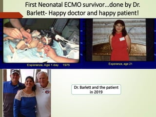
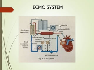
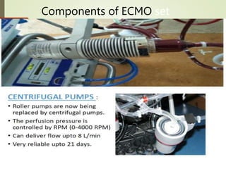
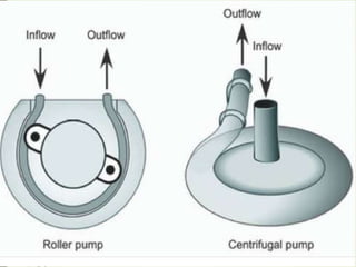
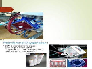
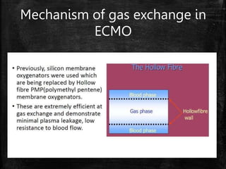
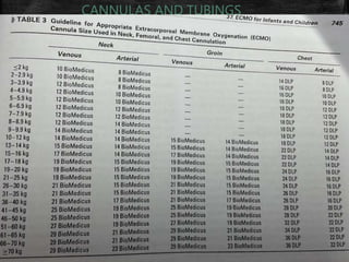
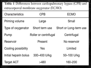
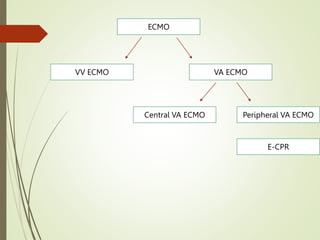
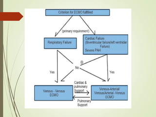
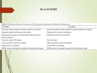
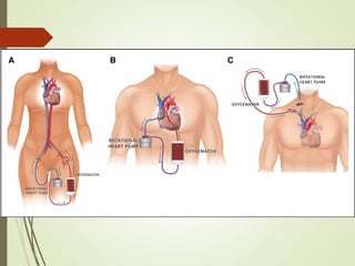
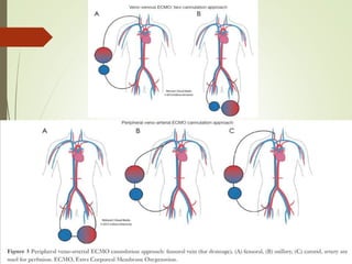
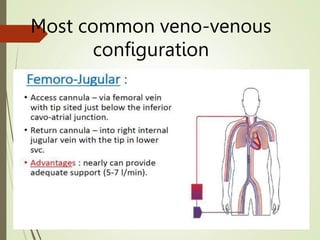
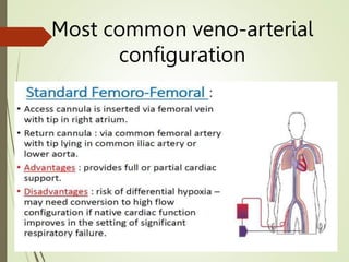
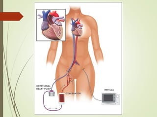
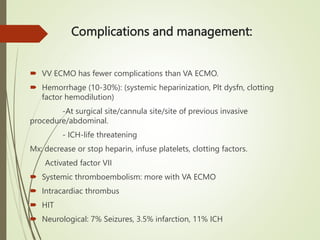
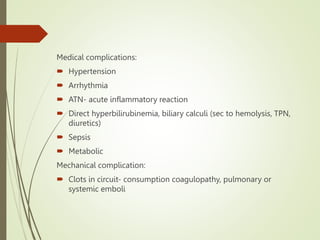
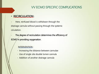
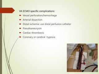
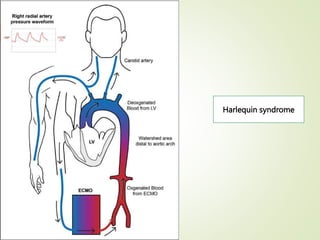
Basics of ECMO
Dr. Sundaram C
What is ECMO?
 Extracorporeal membrane oxygenation
 Rescue therapy in severe respiratory and/or cardiac failure
refractory to conventional treatment.
• Introduction
• History
• Modes of E C M O
• Circuit & components
• Indications
• Contraindications
HISTORY
1950s Development of membrane oxygenator in laboratory
1971 First successful case
1972 First successful paediatric cardiac case
1975 First neonatal case (Esperanza)
1975-89 Trial in ARDS, 10% survival
1990 Standard practice for neonates and pediatrics in some
centers
2000 Standard practice for adults in some centres
2009 Publication of the CESAR trial which led to a significant
growth in the use of ECMO forARDS cases
First successful ECMO
patient in1971
Figure: The first successful extracorporeal life support
patient, treated by J. Donald Hill using the Bramson
oxygenator (foreground), Santa Barbara, 1971.
First Neonatal ECMO survivor…done by Dr.
Barlett- Happy doctor and happy patient!
Dr. Barlett and the patient
in 2019
FROM THIS
TO THIS
ECMO SYSTEM
Components of ECMO set
up
Mechanism of gas exchange in
ECMO
CANNULAS AND TUBINGS
ECMO
VV ECMO VA ECMO
Peripheral VA ECMO
Central VA ECMO
E-CPR
VA vs VV ECMO
Most common veno-venous
configuration
Most common veno-arterial
configuration
VA ECMO
NIRS cerebral oximetry
Rt IJV-12 Fr
Rt CCA-10 Fr
Indications for VV ECMO:
 ARDS
 Airway obstruction, pulmonary contusion, smoke inhalation
 Lung transplant: primary graft failure, bridge to lung transplant
 Lung hyperinflation; status asthmaticus
 Pulmonary hemorrhage/massive hemoptysis
 CDH, meconium aspiration
VA ECMO:
Indications:
 Cardiogenic shock with biventricular failure
-ACS, Arrhythmic storm, Sepsis, Drug toxicity, Myocarditis,
Pulmonary embolism, Isolated cardiac trauma, Acute anaphylaxis
 Post cardiotomy: inability to wean from CPB
 Post heart transplant: primary graft failure
 Chronic cardiomyopathy
 High risk cardiac interventions
 Bridge to transplant
Indications of ECMO in children:
• Low cardiac output syndrome
• Failure to wean off CPB
• PH with circulatory failure
• Refractory arrhythmia
• Cardia arrest unresponsive to conventional resuscitation techniques
• Respiratory failure
• Profound cyanosis due to intracardiac shunting
• Support for intervention in Cath lab
• Bridge to transplant or assist devices
Contraindications:
 Unrecoverable heart and not a transplant candidate
 Disseminated malignancy
 Known severe brain injury
 E-CPR with unknown downtime, unwitnessed, unknown neurological status
 Unrepaired aortic dissection
 Other terminal illness( <2 yrs expected survival)
 Multi-organ system failure, Severe coagulopathy
 Severe AR
 CNS bleed
 VV ECMO contraindicated in cardiogenic failure, severe chronic PH (mPAP>50)
Rt IJV to RA
cannula
Rt Carotid
cannula
ETT
Lt IJV
line
Multiple side
holes
HEMODYNAMICS OF ECMO
Hemodynamic considerations:
 Drains blood from systemic veins: RV preload , peripheral venous
congestion.
 Residual flow through pulmonary circuit, thebesian drainage of CBF,
return of bronchial blood flow to LA.
 With increase in ECMO flow, RAP , Ao pressure , LVEDP , LVESP ,
LV volumes , SV , LAP , PAP , Arterial pulse pressure  Pulmonary edema.
 Ao valve barely opens  Stasis in LV  Thrombosis Stroke,
peripheral/pulmonary emboli.
 LVEDP reduces transcoronary perfusion gradient myocardial ischemia.
Physiologic consideration:
Blood exposure to nonendothelialized surface of ECMO circuit
Activation of innate immune system
Inflammatory response
At risk of LV distension and
pulmonary edema?
 Look at arterial pulse pressure tracing.
 MAP increases, but pulse pressure and SV decrease.
 Echo: Ao valve opening duration and extent (M mode), LV dimension
 Right radial artery or cerebral oximetry (hypoxia of blood exiting LV)
 CXR
 Best: PA catheter, measure PAdP/ PCWP.
Strategies for LV unloading:
Anticoagulation strategy?
 ACT 180-220 s
 aPTT: 50-80s (baseline aPTT prolonged in neonates-difficult
to monitor)
 Anti Xa assay: 0.3-0.7 IU/ml
 AT III functional assay: 80-120%. Repletion if less than 70%
Monitoring
 Paediatric: 150 ml/kg/min
 Flow through lungs- maintained by mechanical ventilator
with high PEEP.
 ACT 180-220s
 APTT 50-80s
 Platelet >50-75000
 NIRS for cerebral oxygenation
 Pre and post membrane pressures: <40mmHg difference
 Target CI >2.2, MAP: 65-80, CVP: 8-12, PCWP <18
Complications and management:
 VV ECMO has fewer complications than VA ECMO.
 Hemorrhage (10-30%): (systemic heparinization, Plt dysfn, clotting
factor hemodilution)
-At surgical site/cannula site/site of previous invasive
procedure/abdominal.
- ICH-life threatening
Mx: decrease or stop heparin, infuse platelets, clotting factors.
Activated factor VII
 Systemic thromboembolism: more with VA ECMO
 Intracardiac thrombus
 HIT
 Neurological: 7% Seizures, 3.5% infarction, 11% ICH
Medical complications:
 Hypertension
 Arrhythmia
 ATN- acute inflammatory reaction
 Direct hyperbilirubinemia, biliary calculi (sec to hemolysis, TPN,
diuretics)
 Sepsis
 Metabolic
Mechanical complication:
 Clots in circuit- consumption coagulopathy, pulmonary or
systemic emboli
• RECIRCULATION:
- Here, reinfused blood is withdrawn through the
drainage cannula without passing through the systemic
circulation.
- The degree of recirculation determines the efficiency of
ECMO in providing oxygenation.
INTERVENTION :
- Increasing the distance between cannulae
- Use of single site double lumen cannula
- Addition of another drainage cannula
VV ECMO SPECIFIC COMPLICATIONS
VA ECMO specific complications:
 Vessel perforation/hemorrhage
 Arterial dissection
 Distal ischemia: use distal perfusion catheter
 Pseudoaneurysm
 Cardiac thrombosis
 Coronary or cerebral hypoxia
Harlequin syndrome
WEANING OFF ECMO
• INDICATIONS :
- For patients with Respiratory failure, improvements in radiographic appearance, pulmonary
compliance and arterial oxyHb saturation.
- With cardiac failure, enhanced aortic pulsatility correlates with improved left
ventricular output.
- One or more trials of taking the patient off of ECMO should be performed prior to
discontinuing ECMO permanently.
Criteria prior to weaning:
 Recovering end organ function
 PaO2/FiO2 > 100
 Vasopressors and inotropes low levels
Daily weaning study:
 Perform every 24 hours.
 Reduce flow rate in increments of 0.5L/min to minimum 2L/min.
 Assess MAP, intracardiac pressures.
 Don’t wean if MAP falls >10-15 or <65mmHg
 significant increases in Rt filling pressure
 ABG, lactate, invasive hemodynamics reassessed.
Bedside assessment for
decannulation:
 Perform after 2L/min for 8 hrs with stable end organ perfusion.
 Decrease flow rate to 1 L/min for 1 min and assess.
 If tolerates, keep at 2 L/min and proceed.
 If fails, keep at 2L/min and reassess after 24 hrs.
Final assessment:
 Perform at operating room.
 Clamp cannulae, blood flow decreased
 Check hemodynamics, ABG
 Echo for BiV function
 If acceptable, decannulate.
 THANK YOU!

ECMO Basics.pptx

  • 1.
  • 2.
    What is ECMO? Extracorporeal membrane oxygenation  Rescue therapy in severe respiratory and/or cardiac failure refractory to conventional treatment.
  • 3.
    • Introduction • History •Modes of E C M O • Circuit & components • Indications • Contraindications HISTORY 1950s Development of membrane oxygenator in laboratory 1971 First successful case 1972 First successful paediatric cardiac case 1975 First neonatal case (Esperanza) 1975-89 Trial in ARDS, 10% survival 1990 Standard practice for neonates and pediatrics in some centers 2000 Standard practice for adults in some centres 2009 Publication of the CESAR trial which led to a significant growth in the use of ECMO forARDS cases
  • 4.
    First successful ECMO patientin1971 Figure: The first successful extracorporeal life support patient, treated by J. Donald Hill using the Bramson oxygenator (foreground), Santa Barbara, 1971.
  • 5.
    First Neonatal ECMOsurvivor…done by Dr. Barlett- Happy doctor and happy patient! Dr. Barlett and the patient in 2019
  • 6.
  • 7.
  • 8.
  • 11.
    Mechanism of gasexchange in ECMO
  • 12.
  • 15.
    ECMO VV ECMO VAECMO Peripheral VA ECMO Central VA ECMO E-CPR
  • 17.
  • 20.
  • 21.
  • 22.
    VA ECMO NIRS cerebraloximetry Rt IJV-12 Fr Rt CCA-10 Fr
  • 23.
    Indications for VVECMO:  ARDS  Airway obstruction, pulmonary contusion, smoke inhalation  Lung transplant: primary graft failure, bridge to lung transplant  Lung hyperinflation; status asthmaticus  Pulmonary hemorrhage/massive hemoptysis  CDH, meconium aspiration
  • 24.
    VA ECMO: Indications:  Cardiogenicshock with biventricular failure -ACS, Arrhythmic storm, Sepsis, Drug toxicity, Myocarditis, Pulmonary embolism, Isolated cardiac trauma, Acute anaphylaxis  Post cardiotomy: inability to wean from CPB  Post heart transplant: primary graft failure  Chronic cardiomyopathy  High risk cardiac interventions  Bridge to transplant
  • 25.
    Indications of ECMOin children: • Low cardiac output syndrome • Failure to wean off CPB • PH with circulatory failure • Refractory arrhythmia • Cardia arrest unresponsive to conventional resuscitation techniques • Respiratory failure • Profound cyanosis due to intracardiac shunting • Support for intervention in Cath lab • Bridge to transplant or assist devices
  • 26.
    Contraindications:  Unrecoverable heartand not a transplant candidate  Disseminated malignancy  Known severe brain injury  E-CPR with unknown downtime, unwitnessed, unknown neurological status  Unrepaired aortic dissection  Other terminal illness( <2 yrs expected survival)  Multi-organ system failure, Severe coagulopathy  Severe AR  CNS bleed  VV ECMO contraindicated in cardiogenic failure, severe chronic PH (mPAP>50)
  • 27.
    Rt IJV toRA cannula Rt Carotid cannula ETT Lt IJV line Multiple side holes
  • 28.
  • 29.
    Hemodynamic considerations:  Drainsblood from systemic veins: RV preload , peripheral venous congestion.  Residual flow through pulmonary circuit, thebesian drainage of CBF, return of bronchial blood flow to LA.  With increase in ECMO flow, RAP , Ao pressure , LVEDP , LVESP , LV volumes , SV , LAP , PAP , Arterial pulse pressure  Pulmonary edema.  Ao valve barely opens  Stasis in LV  Thrombosis Stroke, peripheral/pulmonary emboli.  LVEDP reduces transcoronary perfusion gradient myocardial ischemia.
  • 30.
    Physiologic consideration: Blood exposureto nonendothelialized surface of ECMO circuit Activation of innate immune system Inflammatory response
  • 31.
    At risk ofLV distension and pulmonary edema?  Look at arterial pulse pressure tracing.  MAP increases, but pulse pressure and SV decrease.  Echo: Ao valve opening duration and extent (M mode), LV dimension  Right radial artery or cerebral oximetry (hypoxia of blood exiting LV)  CXR  Best: PA catheter, measure PAdP/ PCWP.
  • 32.
  • 34.
    Anticoagulation strategy?  ACT180-220 s  aPTT: 50-80s (baseline aPTT prolonged in neonates-difficult to monitor)  Anti Xa assay: 0.3-0.7 IU/ml  AT III functional assay: 80-120%. Repletion if less than 70%
  • 36.
    Monitoring  Paediatric: 150ml/kg/min  Flow through lungs- maintained by mechanical ventilator with high PEEP.  ACT 180-220s  APTT 50-80s  Platelet >50-75000  NIRS for cerebral oxygenation  Pre and post membrane pressures: <40mmHg difference  Target CI >2.2, MAP: 65-80, CVP: 8-12, PCWP <18
  • 38.
    Complications and management: VV ECMO has fewer complications than VA ECMO.  Hemorrhage (10-30%): (systemic heparinization, Plt dysfn, clotting factor hemodilution) -At surgical site/cannula site/site of previous invasive procedure/abdominal. - ICH-life threatening Mx: decrease or stop heparin, infuse platelets, clotting factors. Activated factor VII  Systemic thromboembolism: more with VA ECMO  Intracardiac thrombus  HIT  Neurological: 7% Seizures, 3.5% infarction, 11% ICH
  • 39.
    Medical complications:  Hypertension Arrhythmia  ATN- acute inflammatory reaction  Direct hyperbilirubinemia, biliary calculi (sec to hemolysis, TPN, diuretics)  Sepsis  Metabolic Mechanical complication:  Clots in circuit- consumption coagulopathy, pulmonary or systemic emboli
  • 40.
    • RECIRCULATION: - Here,reinfused blood is withdrawn through the drainage cannula without passing through the systemic circulation. - The degree of recirculation determines the efficiency of ECMO in providing oxygenation. INTERVENTION : - Increasing the distance between cannulae - Use of single site double lumen cannula - Addition of another drainage cannula VV ECMO SPECIFIC COMPLICATIONS
  • 41.
    VA ECMO specificcomplications:  Vessel perforation/hemorrhage  Arterial dissection  Distal ischemia: use distal perfusion catheter  Pseudoaneurysm  Cardiac thrombosis  Coronary or cerebral hypoxia
  • 42.
  • 43.
    WEANING OFF ECMO •INDICATIONS : - For patients with Respiratory failure, improvements in radiographic appearance, pulmonary compliance and arterial oxyHb saturation. - With cardiac failure, enhanced aortic pulsatility correlates with improved left ventricular output. - One or more trials of taking the patient off of ECMO should be performed prior to discontinuing ECMO permanently.
  • 44.
    Criteria prior toweaning:  Recovering end organ function  PaO2/FiO2 > 100  Vasopressors and inotropes low levels
  • 45.
    Daily weaning study: Perform every 24 hours.  Reduce flow rate in increments of 0.5L/min to minimum 2L/min.  Assess MAP, intracardiac pressures.  Don’t wean if MAP falls >10-15 or <65mmHg  significant increases in Rt filling pressure  ABG, lactate, invasive hemodynamics reassessed.
  • 46.
    Bedside assessment for decannulation: Perform after 2L/min for 8 hrs with stable end organ perfusion.  Decrease flow rate to 1 L/min for 1 min and assess.  If tolerates, keep at 2 L/min and proceed.  If fails, keep at 2L/min and reassess after 24 hrs.
  • 47.
    Final assessment:  Performat operating room.  Clamp cannulae, blood flow decreased  Check hemodynamics, ABG  Echo for BiV function  If acceptable, decannulate.
  • 49.