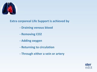
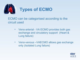
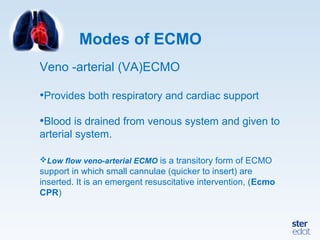
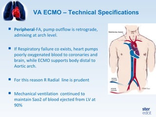
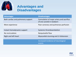
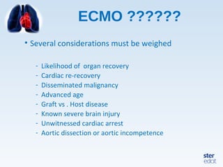
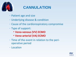
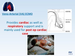
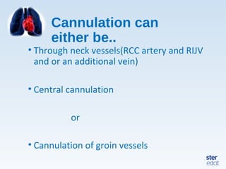
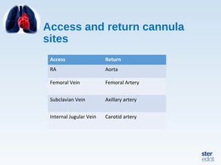
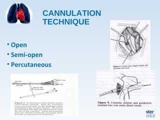
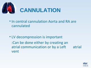
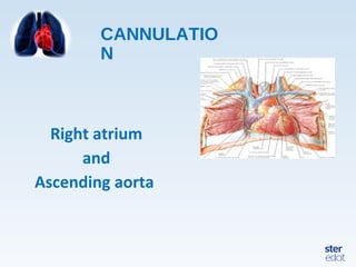
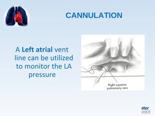
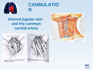
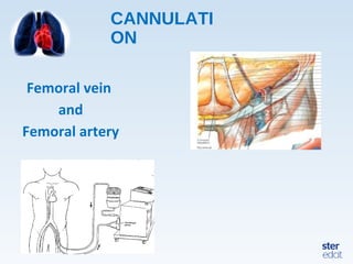
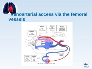
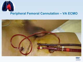
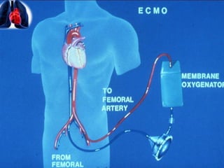
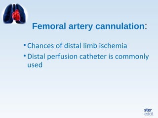
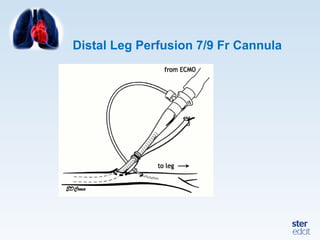
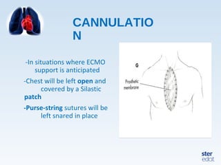
2. Extra corporeal Life Support is achieved by
- Draining venous blood
- Removing CO2
- Adding oxygen
- Returning to circulation
- Through either a vein or artery
3. Types of ECMO
ECMO can be categorised according to the
circuit used
• Veno-arterial - VA ECMO provides both gas
exchange and circulatory support (Heart &
Lung failure)
• Veno-venous –VAECMO allows gas exchange
only (Isolated Lung failure)
4. Modes of ECMO
Veno -arterial (VA)ECMO
•Provides both respiratory and cardiac support
•Blood is drained from venous system and given to
arterial system.
Low flow veno-arterial ECMO is a transitory form of ECMO
support in which small cannulae (quicker to insert) are
inserted. It is an emergent resuscitative intervention, (Ecmo
CPR)
5. VA ECMO – Technical Specifications
Peripheral-FA, pump outflow is retrograde,
admixing at arch level.
If Respiratory failure co exists, heart pumps
poorly oxygenated blood to coronaries and
brain, while ECMO supports body distal to
Aortic arch.
For this reason R Radial line is prudent
Mechanical ventilation continued to
maintain Sao2 of blood ejected from LV at
90%
6. First successful ECMO patient,
1971
J Donald Hill MD and Maury Bramson BME, Santa
Barbara, Ca, 1971. (Courtesy of Robert Bartlett, MD)
10. Advantages and
Disadvantages
Advantages Disadvantages
Both cardiac and pulmonary support Cannulation of major artery and sacrifice
of one carotid in newborn
More experience Poor coronary and pulmonary perfusion
Instant hemodynamic support Systemic thromboembolism
No recirculation Nonpulsatile flow
Right and left heart Myocardial stunning and LV distension
Increased incidence of neurological
events
11. ECMO ??????
• Several considerations must be weighed
- Likelihood of organ recovery
- Cardiac re-recovery
- Disseminated malignancy
- Advanced age
- Graft vs . Host disease
- Known severe brain injury
- Unwitnessed cardiac arrest
- Aortic dissection or aortic incompetence
12. • The physiologic goal is to improve tissue oxygen
delivery , remove CO2 and allow normal aerobic
metabolism whilst the lung rests
• ECMO circulation:
- Dual circulation
- Nonpulsatile flow
13. Dual
circulation
• Native circulation + ECMO circulation
• Sometimes Results in North South syndrome if the
return cannula is in femoral artery
• In most cases ECMO provides 60-80 % of CO
resulting in a discernible pulse contour
14. • Reduces preload
• Increases afterload Myocardial
stunning
• Left sided decompression
• Use of inodilators
15. In Veno Arterial
ECMO
• Perfusate saturation is 100%
• Without lung function LV saturation=RV saturation
• ABG reflects Perfusate+RV saturation
• If 50% blood flows through lungs,50% through
oxygenator –O2 saturation of arterial blood
becomes 90%
16. Increase in Systemic PO2 may
result from..
•Improved lung function at constant
flow
•Increased ECC flow at constant CO
17. CO2
removal..
• Mainly depends on oxygenator surface
area and sweep gas flow rate
• Independent of blood flow
• Moderately depends on inlet CO2
18. During Veno arterial
ECMO..
• O2 consumption decreases ( catecholamines
reduced,less metabolic stress)
• Arterial saturation >95 % and flow adjusted to maintain
arterial/venous saturation
• Bleeding Decreased venous return
Decrease in flow Transfusion
• Without lung function and ejection of heart arterial
saturation decreases
19. • Knowledge of the physiology of ECMO support
the management of ECMO patient
• O2 content is of utmost importance in the
physiologic management of critically ill patients
• DO2/VO2 ratio is reflected by mixed venous
saturation - most important monitor in critically
ill patients
21. CANNULATION
- Patient age and size
- Underlying disease & condition
- Cause of the cardiorespiratory compromise
- Type of support:
• Veno-venous (VV) ECMO
• Veno-arterial (VA) ECMO
- Time of the event in relation to the peri-
operative period
- Location
22. CANNULATI
ON
• For each modality, there are different
kinds and sizes of cannulae that can be
used
• Target ACTshould be accomplished
before ECMO (heparin 100 units/kg)
3 minutes before cannulation.
23. Cannula
Consideration
• Venous cannula should be with the largest
lumen and shortest length possible.
• Venous cannula should have side holes.
• M-number
• Resist kinking
• Smallest double lumen cannula is size 12 Fr
( for V V ecmo in neonate)
27. Cannulation can
either be..
• Through neck vessels(RCC artery and RIJV
and or an additional vein)
• Central cannulation
or
• Cannulation of groin vessels
30. CANNULATION
• In central cannulation Aorta and RA are
cannulated
• LV decompression is important
:Can be done either by creating an
atrial communication or by a Left atrial
vent
41. CANNULATIO
N
-In situations where ECMO
support is anticipated
-Chest will be left open and
covered by a Silastic
patch
-Purse-string sutures will be
left snared in place
42. CANNULATION
PROBLEMS
• Threading the venous catheter
• Vein division
• Proximal vein lost in
mediastinum
• Lack of venous return
• Intrathoracic vein perforation
44. Monitorin
g
• Success lies with vigilant monitoring which helps in early
recognition and diagnosis of problems and timely and
accurate action
• Monitoring includes:
-Clinical parameters
-Biochemical and
Laboratory parameters
-Radiologic monitoring
-Circuit monitoring
45. Clinical
parameters
• Vital parameters(ECG rhythm,Pulse rate,Blood
Pressure ,Temperature , and respiratory rate)
• Arterial and venous saturation(MAP and mixed
venous saturation)
• Daily Echocardiography
• Right hand saturation gives hints regarding
coronary perfusion
46. • Hematological parameters(CBC,HCT 35-
40%,Plasma Free Hemoglobin <0.1g/dl)
• If possible cerebral oxygenation with NIRS
• EtCO2
• RBS : 80-140 mg/dl
• ABG and RBS to be done every 4-6 hours
• Vascular and neurological status
• Urine output and urine colour
47. Coagulation
Monitoring
•Pre ECMO ACT 300 seconds
•On ECMO –Check ACT 2 hourly till
ACT drops to 200 seconds
•Maintenance with ACT 160-200 seconds
•ACT to be repeated every 6-8 hours
•aPTT : 60-70 seconds
48. Circuit
monitoring..
• Circuit blood flow
• Circuit gas flow
• Circuit pressure
• Circuit integrity
These variables should be monitored
continuosly
49. Factors affecting Circuit Blood
flow
Increased Blood flow Decreased Blood flow
Increased RPM Decreased RPM
Decreased Resistance
:Vasodilation
:Improved arterial cannula position
Increased Resistance
:vasoconstriction
:Kinking of tubing
:Improper cannula position
Increased preload
:Increased filling
:Improved venous cannula position
Decreased preload
:Hypovolemia
:Improper cannula position
:Kinking of tubing
50. Circuit
Pressure
• Pre pump pressure: too much negative pressure
results in Hemolysis, cavitation, endothelial
damage of Right atrium and vena cava
• Reasons : Hypovolemia,
Improper catheter placement,
Inadequate cannula size.