Downloaded 442 times


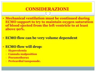

![INSUFFICIENZA
CARDIACA
• Cardiomiopatia end-stage in terapia medica massimale, in
attesa di trapianto cardiaco;
• Infarto miocardico acuto complicato da shock cardiogeno
refrattario
• Miocardite acuta con severa insufficienza d’organo o aritmie
ventricolari subentranti non controllabili con terapia medica e
IABP;
• Embolia polmonare massiva con grave compromissione della
funzionalità ventricolare destra e shock cardiogeno o ACR
• Arresto cardiaco nel paziente giovane adulto con precoce
rianimazione cardiopolmonare (con verosimile ottima
prognosi neurologica) refrattario a terapia rianimatoria
medica ed elettrica. La durata del supporto con ECMO dovrà
essere il più breve possibile (sconsigliato per più di 3 giorni).
[Da associare ad altre strategie di neuroprotezione come
l’ipotermia terapeutica]
• Grave depressione della funzione cardiaca da intossicazione di
farmaci o sepsi;](https://image.slidesharecdn.com/ecmo-140129172444-phpapp02/85/Ecmo-17-320.jpg)
![Indicazioni ECMO
in UTIC
Si deve tener conto:
• della prognosi, in particolare ripresa della
funzionalità dell’organo [bridge-torecovery],
• dell’eleggibilità per un trapianto
cardiaco [bridge-to-transplantation],
• della possibilità di posizionamento di
assistenze meccaniche più o meno a
lunga durata (come Levitronix
CentriMag, Jarvik 2000, Cardiowest)
[bridge-to-bridge].
• Ma anche ……….. bridge to decision.](https://image.slidesharecdn.com/ecmo-140129172444-phpapp02/85/Ecmo-18-320.jpg)


Il documento esamina l'uso dell'ECMO (ossigenazione extracorporea a membrana) per il supporto di pazienti con insufficienza respiratoria e cardiaca, evidenziando esperienze a breve termine e tassi di sopravvivenza variabili tra neonati, bambini e adulti. Viene discusso il meccanismo dell'ECMO, le indicazioni, le controindicazioni e le complicazioni associate al trattamento, nonché l'importanza della valutazione multidisciplinare. Si suggerisce la necessità di un'organizzazione di rete per il supporto ECMO con strutture adeguate e protocolli per il trasferimento dei pazienti.


