MDT
• CT surgeon/vascular surgeon
• Perfusionist
• Nursing team
• Respiratory therapist
• Physiotherapist
• Infection control team
• INTENSIVIST
• Others
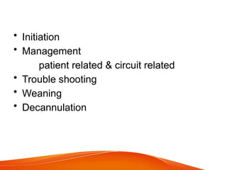
5.
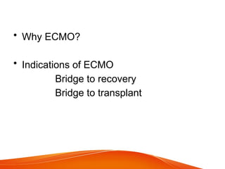
• Why ECMO?
•Indications of ECMO
Bridge to recovery
Bridge to transplant
6.
Indications
One or moreof the following:
• Hypoxemic respiratory failure (PaO2/FiO2
< 80 mm Hg), after optimal medical
management, including, in the absence of
contraindications, a trial of prone
positioning.
• Hypercapneic respiratory failure (pH <
7.25), despite optimal conventional
mechanical ventilation (respiratory rate 35
bpm and plateau pressure [Pplat] ≤ 30 cm
H2O and driving pressures of > 16cm H2O
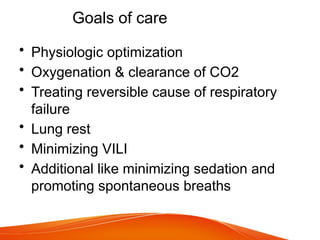
Goals of care
•Physiologic optimization
• Oxygenation & clearance of CO2
• Treating reversible cause of respiratory
failure
• Lung rest
• Minimizing VILI
• Additional like minimizing sedation and
promoting spontaneous breaths
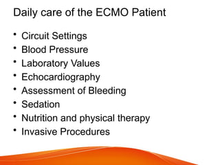
9.
Daily care ofthe ECMO Patient
• Circuit Settings
• Blood Pressure
• Laboratory Values
• Echocardiography
• Assessment of Bleeding
• Sedation
• Nutrition and physical therapy
• Invasive Procedures
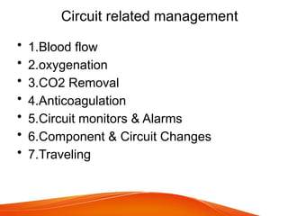
Blood flow
• ForVV ECMO 60-80 cc/kg
• For cardiac support 3L/min/mt2
• Around 60 cc/kg
• Max flow intially lowest possible gradually
• Calcualte DO2/VO2 ratio ratio >3 should
be adequate.
12.
• Oxygenation
• SPO280-85% acceptable
• CO2 removal
• Sweep gas flow usually 1:1
• For only CO2 removal 1:15.
13.
Anticoagulation and ECMO
•Before Initiating - CBC, PT INR, aPTT, and
fibrinogen.
• Initiation of Anticoagulation Heparin 50-
100 units/kg
• Monitoring of Anticoagulation -aPTT / ACT
• HIT (4T score)
• Argatroban / Bivalirudin (direct thrombin
inhibitors)
14.
• Circuit alarmsand monitors
• For safety of patient
• Component change if required
• Transportation and planning
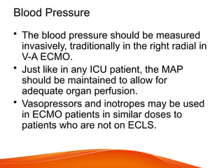
Blood Pressure
• Theblood pressure should be measured
invasively, traditionally in the right radial in
V-A ECMO.
• Just like in any ICU patient, the MAP
should be maintained to allow for
adequate organ perfusion.
• Vasopressors and inotropes may be used
in ECMO patients in similar doses to
patients who are not on ECLS.
17.
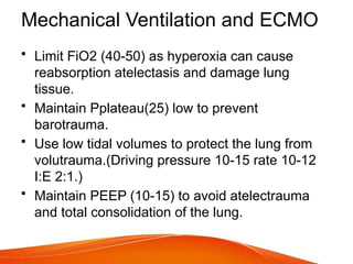
Mechanical Ventilation andECMO
• Limit FiO2 (40-50) as hyperoxia can cause
reabsorption atelectasis and damage lung
tissue.
• Maintain Pplateau(25) low to prevent
barotrauma.
• Use low tidal volumes to protect the lung from
volutrauma.(Driving pressure 10-15 rate 10-12
I:E 2:1.)
• Maintain PEEP (10-15) to avoid atelectrauma
and total consolidation of the lung.
18.
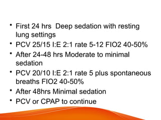
• First 24hrs Deep sedation with resting
lung settings
• PCV 25/15 I:E 2:1 rate 5-12 FIO2 40-50%
• After 24-48 hrs Moderate to minimal
sedation
• PCV 20/10 I:E 2:1 rate 5 plus spontaneous
breaths FIO2 40-50%
• After 48hrs Minimal sedation
• PCV or CPAP to continue
19.
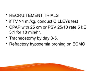
• RECRUITEMENT TRIALS
•if TV >4 ml/kg, conduct CILLEYs test
• CPAP with 25 cm or PSV 25/10 rate 5 I:E
3:1 for 10 min/hr.
• Tracheostomy by day 3-5.
• Refractory hypoxemia proning on ECMO
Assessment of Bleeding
•Intracranial bleeding
• GI Bleeding
• Intrathoracic bleeding
• Pericardial bleeding
• Retroperitoneal bleeding
• Cannulation site bleeding
• Surgical site bleeding
22.
Invasive Procedures
• ArterialLine Insertion
• Central Venous Cahteter Insertion
• Bronchoscopy
• Chest Tube Insertion
• Patients Requiring Surgery
23.
Weaning from ECMO
•The native lungs are providing ≥ 70-80%
of oxygenation.
• Pulmonary compliance and airway
resistance allow for ventilation at
reasonable pressures.
• The FiO2 provided by the ventilator is ≤
50-60%.
• The PaCO2 can be maintained at a near-
normal level within the range of acceptable
ventilator settings.
24.
• The sweepgas flow to the oxygenator is
reduced to zero.
• The patient is observed for signs of
respiratory distress.
• The tidal volumes, minute ventilation and
respiratory rate are measured on the
ventilator.
• The patient's vital signs are monitored.
• Serial blood gases are taken to monitor
the gas exchange of the native lungs.
25.
• Typically patientsare trialled "off sweep"
for a period of 24 hours before a decision
is made to decannulate.
26.
Decannulation
• Heparin tobe off 30-60mt prior to decannulation.
• The VV lines are removed under LA.
• Pursestring sutures are placed by the surgical
team, and the lines are withdrawn sequentially.
• Pressure is placed on the site until bleeding
stops.
• When removing a venous cannula in a
spontaneously breathing patient, there is a risk
of entraining air through the sideholes, so a
valsalva maneuver can be employed to prevent
this.
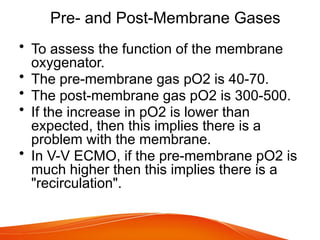
Pre- and Post-MembraneGases
• To assess the function of the membrane
oxygenator.
• The pre-membrane gas pO2 is 40-70.
• The post-membrane gas pO2 is 300-500.
• If the increase in pO2 is lower than
expected, then this implies there is a
problem with the membrane.
• In V-V ECMO, if the pre-membrane pO2 is
much higher then this implies there is a
"recirculation".
29.
Troubleshooting: Chatter
• Chatteris caused by a supply-demand mismatch
between the pump speed and the blood supply
to the pump.
• The pump creates a negative gradient and tries
to "suck" blood from the patient into the circuit.
• If there isn't enough blood available to flow into
the circuit, then the negative pressure relative to
the atmosphere causes the circuit to twist and
straighten.
• It is important to distinguish chatter from normal
pulsations in the circuit from the patient's cardiac
cycle
30.
• Temporarily lowerthe RPM of the pump
until the chatter resolves.
• Administer an intravenous fluid bolus
appropriate for the given clinical scenario
(e.g. crystalloid, albumin, or blood
products).
• Restore the pump speed and see if the
chatter persists.
31.
• A kinkedor malpositioned drainage cannula
causing the vein to collapse, or "suck down"; this
can be identified by an x-ray or
echocardiography.
• This is especially common with an Avalon
catheter, as the flow is exquisitely sensitive to
position and it can chatter very easily.
• Increased intraabdominal pressure.
• Increased intrathoracic pressure.
• Haemorrhage.
32.
Troubleshooting: Dropping Flows
•Low flows can lead to clotting in the circuit.
• Low flows are usually indicative of a
sinister process which needs to be
addressed.
33.
• Make surethe pump speed is at the target
RPM.
• Inspect the circuit for kinks or occlusions.
• Inspect the membrane for large clots.
• Inspect the cannula sites to ensure there
has been no malposition.
• Inspect the circuit for chatter and look for
other signs of fluid responsiveness.
34.
• It isreasonable to try a bolus of fluid if
pt.has intravascular volume depletion.
• If the above measures do not solve the
problem, then consult surgical team.
• Cannula position may be checked with a
chest x ray or echo.
• If a patient is centrally cannulated, then an
echo is extremely important to help rule
out central obstruction.
35.
Troubleshooting: Bleeding Cannula
Site
•Alert the surgical team that inserted the
cannulas.
• Ensure the cannula is positioned properly
at the skin.
• Place absorbent dressings at the site.
• Apply firm sustained manual pressure for
5-15 minutes.
36.
• Heparin mayneed to be temporarily
discontinued.
• Correction of coagulopathy: aim for a
near-normal INR, platelets >50-100,
fibrinogen >1.5gm.
• A surgical repair might be necessary,
either at the bedside or in the operating
room.
37.
Troubleshooting: Hemolysis
• Anemia.
•Increased bilirubin, LDH, and free Hb.
• Pigment nephropathy: dark urine, or high
potassium in CRRT effluent if patient is on
dialysis.
38.
• Relatively highpump speeds.
• Malposition of the drainage cannula
causing a "suck down" effect in the IVC or
SVC.
• Thrombosis of the centrifugal pump.
39.
• Transfuse redblood cells as necessary to
maintain an appropriate hemoglobin level.
• A trial of reduced pump speed may be
warranted.
• Repositioning of the drainage cannula may
be necessary if a "suck down" effect is
seen.
40.
Troubleshooting: Air inthe Circuit
• IF Small amount of air
• This will not cause the circuit to
malfunction, but it should be removed
immediately.
• Try to vent the air by removing the yellow
cap on the venous side of the oxygenator
(this is sometimes called "burping" the
circuit).
• Try to identify the source of the air.
42.
• If largeamount of air in the circuit
• This is an emergency, as an "air lock" can
cause flow to arrest in the circuit.
• Call for help immediately.
• Try to vent the air by "burping the circuit",
but if there is an air lock, this will not help.
• Do not clamp or cut any tubing until a plan
has been made to replace the circuit.
43.
Troubleshooting: Pump Failure
•Before troubleshooting pump failure, call
for help from the perfusionist, ICU staff
and surgical staff immediately
• Insufficient power provided to turn the
motor of the centrifugal pump.
• Malfunction of the motor itself.
• Displacement of the centrifugal impeller.
45.
RESPIRATORY AND VENTILATORY
MANAGEMENT
•Lung protective ventilation – ultra low tv
4ml/kg body wt, rate 10-15, peep 10-15,
plateau <25.
• Bronchoscopy
• Tracheostomy
• Refractory hypoxemia proning on ECMO
• Sedation and analgesia


![Indications
One or more of the following:
• Hypoxemic respiratory failure (PaO2/FiO2
< 80 mm Hg), after optimal medical
management, including, in the absence of
contraindications, a trial of prone
positioning.
• Hypercapneic respiratory failure (pH <
7.25), despite optimal conventional
mechanical ventilation (respiratory rate 35
bpm and plateau pressure [Pplat] ≤ 30 cm
H2O and driving pressures of > 16cm H2O](https://image.slidesharecdn.com/ecmopart-2updated-250317053022-3377dfcc/85/ECMO-part-2-Updated-guidelines-on-life-support-6-320.jpg)