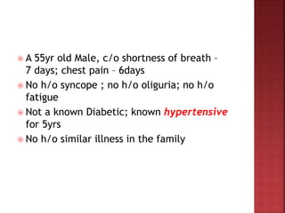
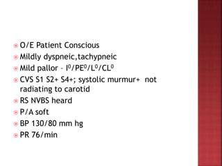
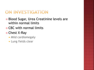
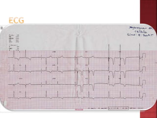
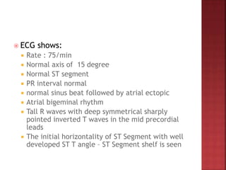
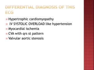
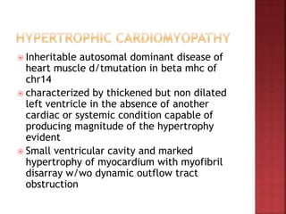
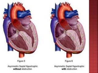
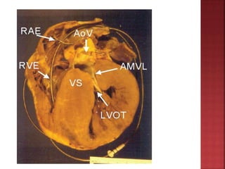
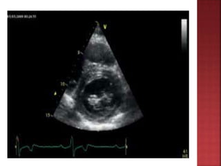
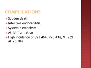
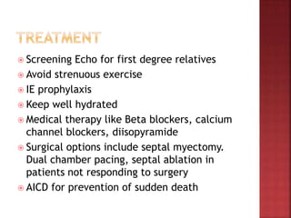
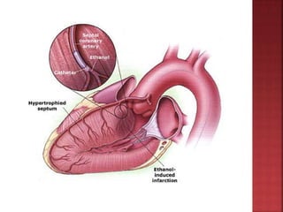
 A 55yr old Male, c/o shortness of breath –
7 days; chest pain – 6days
 No h/o syncope ; no h/o oliguria; no h/o
fatigue
 Not a known Diabetic; known hypertensive
for 5yrs
 No h/o similar illness in the family
 O/E Patient Conscious
 Mildly dyspneic,tachypneic
 Mild pallor – I0/PE0/L0/CL0
 CVS S1 S2+ S4+; systolic murmur+ not
radiating to carotid
 RS NVBS heard
 P/A soft
 BP 130/80 mm hg
 PR 76/min
 Blood Sugar, Urea Creatinine levels are
within normal limits
 CBC with normal limits
 Chest X-Ray
 Mild cardiomegaly
 Lung fields clear
 ECG shows:
 Rate : 75/min
 Normal axis of 15 degree
 Normal ST segment
 PR interval normal
 normal sinus beat followed by atrial ectopic
 Atrial bigeminal rhythm
 Tall R waves with deep symmetrical sharply
pointed inverted T waves in the mid precordial
leads
 The initial horizontality of ST Segment with well
developed ST T angle – ST Segment shelf is seen
 Hypertrophic cardiomyopathy
 lV SYSTOLIC OVERLOAD like hypertension
 Myocardial ischemia
 CVA with qrs st pattern
 Valvular aortic stenosis
 Inheritable autosomal dominant disease of
heart muscle d/tmutation in beta mhc of
chr14
 characterized by thickened but non dilated
left ventricle in the absence of another
cardiac or systemic condition capable of
producing magnitude of the hypertrophy
evident
 Small ventricular cavity and marked
hypertrophy of myocardium with myofibril
disarray w/wo dynamic outflow tract
obstruction
 Most common cause of sudden cardiac death
in young people including trained athelets
 WHO designated with HCM to describe this
unique process of primary muscle
hypertrophy
 M mode echo define ASH
 Myocardial disarray of muscle fibre result in
WHORLING characteristic of HCM
 Diastolic dysfunction
 LV Out flow tract obstruction
 Mitral regurgitation due to elongated mitral
leaflets and chordae
 Intramyocardial ischaemia due to partially
obliterated intra mural coronary arteries
 Arrythymias
 Autonomic dysfunction – systolic BP↓ on
exercise
 MANEUVER PHYSIOL EFFECT HCM AS MR
Valsalva vr,svr,co
squat&hand vr,svr,co
 Grip &phenyl
ephrine
 Amylnitrite vr, dec svr
 Parameters associated with sudden death
 survivor of cardiac arrest
 Sustained VT
 Family history of premature sudden death
 Massive degree of ventricular hypertrophy
 Hypotensive response to exercise
 Myocardial bridging
 Septal thickness > 30mm
Troponin t mutation
Courtesy Braunwald heart diseases & Harrison Medicine
 LV hypertrophy with septum >1.3times
posterior LV wall thickness
 Ground glass appearance of septum
 Spade shaped LV Cavity
 small lv cavity
 SAM of mitral valve
 septal immobility
 premature closure of aortic valve
 resting gradient>30mm
 provocable gradient>50mm
 TYPE 1 ..ANT SEPTUM 10%
 TYPE 2…ANT AND POST SEPTUM 20%
 TYPE 3 ..ANT AND POST SEPTUM INCLUDING
LAT.FREE WALL 52%
 TYPE 4 ..REGION OTHER THAN SEPTUM AND
POST FREE WALL 18%
 Sudden death
 Infective endocarditis
 Systemic embolism
 Atrial fibrillation
 High incidence of SVT 46%, PVC 43%, VT 26%
AF 25-30%
 Screening Echo for first degree relatives
 Avoid strenuous exercise
 IE prophylaxis
 Keep well hydrated
 Medical therapy like Beta blockers, calcium
channel blockers, diisopyramide
 Surgical options include septal myectomy.
Dual chamber pacing, septal ablation in
patients not responding to surgery
 AICD for prevention of sudden death
ECG: Atrial Bigeminy with deep inverted T waves

ECG: Atrial Bigeminy with deep inverted T waves

  • 2.
     A 55yrold Male, c/o shortness of breath – 7 days; chest pain – 6days  No h/o syncope ; no h/o oliguria; no h/o fatigue  Not a known Diabetic; known hypertensive for 5yrs  No h/o similar illness in the family
  • 3.
     O/E PatientConscious  Mildly dyspneic,tachypneic  Mild pallor – I0/PE0/L0/CL0  CVS S1 S2+ S4+; systolic murmur+ not radiating to carotid  RS NVBS heard  P/A soft  BP 130/80 mm hg  PR 76/min
  • 4.
     Blood Sugar,Urea Creatinine levels are within normal limits  CBC with normal limits  Chest X-Ray  Mild cardiomegaly  Lung fields clear
  • 6.
     ECG shows: Rate : 75/min  Normal axis of 15 degree  Normal ST segment  PR interval normal  normal sinus beat followed by atrial ectopic  Atrial bigeminal rhythm  Tall R waves with deep symmetrical sharply pointed inverted T waves in the mid precordial leads  The initial horizontality of ST Segment with well developed ST T angle – ST Segment shelf is seen
  • 7.
     Hypertrophic cardiomyopathy lV SYSTOLIC OVERLOAD like hypertension  Myocardial ischemia  CVA with qrs st pattern  Valvular aortic stenosis
  • 8.
     Inheritable autosomaldominant disease of heart muscle d/tmutation in beta mhc of chr14  characterized by thickened but non dilated left ventricle in the absence of another cardiac or systemic condition capable of producing magnitude of the hypertrophy evident  Small ventricular cavity and marked hypertrophy of myocardium with myofibril disarray w/wo dynamic outflow tract obstruction
  • 11.
     Most commoncause of sudden cardiac death in young people including trained athelets  WHO designated with HCM to describe this unique process of primary muscle hypertrophy  M mode echo define ASH  Myocardial disarray of muscle fibre result in WHORLING characteristic of HCM
  • 12.
     Diastolic dysfunction LV Out flow tract obstruction  Mitral regurgitation due to elongated mitral leaflets and chordae  Intramyocardial ischaemia due to partially obliterated intra mural coronary arteries  Arrythymias  Autonomic dysfunction – systolic BP↓ on exercise
  • 13.
     MANEUVER PHYSIOLEFFECT HCM AS MR Valsalva vr,svr,co squat&hand vr,svr,co  Grip &phenyl ephrine  Amylnitrite vr, dec svr
  • 14.
     Parameters associatedwith sudden death  survivor of cardiac arrest  Sustained VT  Family history of premature sudden death  Massive degree of ventricular hypertrophy  Hypotensive response to exercise  Myocardial bridging  Septal thickness > 30mm Troponin t mutation Courtesy Braunwald heart diseases & Harrison Medicine
  • 15.
     LV hypertrophywith septum >1.3times posterior LV wall thickness  Ground glass appearance of septum  Spade shaped LV Cavity  small lv cavity  SAM of mitral valve  septal immobility  premature closure of aortic valve  resting gradient>30mm  provocable gradient>50mm
  • 16.
     TYPE 1..ANT SEPTUM 10%  TYPE 2…ANT AND POST SEPTUM 20%  TYPE 3 ..ANT AND POST SEPTUM INCLUDING LAT.FREE WALL 52%  TYPE 4 ..REGION OTHER THAN SEPTUM AND POST FREE WALL 18%
  • 18.
     Sudden death Infective endocarditis  Systemic embolism  Atrial fibrillation  High incidence of SVT 46%, PVC 43%, VT 26% AF 25-30%
  • 19.
     Screening Echofor first degree relatives  Avoid strenuous exercise  IE prophylaxis  Keep well hydrated  Medical therapy like Beta blockers, calcium channel blockers, diisopyramide  Surgical options include septal myectomy. Dual chamber pacing, septal ablation in patients not responding to surgery  AICD for prevention of sudden death