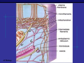
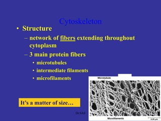
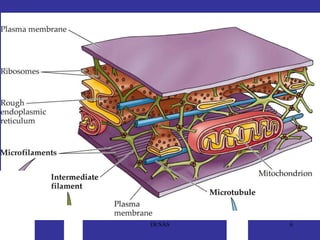
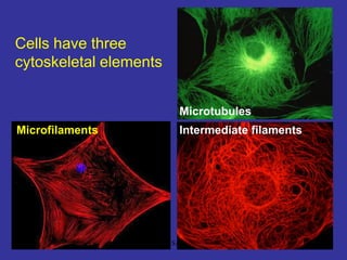
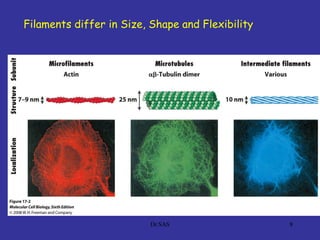
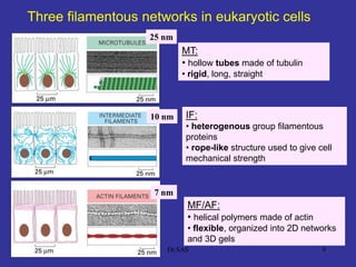
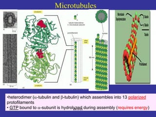
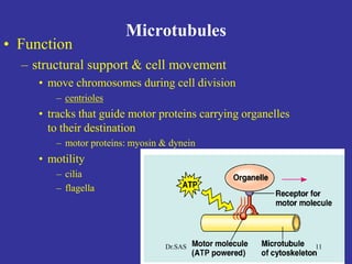
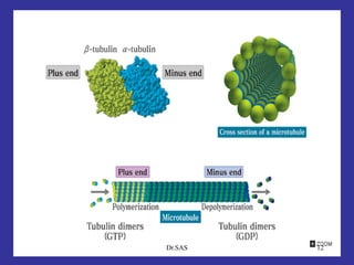
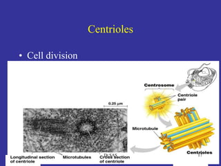
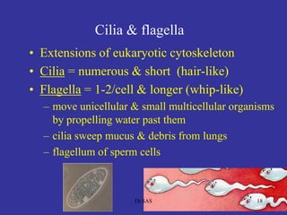
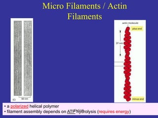
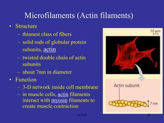
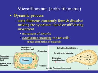
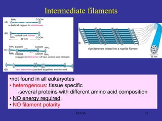
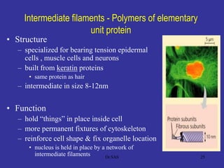
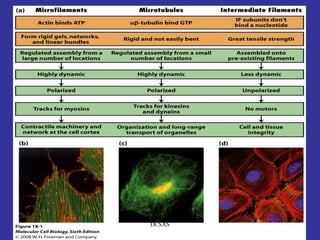
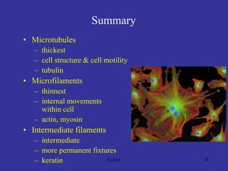
11
CB / U-01 / Cytoskeletal Proteins
Dr.SAS
22
Cytoskeleton: the skeleton of a cell
Cells need a (cyto)skeleton to:
•create shape
•change shape
•allow movement
=
dynamic!
Dr.SAS
AP Biology Dr.SAS
Cytoskeleton
• Function
– structural support
• maintains shape of cell
• provides anchorage for organelles
– motility
• cell locomotion
• cilia, flagella, etc.
– regulation
• organizes structures &
activities of cell
4Dr.SAS
Cytoskeleton
• Structure
– network of fibers extending throughout
cytoplasm
– 3 main protein fibers
• microtubules
• intermediate filaments
• microfilaments
It’s a matter of size…
5Dr.SAS
6Dr.SAS
77
Cells have three
cytoskeletal elements
Microfilaments Intermediate filaments
Microtubules
Dr.SAS
8
Filaments differ in Size, Shape and Flexibility
Dr.SAS
99
Three filamentous networks in eukaryotic cells
MT:
• hollow tubes made of tubulin
• rigid, long, straight
MF/AF:
• helical polymers made of actin
• flexible, organized into 2D networks
and 3D gels
IF:
• heterogenous group filamentous
proteins
• rope-like structure used to give cell
mechanical strength
25 nm
10 nm
7 nm
Dr.SAS
1010
Microtubules
•heterodimer ( -tubulin and -tubulin) which assembles into 13 polarized
protofilaments
• GTP bound to -subunit is hydrolyzed during assembly (requires energy)Dr.SAS
Microtubules
• Function
– structural support & cell movement
• move chromosomes during cell division
– centrioles
• tracks that guide motor proteins carrying organelles
to their destination
– motor proteins: myosin & dynein
• motility
– cilia
– flagella
11Dr.SAS
12Dr.SAS
• Basic arrangement -Tubulin (helical shape with
13 tubulin molecules/turn
• In addition to the tubulin, there are about 20-25
secondary proteins which is called Microtubule
Associated Proteins (MAPs).
• Few other variety of proteins that move along
them, form cross-bridges or influence their state
of polymerization - Kinesin and Cytoplasmic
dynein
13Dr.SAS
Microtubule Motor Protein
Kinesin:
• Move from – end to + end
• Transport organelles
• Mitosis and meiosis transport
• Synaptic vesicles along axons
Dynein :
• Move from + end to – end
• Cilia dynein – cilia movement
• Cytoplasmic dynein – organelle transport, mitosis
14Dr.SAS
• ‘Molecular motor or motor molecule’ in cells which converts the
chemical energy contained in ATP into the mechanical energy of
movement.
• Dynein transports various cellular cargo by "walking"
along cytoskeletal microtubules towards the minus-end of the
microtubule, which is usually oriented towards the cell center.
• Thus, they are called "minus-end directed motors."
• This form of transport is known as retrograde transport.
• In CONTRAST, kinesins are motor proteins that move toward
the microtubules' plus end, are called plus-end directed motors.
Dynein is a motor protein
15Dr.SAS
Microtubule -Function
1. Mechanical function
2. Cell membrane movement
3. Movement of cilia and flagella
4. Chromosomal movement during cell
division
5. Circulation and transport
16Dr.SAS
Centrioles
• Cell division
– in animal cells, pair of centrioles
organize microtubules guiding chromosomes in
cell division
17Dr.SAS
Cilia & flagella
• Extensions of eukaryotic cytoskeleton
• Cilia = numerous & short (hair-like)
• Flagella = 1-2/cell & longer (whip-like)
– move unicellular & small multicellular organisms
by propelling water past them
– cilia sweep mucus & debris from lungs
– flagellum of sperm cells
18Dr.SAS
1919
• a polarized helical polymer
• filament assembly depends on ATP hydrolysis (requires energy)
Micro Filaments / Actin
Filaments
Dr.SAS
Microfilaments (Actin filaments)
• Structure
– thinnest class of fibers
– solid rods of globular protein
subunits, actin
– twisted double chain of actin
subunits
– about 7nm in diameter
• Function
– 3-D network inside cell membrane
– in muscle cells, actin filaments
interact with myosin filaments to
create muscle contraction
20Dr.SAS
Microfilaments (actin filaments)
• Dynamic process
– actin filaments constantly form & dissolve
making the cytoplasm liquid or stiff during
movement
• movement of Amoeba
• cytoplasmic streaming in plant cells
– speeds distribution of materials
21Dr.SAS
Microfilaments -Function
• Shape – Cytoskeleton
• Muscle Contraction
• Amoeboid movement
• Cytoplasmic streaming
• Cytokinesis
• Biconcave shape for RBC is by actin
filaments
22Dr.SAS
2323
•not found in all eukaryotes
• heterogenous: tissue specific
-several proteins with different amino acid composition
• NO energy required,
• NO filament polarity
Intermediate filaments
Dr.SAS
2424
Higher order organization of IF has functional
consequences
Monomers
Polymers
(protofilament)
Filaments
Dr.SAS
Intermediate filaments - Polymers of elementary
unit protein
• Structure
– specialized for bearing tension epidermal
cells , muscle cells and neurons
– built from keratin proteins
• same protein as hair
– intermediate in size 8-12nm
• Function
– hold “things” in place inside cell
– more permanent fixtures of cytoskeleton
– reinforce cell shape & fix organelle location
• nucleus is held in place by a network of
intermediate filaments 25Dr.SAS
The polymerization steps of
intermediate filaments
• two basic units (i.e., monomers) associate in
the same direction to form a dimer.
•Two dimers running in
the reverse direction are line up
to form a tetramer, with the two strands
slightly offset in their opposite directions.
•Tetramers are arranged side by side to form a
protofilament.
•Eight protofilaments are
bundled to form an intermediate filament with
a diameter of approximately 10 nm.
Lack of polarity/Does not require GTP or ATP
26Dr.SAS
27Dr.SAS
Summary
• Microtubules
– thickest
– cell structure & cell motility
– tubulin
• Microfilaments
– thinnest
– internal movements
within cell
– actin, myosin
• Intermediate filaments
– intermediate
– more permanent fixtures
– keratin 28Dr.SAS

Dr.sas cytoskeletal proteins

  • 1.
    11 CB / U-01/ Cytoskeletal Proteins Dr.SAS
  • 2.
    22 Cytoskeleton: the skeletonof a cell Cells need a (cyto)skeleton to: •create shape •change shape •allow movement = dynamic! Dr.SAS
  • 3.
  • 4.
    Cytoskeleton • Function – structuralsupport • maintains shape of cell • provides anchorage for organelles – motility • cell locomotion • cilia, flagella, etc. – regulation • organizes structures & activities of cell 4Dr.SAS
  • 5.
    Cytoskeleton • Structure – networkof fibers extending throughout cytoplasm – 3 main protein fibers • microtubules • intermediate filaments • microfilaments It’s a matter of size… 5Dr.SAS
  • 6.
  • 7.
    77 Cells have three cytoskeletalelements Microfilaments Intermediate filaments Microtubules Dr.SAS
  • 8.
    8 Filaments differ inSize, Shape and Flexibility Dr.SAS
  • 9.
    99 Three filamentous networksin eukaryotic cells MT: • hollow tubes made of tubulin • rigid, long, straight MF/AF: • helical polymers made of actin • flexible, organized into 2D networks and 3D gels IF: • heterogenous group filamentous proteins • rope-like structure used to give cell mechanical strength 25 nm 10 nm 7 nm Dr.SAS
  • 10.
    1010 Microtubules •heterodimer ( -tubulinand -tubulin) which assembles into 13 polarized protofilaments • GTP bound to -subunit is hydrolyzed during assembly (requires energy)Dr.SAS
  • 11.
    Microtubules • Function – structuralsupport & cell movement • move chromosomes during cell division – centrioles • tracks that guide motor proteins carrying organelles to their destination – motor proteins: myosin & dynein • motility – cilia – flagella 11Dr.SAS
  • 12.
  • 13.
    • Basic arrangement-Tubulin (helical shape with 13 tubulin molecules/turn • In addition to the tubulin, there are about 20-25 secondary proteins which is called Microtubule Associated Proteins (MAPs). • Few other variety of proteins that move along them, form cross-bridges or influence their state of polymerization - Kinesin and Cytoplasmic dynein 13Dr.SAS
  • 14.
    Microtubule Motor Protein Kinesin: •Move from – end to + end • Transport organelles • Mitosis and meiosis transport • Synaptic vesicles along axons Dynein : • Move from + end to – end • Cilia dynein – cilia movement • Cytoplasmic dynein – organelle transport, mitosis 14Dr.SAS
  • 15.
    • ‘Molecular motoror motor molecule’ in cells which converts the chemical energy contained in ATP into the mechanical energy of movement. • Dynein transports various cellular cargo by "walking" along cytoskeletal microtubules towards the minus-end of the microtubule, which is usually oriented towards the cell center. • Thus, they are called "minus-end directed motors." • This form of transport is known as retrograde transport. • In CONTRAST, kinesins are motor proteins that move toward the microtubules' plus end, are called plus-end directed motors. Dynein is a motor protein 15Dr.SAS
  • 16.
    Microtubule -Function 1. Mechanicalfunction 2. Cell membrane movement 3. Movement of cilia and flagella 4. Chromosomal movement during cell division 5. Circulation and transport 16Dr.SAS
  • 17.
    Centrioles • Cell division –in animal cells, pair of centrioles organize microtubules guiding chromosomes in cell division 17Dr.SAS
  • 18.
    Cilia & flagella •Extensions of eukaryotic cytoskeleton • Cilia = numerous & short (hair-like) • Flagella = 1-2/cell & longer (whip-like) – move unicellular & small multicellular organisms by propelling water past them – cilia sweep mucus & debris from lungs – flagellum of sperm cells 18Dr.SAS
  • 19.
    1919 • a polarizedhelical polymer • filament assembly depends on ATP hydrolysis (requires energy) Micro Filaments / Actin Filaments Dr.SAS
  • 20.
    Microfilaments (Actin filaments) •Structure – thinnest class of fibers – solid rods of globular protein subunits, actin – twisted double chain of actin subunits – about 7nm in diameter • Function – 3-D network inside cell membrane – in muscle cells, actin filaments interact with myosin filaments to create muscle contraction 20Dr.SAS
  • 21.
    Microfilaments (actin filaments) •Dynamic process – actin filaments constantly form & dissolve making the cytoplasm liquid or stiff during movement • movement of Amoeba • cytoplasmic streaming in plant cells – speeds distribution of materials 21Dr.SAS
  • 22.
    Microfilaments -Function • Shape– Cytoskeleton • Muscle Contraction • Amoeboid movement • Cytoplasmic streaming • Cytokinesis • Biconcave shape for RBC is by actin filaments 22Dr.SAS
  • 23.
    2323 •not found inall eukaryotes • heterogenous: tissue specific -several proteins with different amino acid composition • NO energy required, • NO filament polarity Intermediate filaments Dr.SAS
  • 24.
    2424 Higher order organizationof IF has functional consequences Monomers Polymers (protofilament) Filaments Dr.SAS
  • 25.
    Intermediate filaments -Polymers of elementary unit protein • Structure – specialized for bearing tension epidermal cells , muscle cells and neurons – built from keratin proteins • same protein as hair – intermediate in size 8-12nm • Function – hold “things” in place inside cell – more permanent fixtures of cytoskeleton – reinforce cell shape & fix organelle location • nucleus is held in place by a network of intermediate filaments 25Dr.SAS
  • 26.
    The polymerization stepsof intermediate filaments • two basic units (i.e., monomers) associate in the same direction to form a dimer. •Two dimers running in the reverse direction are line up to form a tetramer, with the two strands slightly offset in their opposite directions. •Tetramers are arranged side by side to form a protofilament. •Eight protofilaments are bundled to form an intermediate filament with a diameter of approximately 10 nm. Lack of polarity/Does not require GTP or ATP 26Dr.SAS
  • 27.
  • 28.
    Summary • Microtubules – thickest –cell structure & cell motility – tubulin • Microfilaments – thinnest – internal movements within cell – actin, myosin • Intermediate filaments – intermediate – more permanent fixtures – keratin 28Dr.SAS