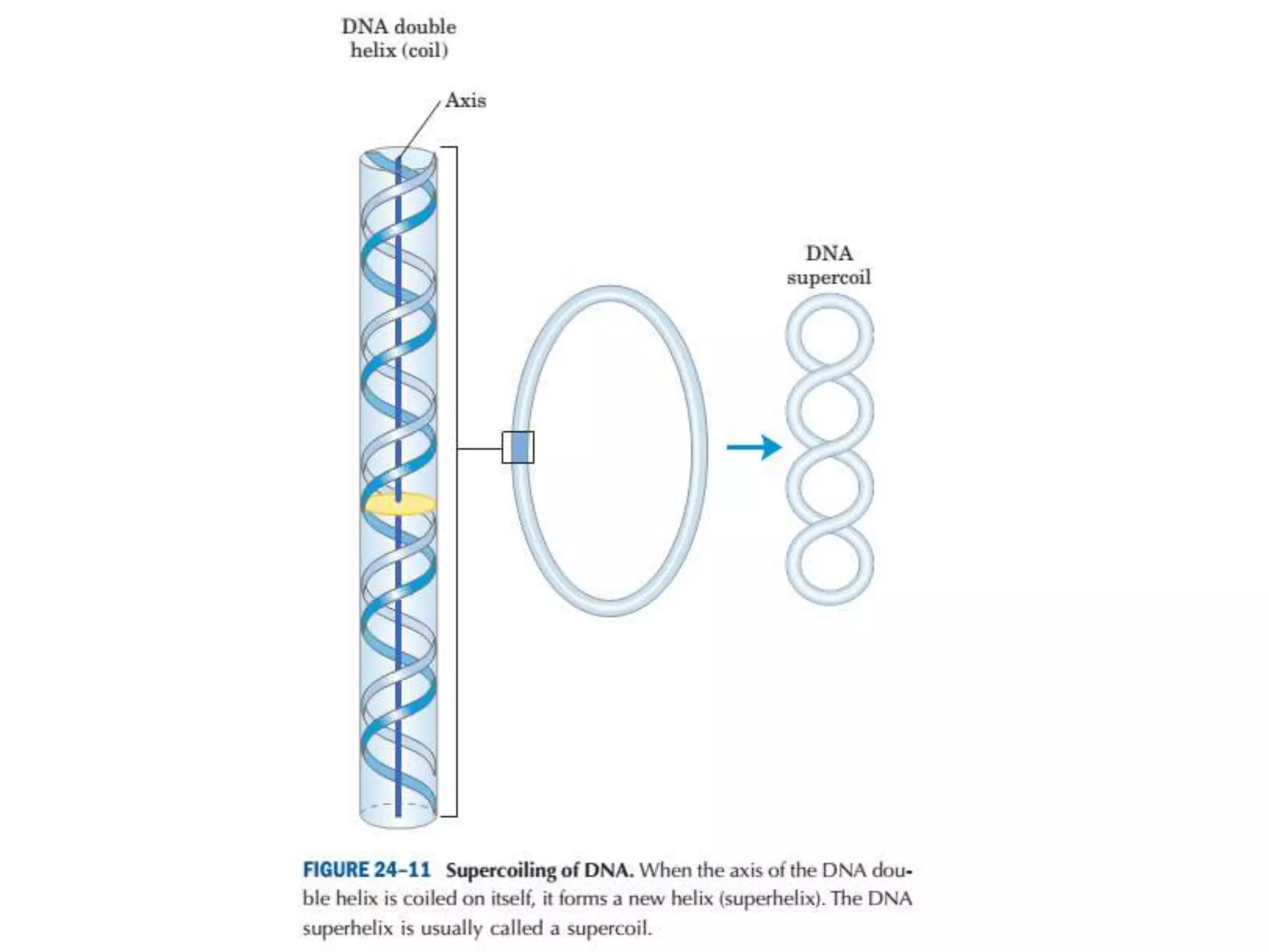
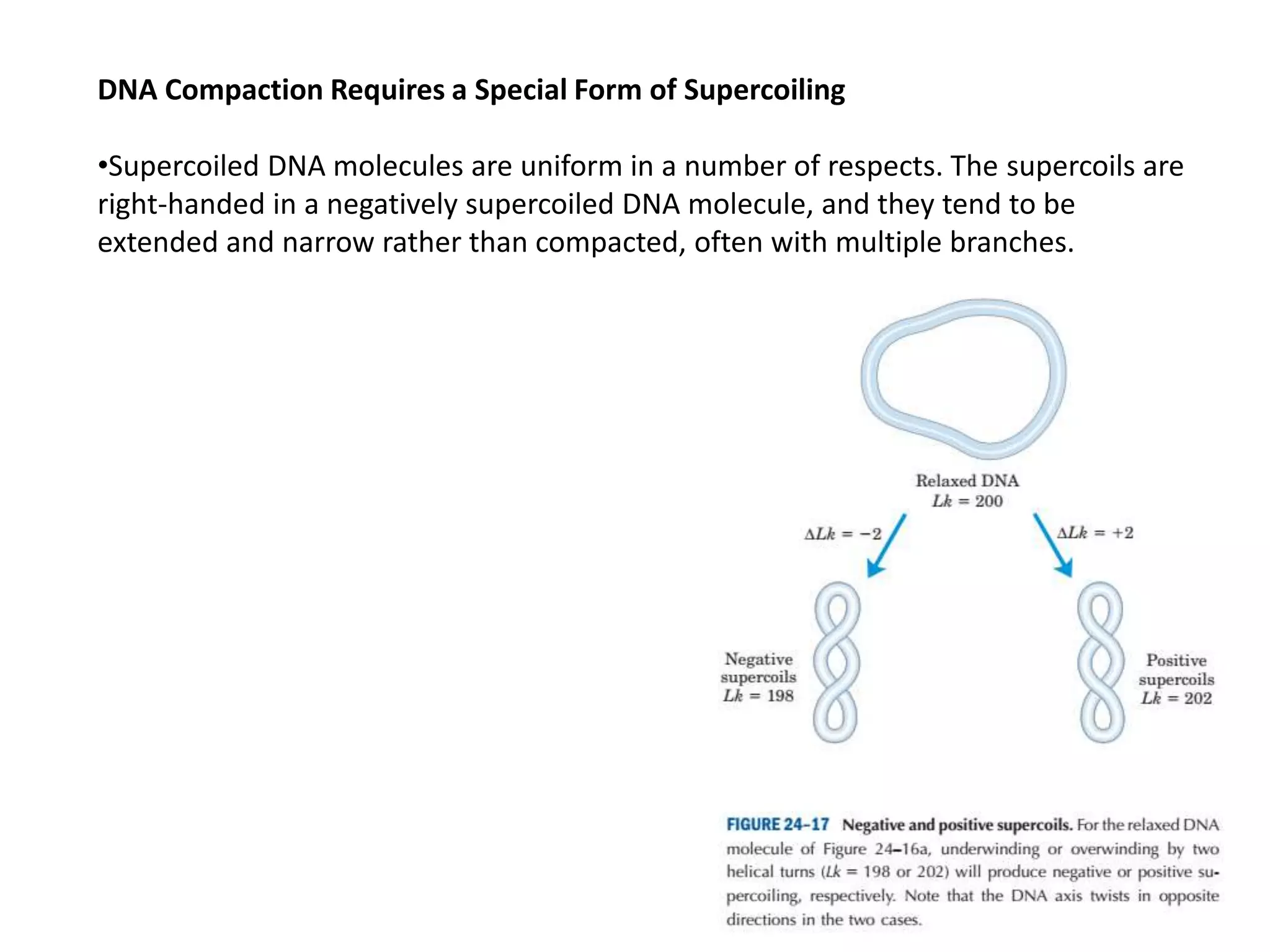
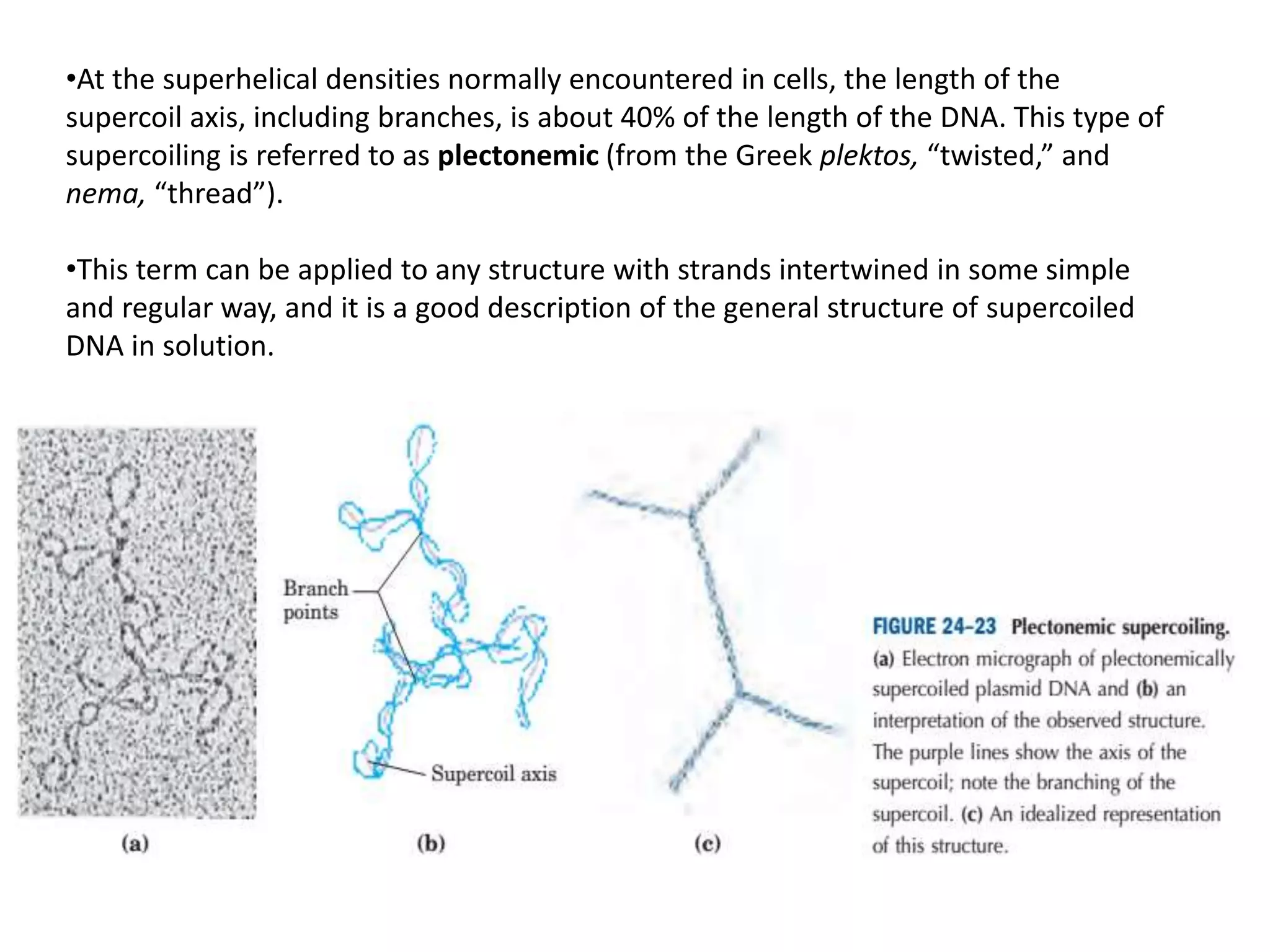
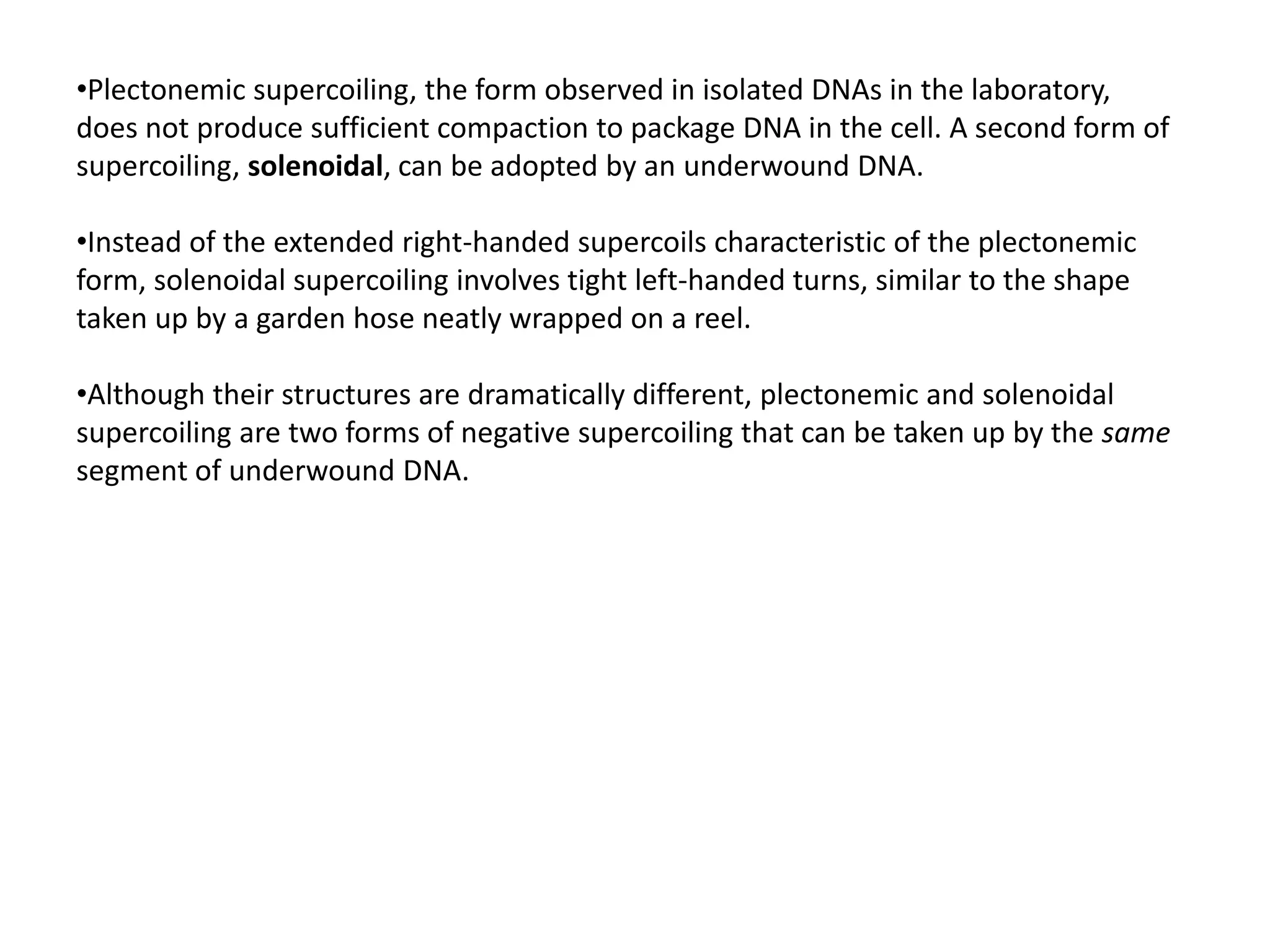
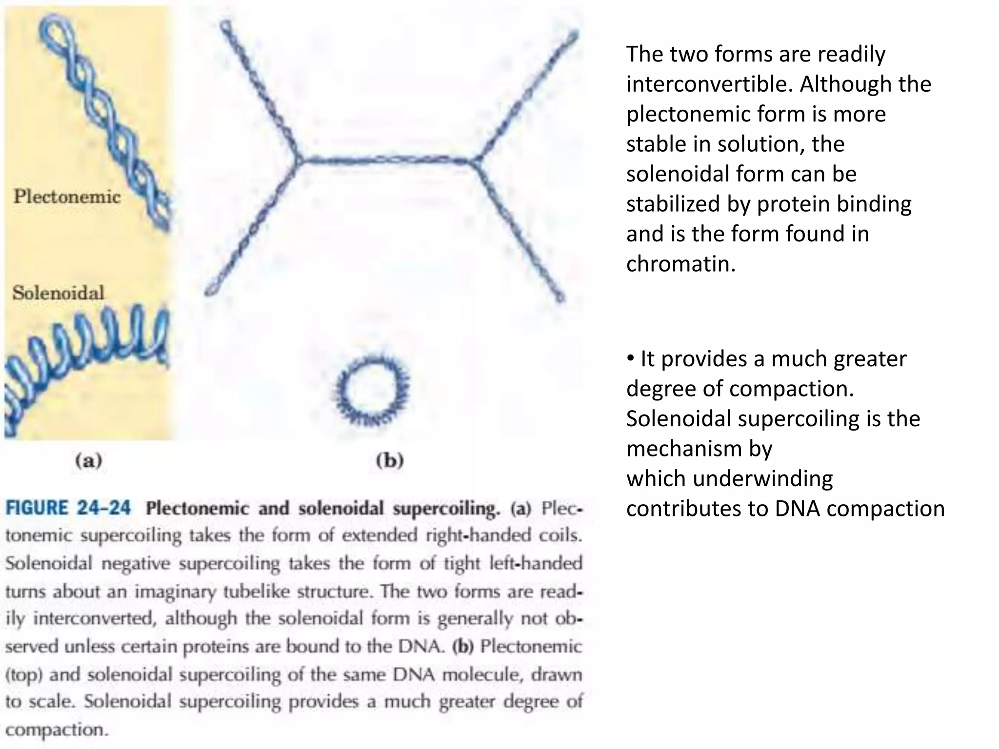
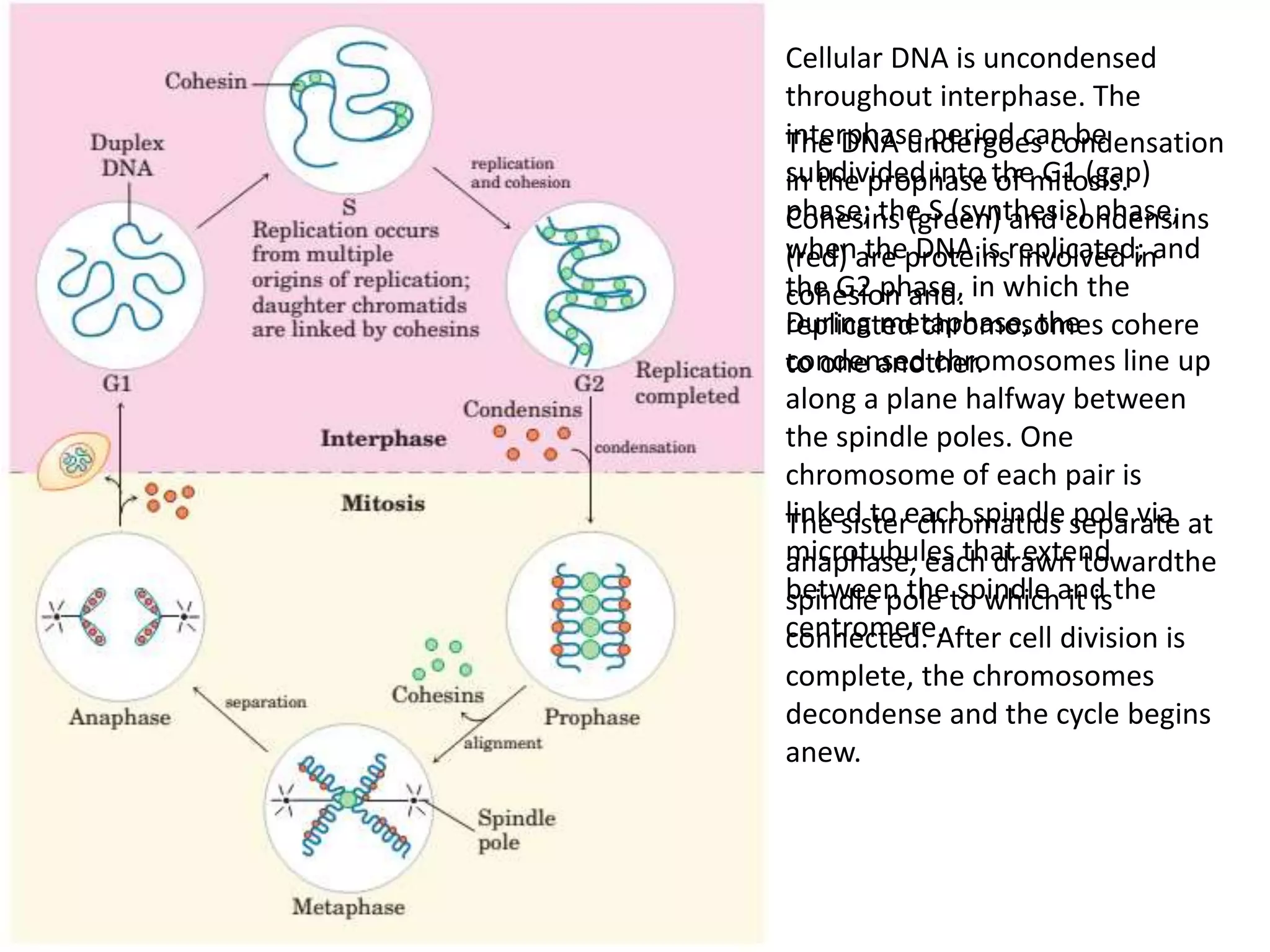
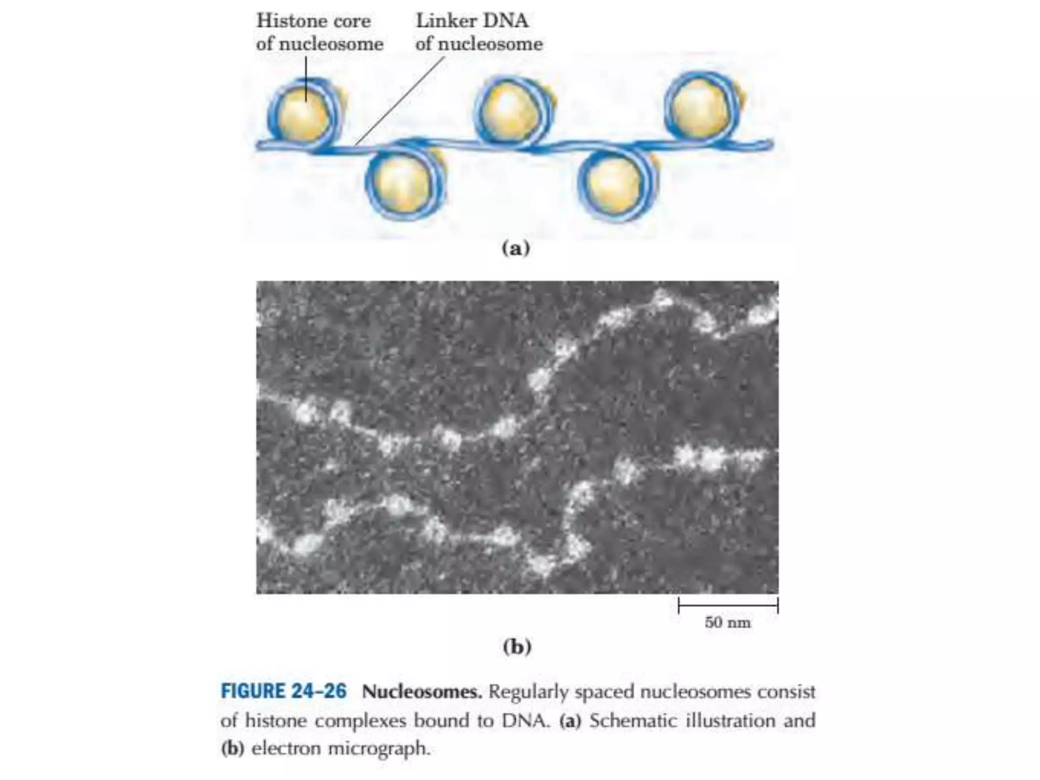
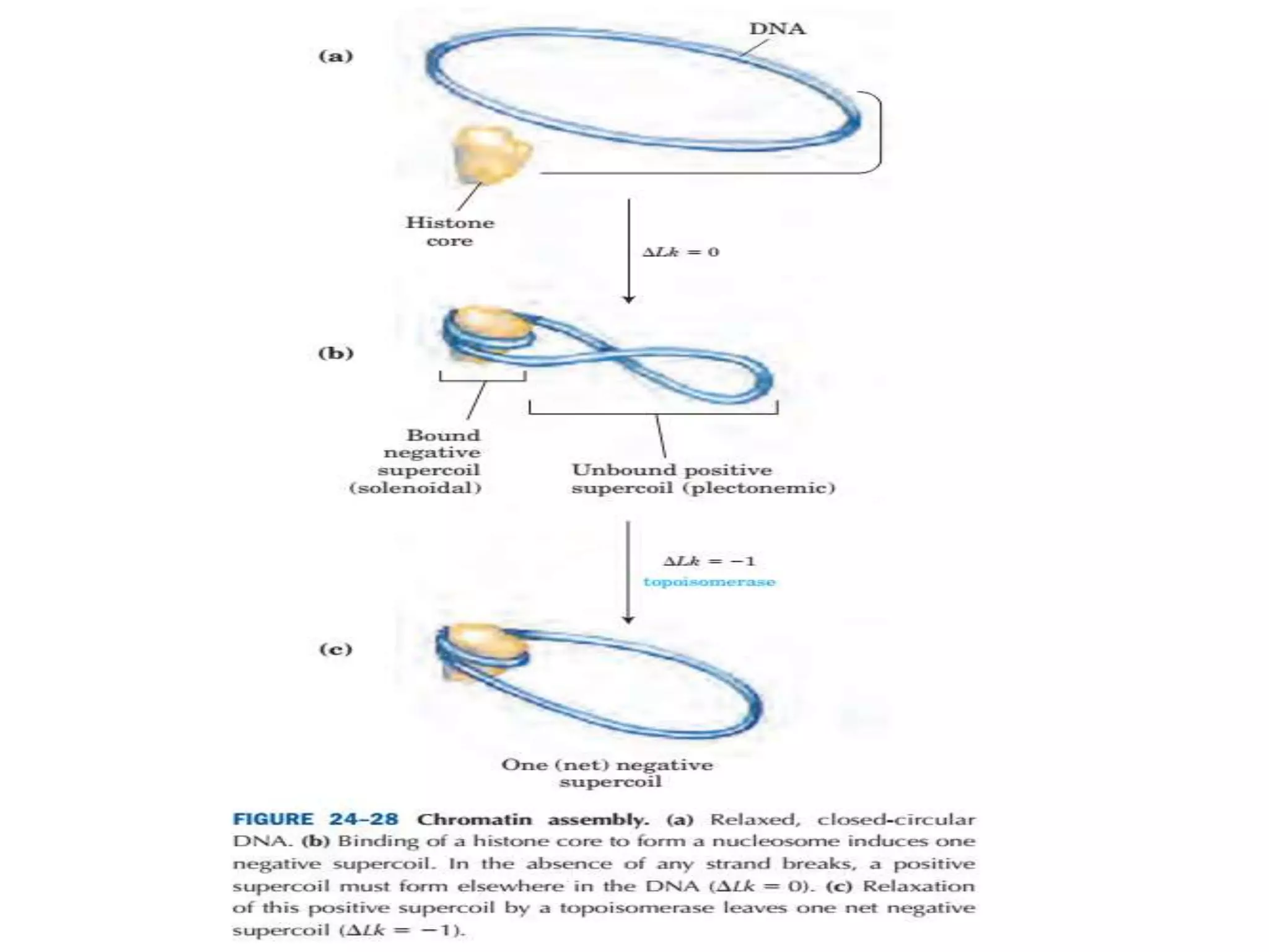
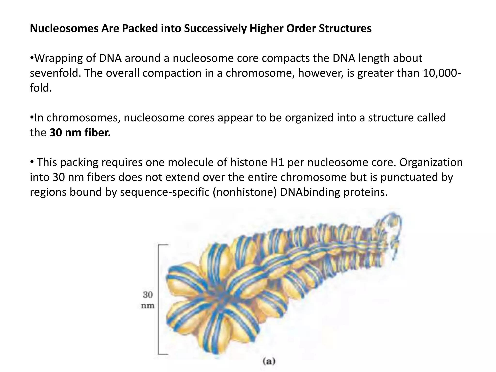
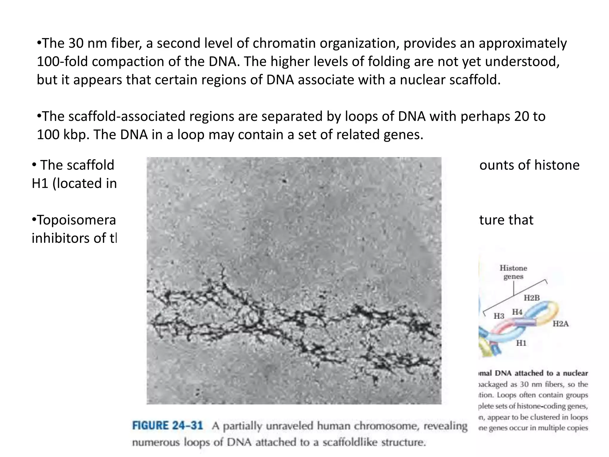
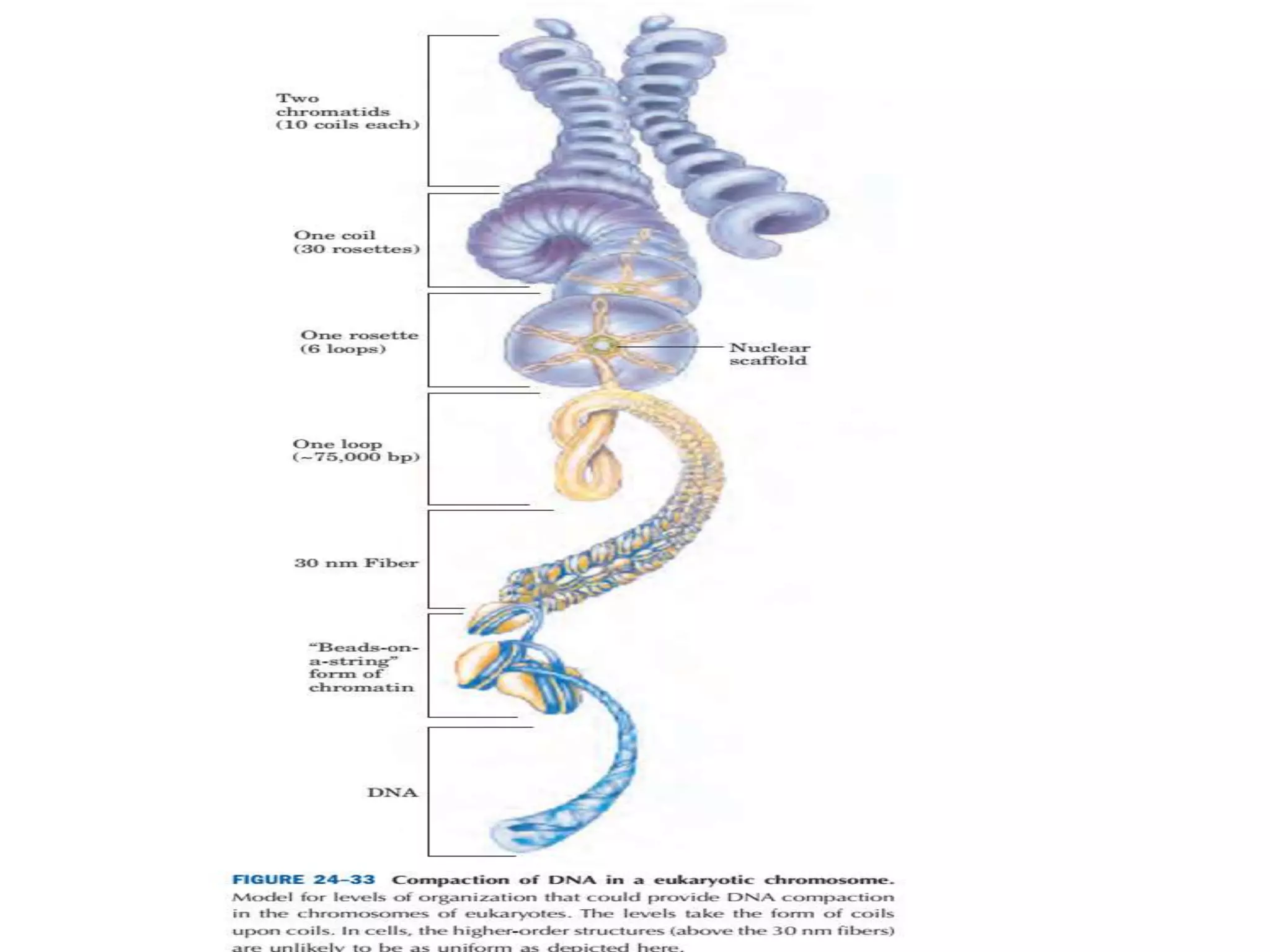
2. DNA Supercoiling
•Cellular DNA, is extremely compacted, implying a high degree of structural
organization. The folding mechanism must not only pack the DNA but also permit
access to the information in the DNA.
•DNA is coiled in the form of a double helix, with both strands of the DNA coiling
around an axis. The further coiling of that axis upon itself produces DNA supercoiling.
•DNA supercoiling is generally a manifestation of structural strain. When there is no
net bending of the DNA axis upon itself, the DNA is said to be in a relaxed state
3.
4. DNA Compaction Requires a Special Form of Supercoiling
•Supercoiled DNA molecules are uniform in a number of respects. The supercoils are
right-handed in a negatively supercoiled DNA molecule, and they tend to be
extended and narrow rather than compacted, often with multiple branches.
5. •At the superhelical densities normally encountered in cells, the length of the
supercoil axis, including branches, is about 40% of the length of the DNA. This type of
supercoiling is referred to as plectonemic (from the Greek plektos, “twisted,” and
nema, “thread”).
•This term can be applied to any structure with strands intertwined in some simple
and regular way, and it is a good description of the general structure of supercoiled
DNA in solution.
6. •Plectonemic supercoiling, the form observed in isolated DNAs in the laboratory,
does not produce sufficient compaction to package DNA in the cell. A second form of
supercoiling, solenoidal, can be adopted by an underwound DNA.
•Instead of the extended right-handed supercoils characteristic of the plectonemic
form, solenoidal supercoiling involves tight left-handed turns, similar to the shape
taken up by a garden hose neatly wrapped on a reel.
•Although their structures are dramatically different, plectonemic and solenoidal
supercoiling are two forms of negative supercoiling that can be taken up by the same
segment of underwound DNA.
7. The two forms are readily
interconvertible. Although the
plectonemic form is more
stable in solution, the
solenoidal form can be
stabilized by protein binding
and is the form found in
chromatin.
• It provides a much greater
degree of compaction.
Solenoidal supercoiling is the
mechanism by
which underwinding
contributes to DNA compaction
8. The Structure of Chromosomes
•The term “chromosome” is used to refer to a nucleic acid molecule that is the
repository of genetic information in a virus, a bacterium, a eukaryotic cell, or an
organelle.
• It also refers to the densely colored bodies seen in the nuclei of dye-stained
eukaryotic cells, as visualized using a light microscope.
•The eukaryotic cell cycle produces remarkable changes in the structure of
chromosomes.
• In nondividing eukaryotic cells (in G0) and those in interphase (G1, S, and G2),
the chromosomal material, chromatin, is amorphous and appears to be randomly
dispersed in certain parts of the nucleus.
9. Cellular DNA is uncondensed
throughout interphase. The
interphase period can be
subdivided into the G1 (gap)
phase; the S (synthesis) phase,
when the DNA is replicated; and
the G2 phase, in which the
replicated chromosomes cohere
to one another.
The DNA undergoes condensation
in the prophase of mitosis.
Cohesins (green) and condensins
(red) are proteins involved in
cohesion and.
During metaphase, the
condensed chromosomes line up
along a plane halfway between
the spindle poles. One
chromosome of each pair is
linked to each spindle pole via
microtubules that extend
between the spindle and the
centromere.
The sister chromatids separate at
anaphase, each drawn towardthe
spindle pole to which it is
connected. After cell division is
complete, the chromosomes
decondense and the cycle begins
anew.
10. Chromatin
•Chromatin consists of fibers containing protein and DNA in approximately equal
masses, along with a small amount of RNA.
•The DNA in the chromatin is very tightly associated with proteins called histones, which
package and order the DNA into structural units called nucleosomes.
• Also found in chromatin are many non-histone proteins, some of which help maintain
chromosome structure, others that regulate the expression of specific genes.
•Beginning with nucleosomes, eukaryotic chromosomal DNA is packaged into a
succession of higher-order structures that ultimately yield the compact chromosome
seen with the light microscope.
11.
12. Histones Are Small, Basic Proteins
•Found in the chromatin of all eukaryotic cells, histones have molecular weights
between 11,000 and 21,000 and are very rich in the basic amino acids arginine and
lysine.
•All eukaryotic cells have five major classes of histones, differing in molecular weight
and amino acid composition. The H3 histones are nearly identical in amino acid
sequence in all eukaryotes, as are the H4 histones, suggesting strict conservation of
their functions.
• For example, only 2 of 102 amino acid residues differ between the H4 histone
molecules of peas and cows, and only 8 differ between the H4 histones of humans and
yeast.
•Histones H1, H2A, and H2B show less sequence similarity among eukaryotic species.
13.
14. Nucleosomes Are the Fundamental Organizational Units of Chromatin
•Subjection of chromosomes to treatments that partially unfold them reveals a
structure in which the DNA is bound tightly to beads of protein, often regularly
spaced.
• The beads in this “beads-on-a-string” arrangement are complexes of histones and
DNA. The bead plus the connecting DNA that leads to the next bead form the
nucleosome, the fundamental unit of organization upon which the higher-order
packing of chromatin is built.
15. •The bead of each nucleosome contains eight histone molecules: two copies each of H2A,
H2B, H3, and H4.
•The spacing of the nucleosome beads provides a repeating unit typically of about 200 bp,
of which 146 bp are bound tightly around the eight-part histone core and the remainder
serve as linker DNA between nucleosome beads.
• Histone H1 binds to the linker DNA. Brief treatment of chromatin with enzymes that
digest DNA causes preferential degradation of the linker DNA, releasing histone particles
containing 146 bp of bound DNA that have been protected from digestion.
•Researchers have crystallized nucleosome cores obtained in this way, and x-ray diffraction
analysis reveals a particle made up of the eight histone molecules with the DNA wrapped
around it in the form of a left-handed solenoidal supercoil.
16. •The tight wrapping of DNA around the histone core requires the removal of about
one helical turn in the DNA. When the protein core of a nucleosome binds in vitro to a
relaxed, closed-circular DNA, the binding introduces a negative supercoil.
• Because this binding process does not break the DNA or change the linking number,
the formation of a negative solenoidal supercoil must be accompanied by a
compensatory positive supercoil in the unbound region of the DNA (Fig. 24–28).
• Eukaryotic topoisomerases can relax positive supercoils. Relaxing the unbound
positive supercoil leaves the negative supercoil fixed (through its binding to the
nucleosome histone core) and results in an overall decrease in linking number.
• Indeed, topoisomerases have proved necessary for assembling chromatin from
purified histones and closed-circular DNA in vitro.
17.
18. Nucleosomes Are Packed into Successively Higher Order Structures
•Wrapping of DNA around a nucleosome core compacts the DNA length about
sevenfold. The overall compaction in a chromosome, however, is greater than 10,000-
fold.
•In chromosomes, nucleosome cores appear to be organized into a structure called
the 30 nm fiber.
• This packing requires one molecule of histone H1 per nucleosome core. Organization
into 30 nm fibers does not extend over the entire chromosome but is punctuated by
regions bound by sequence-specific (nonhistone) DNAbinding proteins.
19. •The 30 nm fiber, a second level of chromatin organization, provides an approximately
100-fold compaction of the DNA. The higher levels of folding are not yet understood,
but it appears that certain regions of DNA associate with a nuclear scaffold.
•The scaffold-associated regions are separated by loops of DNA with perhaps 20 to
100 kbp. The DNA in a loop may contain a set of related genes.
• The scaffold itself appears to contain several proteins, notably large amounts of histone
H1 (located in the interior of the fiber) and topoisomerase II.
•Topoisomerase II is so important to the maintenance of chromatin structure that
inhibitors of this enzyme can kill rapidly dividing cells.
20. Additional layers of organization
•Evidence exists for additional layers of organization in eukaryotic chromosomes, each
dramatically enhancing the degree of compaction.
• One model for achieving this compaction is illustrated in Figure. Higher order chromatin
structure probably varies from chromosome to chromosome, from one region to the next
in a single chromosome, and from moment to moment in the life of a cell.
• No single model can adequately describe these structures. Nevertheless, the principle is
clear: DNA compaction in eukaryotic chromosomes is likely to involve coils upon coils upon
coils…