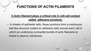
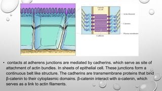
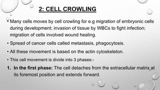
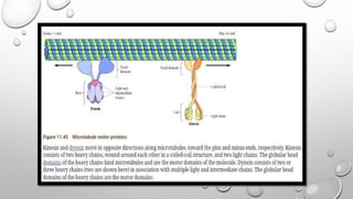
Cell movement is accomplished through the cytoskeleton, which is composed of three main types of fibers: actin filaments, microtubules, and intermediate filaments. Actin filaments interact with motor proteins like myosin to generate movement and play roles in cell adhesion, crawling, and contraction. Microtubules and their motor proteins kinesin and dynein facilitate intracellular transport and movement of cilia and flagella. Intermediate filaments provide structural support to cells but are not involved in motility. The three fiber types work together with associated proteins to control cell shape, division, transport, and movement.
A membrane protein is a protein molecule that is attached to, or associated with the membrane of a cell or an organelle.
More than half of all proteins interact with membranes.
Structure and functions of endoplasmic reticulumICHHA PURAK
The presentation consists of 57 slides,describes following heads
• DISCOVERY
• INTRODUCTION
• BIOGENESIS OF ER
• ISOLATION OF MICROSOMES FROM E R
• STRUCTURE
• COMPONENTS OF ER
CISTERNAE
VESICLES
TUBULES
• MAIN FUNCTION OF ER
• TYPES OF ENDOPLASMIC RETICULUM
• SMOOTH ENDOPLASMIC RETICULUM (SER)
• FUNCTIONS OF SER
• ROUGH ENDOPLASMIC RETICULUM (RER)
• FUNCTIONS OF RER
• SUMMARY
• REFERENCES
• QUESTIONS
Cytoskeleton - microtubules ,microfilaments and intermediate filamentsBIOTECH SIMPLIFIED
The cytoskeleton is a structure that helps cells maintain their shape and internal organization, and it also provides mechanical support that enables cells to carry out essential functions like division and movement. There is no single cytoskeletal component. Rather, several different components work together to form the cytoskeleton.
Introduction
About Drosophila
Genome of Drosophila
Life cycle
Differentiation
Development of Drosophila
* Embryonic development
* Dorsal -ventral and
* Anterior posterior development
* Body segmentation
* Homeotic gene
Conclusion
Reference
Slides about Cell Fate, Cell Potency, Differentiation, Specification, Modes of Specification, Role of Cytoplasm. Cell Interactions, Regulation in Development
INTRODUCTION
Definition
history
DIFFERENT PHASE
G0 PHASE
INTERPHASE
M PHASE
CHECKPOINT
HOW DOES IT WORK
Inhibitors
Mechanism of action
Function
CONCLUSION
references
Muscle physiology in orthodontics/certified fixed orthodontic courses by Ind...Indian dental academy
The Indian Dental Academy is the Leader in continuing dental education , training dentists in all aspects of dentistry and
offering a wide range of dental certified courses in different formats.for more details please visit
www.indiandentalacademy.com
Safalta Digital marketing institute in Noida, provide complete applications that encompass a huge range of virtual advertising and marketing additives, which includes search engine optimization, virtual communication advertising, pay-per-click on marketing, content material advertising, internet analytics, and greater. These university courses are designed for students who possess a comprehensive understanding of virtual marketing strategies and attributes.Safalta Digital Marketing Institute in Noida is a first choice for young individuals or students who are looking to start their careers in the field of digital advertising. The institute gives specialized courses designed and certification.
for beginners, providing thorough training in areas such as SEO, digital communication marketing, and PPC training in Noida. After finishing the program, students receive the certifications recognised by top different universitie, setting a strong foundation for a successful career in digital marketing.
Francesca Gottschalk - How can education support child empowerment.pptxEduSkills OECD
Francesca Gottschalk from the OECD’s Centre for Educational Research and Innovation presents at the Ask an Expert Webinar: How can education support child empowerment?
Biological screening of herbal drugs: Introduction and Need for
Phyto-Pharmacological Screening, New Strategies for evaluating
Natural Products, In vitro evaluation techniques for Antioxidants, Antimicrobial and Anticancer drugs. In vivo evaluation techniques
for Anti-inflammatory, Antiulcer, Anticancer, Wound healing, Antidiabetic, Hepatoprotective, Cardio protective, Diuretics and
Antifertility, Toxicity studies as per OECD guidelines
2. CONTENTS
• What is cell movement?
• Introduction of cytoskeleton
• Actin & its role in cell movement
• Microtubule & its role in cell movement
• Intermediate filament & its role in cell movement
• References
3. CELL MOVEMENT
• Cell movement is necessary function in organisms. Without the
ability to move, cell could not grow and divide or migrate to
areas where they needed. The cytoskeleton is the component
of the cell that makes cell movement possible.
•What is involved in cell movement?
• In addition to playing this structural role, the cytoskeleton is
responsible for cell movements. This include not only the
movement of entire cells, but also the transport of organells &
other structures through cytoplasm.
4. • Cell movement is a complex process.
• Cell motility is accomplished through the activity
of cytoskeleton fibers. The cytoskeleton is a network which is
composed of 3 distinct biopolymer:
1) Actin
2) Microtubules
3) Intermediate filament
• They are differentiated by their rigidity.
5. 1) ACTIN
• Actin filaments are semiflexible polymer(Lp~17µm).
• They are 7nm in diameter. They are built from dimer pairs of
globular actin monomers & are polar in nature i.e they have
two distinct ends: one is fast growing end & other is slow
growing end.
• Fast growing end denotes as plus(+) end & slow end denotes
as negative(-) end.
• The minus end has a critical actin monomers concentration i.e
~6 times higher than at plus end.
6.
7. • when the end of an actin filament is exposed to a concentration
of monomeric actin that is above it’s critical concentration, the
monomers bind at filament end & grows by polymerization &
viceversa.
• So, due to different critical concentration value at both end AFs
grows assymetrically. When the monomers concentration lies
between two value only the + end will grows & - end will
shrinks. So, length of filament remain constant but monomer
elongates forward due to asymmetric + end polymerization
which is known as treadmilling.
8. ACTIN & THEIR MOTOR PROTEINS(MYOSINE)
• Actin filaments also generate motility forces by interaction with myosin
motors.
• motors are made up of head, neck & tail region.
• The head region is responsible for attachment & force production. While
tail region is used for connecting cargo such as vesicle, filament or other
myosines.
• Myosin motors work on actin filaments through 3 step process:
binding, power stroke & unbinding.
9.
10. FUNCTIONS OF ACTIN FILAMENTS
1) Actin filament plays a critical role in cell-cell contact
called adherens junctions.
• In sheets of epithelial cells, these junctions form a continuous
belt like structure (called an adhesion belt) around each cell in
which an underlying contractile bundle of actin filaments is
linked to plasma membrane.
11. • contacts at adherens junctions are mediated by cadherins. which serve as site of
attachment of actin bundles. In sheets of epithelial cell. These junctions form a
continuous belt like structure. The cadherins are transmembrane proteins that bind
β-catenin to their cytoplasmic domains. β-catenin interact with α-catenin, which
serves as a link to actin filaments.
12. 2: CELL CROWLING
• Many cells moves by cell crowling for e.g migration of embryonic cells
during development; invasion of tissue by WBCs to fight infection;
migration of cells involved wound healing.
• Spread of cancer cells called metastasis, phagocytosis.
• All these movement is based on the actin cytoskeleton.
• This cell movement is divide into 3 phases:-
1. In the first phase: The cell detaches from the extracellular matrix at
its foremost position and extends forward.
13. 2) In the second phase: the detached portion of the cell moves
forward and re-attaches at a new forward position. The rear
portion of the cell also detaches from the extracellular matrix.
3) In the third phase: the cell is pulled forward to a new
position by the motor protein myosin. Myosin utilizes the energy
derived from ATP to move along actin filaments.
Each of these steps requires physical forces which is produced
by actin filaments.
14.
15.
16.
17. 3: Actin myosin contraction in non muscle cells
It is useful for cytokinesis (the division of cell into two daughter
cells at the end of mitosis.)
Toward the end of mitosis in animal cells, a contractile ring is
assembles which contain actin filaments & myosin2 just
underneath the plasmamembrane.
Its contraction pulls the plasmamembrane inward side &
pinching in two separate daughter cell.
As cell division completed ring totally gets disappear.
Actin filaments also play an important role in muscle
contraction also.
18.
19. 2) MICROTUBULES
• Microtubules are the stiffest biopolymers with Lp ranging from
100-5000µm. It is an rod like polymers with an outer diameter
of ~25nm.
• It is made up of tubulin protein/monomer.
• Tubulin monomers associate assemble into protofilament & 13
of these protofilaments align to form a hollow tube.
• They are also polar, treadmill & can impart a force through
polymerization.
20.
21. MTs & their motorproteins (kinesins & dynein)
• MTs are responsible for a variety of cell movements such as
intracellular transport of membrane vesicle, the separation of
chromosomes at mitosis, movement of cilia and flagella.
• This action of MTs is based on motor proteins which is utilize the
energy from ATP hydrolysis to produce force & movement.
• Motor proteins are kinesins & dyneins which are responsible for
movement in which microtubules are participate.
• The kinesin move toward the plus end & dynein moves towards the
minus end when they are bind to MTs
• The first motor protein is identified is dynein.
22.
23. • Kinesin is similar as myosin. In structure the tail portion of the kinesin
molecule consists of the light chain & a heavy chain.
• This portion of the kinesin is responsible for binding to other organelles
that are transported along MTs. The amino terminal globular head
domains of heavy chains which bind to both microtubules & ATP.
• This ATP hydrolysis provides energy for movement.
• Dynein is also contain head & tail portion. Head binds with MTs & ATP &
tail binds to cargo membrane enclosed organelles such as the ER,
Golgi apparatus, Mitochondria within the cell.
• For e.g the ER is extend to the periphery of the cell in association with
microtubules.
24. • Drug(Demecolcine & Nocodazole) that depolymerizes the MTs cause
ER extends to retracts toward the cell center, indicating that
association with MTs is required to maintain the ER in its extended
state.
• This positioning of ER occur due to involvement of action of kinesin
which pulls the ER along microtubules in the plus end direction,
towards the cell periphery.
• Same as dynein is play a role in positioning of Golgi apparatus. The
Golgi vesicles transported to the center of the cell (towards the minus
nd of MTs) by dynein.
25. CILIA & FLAGELLA
• Cilia &flagella are microtubule based projection of plasma membrane
which are responsible for movement of eukaryotic cell.
• Many bacteria also have flagella, but these prokaryotic Flagella are
made up of protein filaments which is projecting from the cell surface,
rather than plasma membrane supported by MTs in eukaryotes.
• In short the cilia and flagella are made up of MTs & their associated
motor proteins.
• Cilia are 10µm in length. They move in a coordinate back & forth
motion. which moves fluid over the surface of the cell. It act as a
wiper which is wipe the fluid from the surface of the cell.
26. • Flagella is more in length(~20nm).
• They moves in a wave like pattern. Cells usually have only one
or two flagella which are responsible for locomotion of a cell
such as sperm cell.
27. 3) INTERMEDIATE FILAMENTS
• They are much more flexible than AFs & MTs(Lp~0.3-1.0µm)
• The diameter is 8-12 nm.
• IFs has different classes which are present in different cells.
e.g vimetin, desmin, keratin(hair & nails), lamin etc.
• IFs are not polarized, not treadmill.
• They generally do not depolymerized under physiological
condition. Once get polymerized it can not undergo in
depolymerization under normal physiological condition.
28.
29. They are more static in nature than AFs and MTs.
These three kinds of biopolymers build an internal cellular
scaffold k/a cytoskeleton.
These network also have some proteins. This network work
together for cell function such as shape, division & organelle
transport & cell motility.
30. FUNCTIONS OF INTERMEDIATE FILAMENTS
The main function of IFs is to provide structural support & to organize
cells into tissue.
The supportive role of IFs is carried to an extreme in claws & hair
which are composed of IF.
It provides mechanical support to the plasma membrane.
IFs are not participate in cell motility.
31.
32. REFERENCES
The journal of cell biology
NCBI Book shelf
The cell: A Molecular approach. 2nd edition by Cooper
Wikipedia