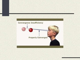
This document discusses convergence insufficiency (CI), including its definition, causes, symptoms, diagnosis, and treatment. CI is characterized by a weak ability to maintain proper binocular eye alignment for near vision tasks. It is often associated with exophoria and reduced fusional convergence. Symptoms include eyestrain, headaches, and blurred vision when reading. Treatment involves orthoptic exercises to strengthen convergence, as well as base-in prisms or surgery in severe cases. The document provides detailed explanations of CI and outlines goals and aspects of its medical, surgical, and follow-up management.
Diplopia charting involves recording the separation of double images seen by a patient in nine positions of gaze. The examiner uses a red glass over one eye while holding a light source in different positions. The patient reports if they see one image, two separate images, or a tilted image. The separation is greatest in the direction of an underacting or paralyzed eye muscle. When recording the results, the patient's right and left sides are noted along with the distance of any separation between images and any tilting. The diplopia chart should be interpreted along with a clinical exam and Hess chart.
This document provides guidelines for prescribing glasses in children. It discusses that the pediatric eye is different from the adult eye in terms of axial length, corneal curvature, and lens power. The goals of prescribing glasses in children are to provide a focused retinal image and achieve optimal balance between accommodation and convergence. It is more difficult to prescribe glasses for children due to lack of subjective response and poor attention. American guidelines provide recommendations on refractive errors that warrant correction at different ages. Factors like emmetropization, amblyopia risk, and presence of strabismus are considered. Frame selection depends on the child's condition and age, aiming for correct fit, comfort, safety, and not hindering nasal development.
Contact lens for congenital aphakia and other eye conditions for infants and toddlers. The slide presentation encompasses indications for CL fitting in paediatric, contact lens options, fitting techniques, challenges and contact lens as myopia control.
1. The document discusses the benefits of disposable contact lenses and frequent replacement programs (FRP) for providing healthier contact lens wear. It notes that consumers prioritize healthier lenses that do not cause infections or complications.
2. Disposable and FRP lenses are presented as ways to eliminate obstacles to successful contact lens wear like deposits, films, irritation and reduced comfort by replacing lenses frequently. They provide benefits like superior comfort, hygiene, and convenience.
3. The document outlines different types of disposable and FRP lenses available based on modality, wearing time, material, correction, and discusses their advantages over traditional lenses in promoting ocular health, satisfaction, and retention of contact lens wearers.
Types of pediatric contact lens [autosaved]Bipin Koirala
This document discusses pediatric contact lens fitting and evaluation. It begins by outlining the advantages of contact lenses over glasses for children, including a wider field of view. Key considerations for fitting include small eye size, tear production, and compliance. Conditions that may require lenses include refractive errors, amblyopia treatment, and aphakia following cataract surgery. Evaluations include testing visual acuity and ocular health. Lens options discussed are silicone, hydrogel, and rigid gas permeable lenses. Special fitting considerations for aphakic children include initially high powers of +20D to +35D, depending on age.
This document discusses amblyopia, including its classification, pathophysiology, clinical characteristics, evaluation, and management. It defines amblyopia and outlines its prevalence. Amblyopia can be classified as functional or organic, and further divided into types such as strabismic, anisometropic, and stimulus deprivation amblyopia. Clinical signs may include reduced visual acuity, abnormal fixation, impaired stereopsis, and altered contrast sensitivity. Evaluation involves assessing visual acuity, fixation, binocularity, and refractive error. Management is focused on occlusion therapy to treat the amblyopic eye.
This document discusses rigid gas permeable (RGP) contact lenses. It notes that RGP lenses are made of oxygen permeable materials and are better than soft lenses for vision, durability, correcting astigmatism, eye health, and ease of care. RGP lenses are recommended for conditions like keratoconus or high refractive errors. The fitting process involves screening patients, measuring the eye, trial fittings, and dynamic and static assessments. Proper care and maintenance of RGP lenses is also discussed.
What are the tests for binocular vision?
During a Binocular Vision Assessment, the eye doctor evaluates both binocular vision functioning and visual perceptual skills:
Accommodation.
Convergence.
Depth perception (3D)
Fusion.
Ocular motility.
Ocular posture.
Presence of conditions that affect binocular vision functioning.
Spatial awareness / planning.
This document discusses eccentric fixation (EF), a condition where an eye fails to fixate with the fovea and instead fixates at another retinal point. It describes several theories for the cause of EF, including suppression, anomalous correspondence, motor, and sensory motor theories. It outlines methods for investigating EF, such as ophthalmoscopy and visuscopy. Treatment options discussed include occlusion therapy and pleoptic treatment to encourage foveal fixation, though EF is often difficult to fully correct once established. The document also discusses microtropia, a small-angle strabismus associated with EF and amblyopia.
The synaptophore is an orthoptic instrument used for both diagnostic and therapeutic purposes in optometry. It works using the haploscopic principle to divide visual space into two separate areas visible to only one eye each. Slides can be used for simultaneous perception, fusion, stereopsis, and other tests. Diagnostic uses include measuring deviations, retinal correspondence, and fusional reserves. Therapeutic uses treat suppression, amblyopia, and heterophorias. Proper adjustment and preliminary settings are required before administering tests to accurately diagnose and manage binocular vision anomalies.
Pattern strabismus occurs when there is a change in the magnitude of horizontal deviation between up and down gaze. The most common types are A pattern (convergence in up gaze) and V pattern (divergence in up gaze). Pattern strabismus can be caused by abnormalities of vertical or horizontal muscle action, anatomical anomalies, disorders of muscle innervation, or anomalous muscle insertions. Evaluation involves measuring the deviation in different gazes using cover-uncover testing and Hess screening. Management may involve adaptation or surgery tailored to the specific pattern, which aims to improve alignment and binocular function.
The document discusses patterns of strabismus, specifically the A pattern and V pattern. The A pattern involves relative convergence on upgaze and divergence on downgaze, while the V pattern is the opposite with relative divergence on upgaze and convergence on downgaze. Variants include the X, Y, lambda, and diamond patterns. The etiology of these patterns involves dysfunction of the horizontal, vertical, or oblique eye muscles. Clinical features may include anomalous head posture, amblyopia, and abnormal retinal correspondence. Diagnosis involves measuring alignment in upgaze and downgaze while preventing accommodation.
This document discusses retinal correspondence and abnormal retinal correspondence. It defines retinal correspondence as the relationship between paired retinal visual cells in the two eyes that allows for single binocular vision. Abnormal retinal correspondence occurs when the fovea of one eye corresponds to an extrafoveal area in the other eye, resulting in eccentric fixation but maintained binocular vision. The document describes tests to assess normal versus abnormal retinal correspondence, including the Bagolini striated glasses test, red filter test, and Hering-Bielschowsky after-image test.
This document discusses various tests used to evaluate strabismus and retinal correspondence, including Bagolini's striated glasses test, Worth's four dot test, synaptophore testing, and progressive prism adaptation testing. It also describes techniques for stimulating the retina of a deviated eye, such as macular massage and bi-kinetic retinal stimulation, with the goal of achieving normal retinal correspondence. Surgical correction of strabismus is not recommended until tests confirm the development of normal correspondence and some degree of fusion.
The term ‘‘aniseikonia” comes from the Greek words ‘‘an” (not) ‘‘is” (equal) & ‘‘eikon” (icon or image) so aniseikonia is a binocular condition in which the apparent sizes of the images seen with the two eyes are unequal.
Whenever refractive ametropias in the two eyes of a person are different (i.e., when there is an anisometropia), the corrected retinal images of the two eyes, and consequently the two visual images, differ in size.
This condition has been termed aniseikonia
Optical aniseikonia
Retinal aniseikonia
Cortical aniseikonia
Insertion and removal of rgp contact lens.Anandhan K
This document provides instructions for inserting, centering, and removing rigid gas-permeable (RGP) contact lenses. It describes RGP lenses as semi-soft lenses that allow oxygen to pass to the eye. For insertion, it details cleaning and handling the lens properly before placing it on the eye and blinking to center it. Centering involves using fingertips to nudge the lens into position while looking in different directions. For removal, it outlines either using a blink method by pulling the eyelid tight and blinking, or a two-finger method of pressing the eyelids together to dislodge the lens.
This document summarizes a presentation on esodeviations (convergent strabismus). It discusses various types of esotropia including infantile, accommodative, acquired non-accommodative, and incomitant esotropia. Infantile esotropia presents in the first 6 months of life and requires early surgery. Accommodative esotropia is associated with hyperopia and convergence accommodation. Acquired non-accommodative esotropia develops after 6 months without hyperopia. Incomitant esotropia involves limitations in eye movement. Treatment depends on the type but may include glasses, patching, prisms, botulinum toxin injections, or strabismus surgery
The document discusses rigid gas permeable contact lenses, including their benefits, applications, fitting process, and lens design considerations. Some key points covered include:
1. RGP lenses can automatically correct astigmatism, provide good vision and eye health benefits like increased oxygen transmission.
2. The fitting process involves evaluating the lens-cornea relationship using fluorescein dye to identify any bearing, clearance or sealing issues.
3. Important lens design factors are the overall diameter, optical zone size, base curve, thickness, and peripheral curve to achieve a proper alignment fit.
The optical center of a lens is the point where light rays pass through without deviation. It is important for the optical center to be directly in front of the pupil for optimum vision. Decentering a lens, or moving it so the optical center is no longer in front of the pupil, introduces a prismatic effect. The amount of prismatic effect, measured in prism diopters, is calculated by multiplying the distance the lens is decentered in centimeters by the lens power in diopters. Decentering a lens with a spherical prescription or cylinder introduces different prismatic effects depending on the orientation of the cylinder axis relative to the direction of decentration.
This document discusses pediatric refraction and various techniques used for refracting children. Pediatric refraction is different from adult refraction due to active accommodation in children. Cycloplegic refraction is preferable to paralyze accommodation. Different techniques are used based on the age of the child, including near retinoscopy, dynamic retinoscopy, and book retinoscopy. Cycloplegics help obtain an accurate refraction by paralyzing accommodation.
This document discusses measuring and classifying accommodative convergence/accommodation (AC/A) ratios. It defines the AC/A ratio as the change in accommodative convergence per diopter of accommodation. Abnormal AC/A ratios can cause strabismus. There are several methods described for measuring the AC/A ratio clinically, including the heterophoria, gradient, and graphical methods. The document outlines treatments for different AC/A ratio abnormalities like convergence excess, convergence insufficiency, divergence excess, and divergence insufficiency.
Scleral lens is a large rigid contact lens with a diameter range of 15mm to 25mm. Its resting point is beyond the
corneal borders, and are believed to be among the best vision correction options for irregular corneas. Wearing scleral lens also can postpone or even prevent surgical intervention as well as decrease the risk of corneal scarring.
Non - surgical treatment of squint i.e. all types of squint have some modalities of treatment [ optical treatment, orthoptic treatment, Prismo-therapy, and pharmacological treatment] except surgical treatment.
1. OPTICAL TREATMENT -
in optical treatment, it should be include correction of refractive error and prismotherapy.
SPECTACLES should be prescribed in every cases.
It may correct to squint partially or completely.
IN PRISMOTHERAPY, for correction of squint, This is light weight, and easy to apply on the back surface of glass.
It is useful in heterophoria, nystagmus, convergence insufficiency, managing diplopia and maintain binocular single vision.
IN PHARMACOLOGICAL TREATMENT, miotics, atropine and botulinum toxin are prescribed in some types of cases of strabismus.
IN ORTHOPTIC TREATMENT, means straight eyes.
It is used as a diagnostic purpose and therapeutic purposes.
- to increase fusion amplitude.
- anti suppression exercises.
- treatment of amblyopia.
- treatment of abnormal retinal correspondance.
- to control deviations.
ORDER OF ORHOPTIC TREATMENT -
. amblyopia is treated firstly.
. anti- suppression therapy.
- diplopia training.
- amplitude improvement.
Aniseikonia refers to an unequal apparent size of images seen by the two eyes. It can result from differences in refractive errors between the eyes (refractive aniseikonia) or differences in the distribution of retinal elements (basic aniseikonia). Symptoms include headaches, asthenopia, and difficulties with mobility or fusion. Aniseikonia is usually caused by anisometropia above 1.50-2.00 diopters and analyzing ocular components can help determine if it is due to refractive or axial differences.
This document discusses low vision aids and their use for people with visual impairments. It defines low vision according to the WHO and describes common causes of visual dysfunction like macular degeneration and glaucoma. The goals of low vision rehabilitation are to maintain and improve visual function through clinical assessment and optometric intervention. Low vision aids can be optical devices like magnifying glasses, telescopes, or non-optical devices that alter lighting, contrast and size of objects. Common optical devices discussed include magnifying spectacles, hand magnifiers, stand magnifiers, and telescopes.
Vergence refers to the simultaneous movement of the eyes in opposite directions to maintain binocular vision. There are different types of vergence including fusional vergence, proximal vergence, tonic vergence, and accommodative vergence. Vergence is measured through tests such as near point of convergence and AC/A ratio. Common vergence anomalies include convergence insufficiency, convergence excess, divergence insufficiency, and divergence excess, each characterized by specific symptoms and treatments.
This document discusses various tests used to evaluate binocular vision, including cover tests, Hess charting, and diplopia charting. Cover tests are used to detect manifest or latent strabismus and determine deviation direction. Hess charting maps eye positions in 9 gazes using colored lenses to dissociate vision between eyes. It identifies muscle under or overaction. Diplopia charting records double vision separation in 9 gazes to localize affected muscles. These objective tests evaluate binocular function and strabismus type and localization.
Binocular vision assessment involves evaluating sensory and motor fusion through tests of phoria, vergence, accommodation, and stereopsis. Key tests include near point of convergence, vergence ranges, and accommodative response. Assessing binocular vision helps diagnose problems like convergence insufficiency, accommodative insufficiency, and other issues that can cause symptoms like eyestrain, headaches, and blurred vision. Referral for further orthoptic evaluation is recommended for patients presenting with these types of symptoms.
Convergence is the inward movement of the eyes to maintain single binocular vision. It can be symmetrical or asymmetrical depending on the position of the fixation point. The near point of convergence is the closest point an object can be focused on binocularly and is measured clinically. Convergence insufficiency is the most common convergence anomaly and causes eye strain. Other anomalies include convergence paralysis which is a total lack of convergence ability due to brain lesions. Convergence spasm involves intermittent periods of excessive convergence that can cause diplopia. Exercises are used to treat convergence insufficiency while prisms are used for convergence paralysis.
Convergence is the simultaneous inward rotation of the eyes to maintain single binocular vision at near distances. Convergence insufficiency is a common cause of eye strain and headaches in people who do intensive near work. It involves reduced ability to converge the eyes voluntarily. Symptoms include blurred vision, eye fatigue, and headaches. Assessment involves measuring near point of convergence, fusional vergence ranges, and near-distance exophoria. Treatment focuses on exercises to improve convergence ability, base-in prism reading glasses, and in severe cases surgery may be considered.
This document discusses suppression, which is one of the three mechanisms of sensory adaptation that occurs in patients with strabismus. Suppression refers to the active inhibition of the image from the deviated eye to avoid diplopia. There are different types of suppression depending on factors such as etiology, retinal area involved, constancy, and the eye affected. Several tests are used to diagnose suppression including the Worth four dot test, Bagolini striated glass test, and visual acuity testing. Treatment involves refractive correction, occlusion therapy, eye alignment procedures, and anti-suppression exercises.
This document discusses different types of esotropia (inward eye turning), including accommodative esotropia caused by uncorrected refractive errors, convergence excess esotropia caused by a high AC/A ratio, and early onset esotropia appearing before age 6 months. Accommodative esotropia is treated initially with corrective lenses, while convergence excess may require bifocals or surgery. Early onset esotropia often requires surgery by age 1-2 years to align the eyes, with risks of undercorrection, amblyopia, or development of dissociated vertical deviation. The document provides details on diagnostic criteria and management approaches for various forms of esotropia.
This document provides an overview of esotropia, or convergent strabismus, including its classification and types. It discusses accommodative esotropia in more detail. Accommodative esotropia can be refractive, caused by hyperopia, or non-refractive, caused by a high AC/A ratio. Treatment involves correcting refractive errors with glasses and potentially bifocals. Surgery such as medial rectus recession may be considered if the deviation is not fully corrected with optical correction alone. The document outlines different types of accommodative esotropia and their typical treatments.
1. Monocular elevation deficiency (MED), also known as double elevator palsy, is characterized by an inability to elevate one eye in all fields of gaze, resulting in hypotropia of the affected eye.
2. The condition can be congenital or acquired, with causes including superior rectus palsy, inferior rectus restriction, and supranuclear lesions.
3. Surgical management of MED depends on forced duction testing and may include inferior rectus recession, superior rectus resection, or Knapp's procedure to improve eye alignment and increase binocular vision.
1. Monocular elevation deficiency (MED), also known as double elevator palsy, is characterized by an inability to elevate one eye in all fields of gaze, resulting in hypotropia of the affected eye.
2. The condition can be congenital or acquired, with causes including superior rectus palsy, inferior rectus restriction, and supranuclear lesions.
3. Surgical management of MED depends on forced duction test results and may include inferior rectus recession, superior rectus resection, or Knapp's procedure to transpose the horizontal rectus muscles. The goal is to improve eye position and increase binocular vision.
This document discusses the evaluation and management of esotropia in children. It begins by defining esotropia as inward turning of one or both eyes. It then describes the different types of esotropia including esophoria, intermittent esotropia, and constant esotropia. For evaluation, it recommends assessing visual acuity, stereopsis, ocular alignment and motility. Management involves treating any amblyopia or refractive error first through non-surgical means such as glasses, patching, or botulinum toxin injection. For persistent esotropia, surgical options include recession or resection of the medial or lateral rectus muscles. The document outlines approaches for different types of esotropia
1. Accommodation is the process by which the eye increases optical power to maintain clear focus on nearby objects. During accommodation, the ciliary muscles contract and relax zonular tension on the lens, allowing the lens to become more convex.
2. Presbyopia is the age-related loss of accommodation due to hardening of the lens and weakening of the ciliary muscles. It results in difficulty seeing objects close up and is treated with convex lenses.
3. Other abnormalities of accommodation include insufficiency, paralysis, and spasm. Insufficiency is reduced accommodation ability, paralysis is a sudden loss of accommodation ability, and spasm is excessive accommodation exertion causing induced myopia.
This document discusses esotropia, which is an inward turning of one or both eyes. It defines esotropia and describes the different types including accommodative esotropia, congenital esotropia, and microtropia. It outlines the causes, characteristics, diagnosis, and management of each type of esotropia. Some key points covered include the role of accommodation and refractive error in accommodative esotropia, the importance of early treatment for congenital esotropia to prevent amblyopia, and the use of occlusion therapy, refractive correction, surgery, and botulinum toxin injection in the management of esotropia.
Non-surgical treatments such as prescription lenses and occlusion therapy can effectively manage certain types of strabismus. Prescribing lenses aims to provide a clear retinal image while balancing accommodation and convergence. For children, the full refractive error is typically prescribed from infancy to preschool age. Occasionally, hyperopic corrections may be reduced in older children or esotropic patients. Occlusion therapy and minus lenses are common non-surgical approaches for intermittent exotropia. Prisms can also effectively treat strabismus, diplopia, and symptomatic heterophoria in some cases.
In children, spectacle prescriptions must consider the ongoing development of refractive errors and vision. Objective and subjective testing is used to refine optical corrections appropriately for each age. Spectacles are indicated for high refractive errors that could cause amblyopia, or when refractive errors are associated with other eye conditions. The child should return for follow up to ensure proper adaptation to new spectacles. Considerations for specific refractive errors like hyperopia, myopia, and astigmatism include addressing amblyopia risks and a child's visual needs based on their age and activities. Anisometropia and strabismus may also impact spectacle prescriptions.
Esotropia , classification , diagnosis and managementDrAzmat Ali
This document provides information on various types of esotropia (convergent strabismus), including:
- Accommodative esotropia caused by uncorrected hyperopia or a high AC/A ratio
- Partially accommodative esotropia with both accommodative and non-accommodative components
- Non-accommodative esotropia including convergence excess, cyclical esotropia, and acquired forms
- Esotropia associated with high myopia or nystagmus is also discussed
Treatment options including refractive correction, orthoptic exercises, prisms, botulinum toxin, and surgery are mentioned for different types of esotropia.
Unit IV 4.3 & 4.4 Management Strategy and Treatment Options in Pediatric Pat...RhezaMarisseBadon
Management strategy and Treatment Options of a pediatric patient
Reference by:
Visual development, diagnosis and treatment of the pediatric patient, 2nd edition by Pamela H. Schnell, Marc B. Taub and Robert H. Duckman
Convergence insufficiency is the inability to maintain binocular convergence without undue effort. It is the most common cause of eyestrain. It can be caused by refractive errors, presbyopia, muscle imbalances, or other factors like wide pupil distance. Clinical features include eyestrain in desk workers and blurred near vision. Diagnosis involves measuring near point of convergence over 10cm and difficulty maintaining 30 degrees of convergence. Treatment includes optical correction, orthoptic exercises to improve near point convergence and fusional vergence, relaxation exercises, and prism therapy. Surgical treatment is a last resort.
This document provides an overview of esotropia, including its types, causes, and clinical features. Esotropia is a convergent misalignment of the eyes. There are two main types - infantile esotropia, which presents in the first 6 months of life, and accommodative esotropia, which is usually caused by hyperopia. Accommodative esotropia can be fully, partially, or non-refractive. Clinical assessment of esotropia involves measuring visual acuity and the angle of deviation using tests such as cover-uncover and prism cover tests. Treatment involves correcting refractive errors with glasses and potentially strabismus surgery.
The document discusses the pathophysiology, classification, and management of amblyopia, noting that it is a decreased vision in an otherwise normal eye due to visual deprivation or abnormal binocular interaction during visual development. It describes the different types of amblyopia including strabismic, anisometric, and form vision deprivation and recommends early detection and treatment with refractive correction, occlusion or penalization of the better eye, and visual stimulation activities to improve vision in the amblyopic eye. Prognosis is best for strabismic amblyopia and when treatment begins at a younger age.
This document discusses esotropia, or inward eye deviation. It defines concomitant and inconcomitant esotropia and describes various types including accommodative, refractive, and early onset esotropia. Accommodative esotropia is caused by excess convergence during accommodation. Refractive accommodative esotropia involves excessive hyperopia. Early onset esotropia develops within 6 months and involves a large, stable deviation. The document outlines signs, etiologies, and management including optical correction, miotics, and surgery for different types of esotropia.
This document provides information on various community services available for low vision patients. It describes organizations that provide services such as counseling, orientation and mobility training, rehabilitation teaching, social services, and vocational rehabilitation. It also outlines government agencies, nonprofit organizations, and their roles in assisting the low vision community through services like healthcare, education, family support, transportation assistance, and more. The overall goal is to facilitate independence and improve quality of life for those with visual impairments.
Low Vision Managment, Age Related Macular Degeneration ARMDmahendra singh
This document discusses age-related macular degeneration (ARMD), including its definition, types, risk factors, functional implications, evaluation, and management. ARMD is the leading cause of vision loss in people over 65 and has two main types - dry (atrophic) and wet (neovascular). Dry ARMD is characterized by drusen and geographic atrophy, while wet ARMD involves choroidal neovascularization and scarring. Evaluation includes visual acuity testing, Amsler grid, and visual fields. Management focuses on magnification, illumination, refraction, and non-optical devices to aid reading and daily tasks. Counseling and low vision rehabilitation are also important parts of ARMD management.
This document discusses various antisuppression exercises used to treat conditions involving suppression such as amblyopia and strabismus. It describes exercises like Brock string, tube and hand, cheiroscope, and use of an amblyoscope. The goals are to make the patient aware of diplopia, establish sensory and motor fusion, and improve binocular vision skills. A variety of targets incorporating attributes like brightness, size, motion, and color are used. Lenses, prisms, occlusion therapy may also be incorporated based on the patient's needs.
Amblyopia is a decrease in best corrected visual acuity that results from abnormal visual development during a critical period in childhood. It is usually unilateral and not correctable by refractive means alone. There are several types of amblyopia including strabismic amblyopia caused by strabismus, anisometropic amblyopia caused by unequal refractive error between eyes, and stimulus deprivation amblyopia caused by lack of visual input. Amblyopia can be classified as functional and reversible or organic and irreversible. Treatment depends on the type of amblyopia and may include refractive correction, patching or blurring of the better-seeing eye, and visual stimulation therapies.
This document provides information about albinism, a genetic disorder affecting pigmentation. It begins by defining albinism and describing its effects on the eyes, skin, and hair. It then discusses the causes, classifications, and symptoms of different types of albinism. The rest of the document details characteristics of different albinism types and treatments focused on sun protection, glare control, refractive correction, low vision aids, contact lenses, surgery, and genetic counseling.
- Aging causes many changes to the structures of the eye that can affect vision and eye comfort. These include yellowing of the sclera, decreased tear production, hardening and clouding of the lens, thinning of the vitreous humor, and loss of peripheral vision. However, central vision can typically be maintained with correction.
- Common age-related visual changes are presbyopia requiring reading glasses, decreased contrast sensitivity and dark adaptation, increased floaters, and reduced color discrimination especially of blues. These changes are usually not medically significant.
- Eye discomfort from dryness or strain may occur but headaches should be evaluated, as in elderly they could signal an ocular emergency like acute glaucoma needing prompt
The document discusses the AC/A ratio, which is the ratio of accommodative convergence to accommodation. It defines the AC/A ratio and discusses various methods for measuring it, including the heterophoria method, gradient method, major amblyoscope method, graphic method, and fixation disparity method. It notes that the normal range is 3:1 to 5:1. Formulas for calculating the AC/A ratio using different methods are provided along with examples. The significance of measuring the AC/A ratio and how it can be influenced by treatment methods like lenses, miotics, and surgery are also summarized.
Direct ophthalmoscopy involves examining the retina using an ophthalmoscope held close to the patient's eye, providing a magnified inverted image. Indirect ophthalmoscopy uses a condensing lens placed near the eye to form an erect magnified image, allowing a wider field of view but is more difficult to perform. The document describes the techniques, advantages, and disadvantages of direct and indirect ophthalmoscopy for examining the interior of the eye.
Direct ophthalmoscopy involves examining the retina using an ophthalmoscope held close to the patient's eye, providing a magnified inverted image. Indirect ophthalmoscopy uses a condensing lens placed near the eye to render it highly myopic, producing an upright magnified image seen by the examiner. Both methods allow assessment of the retina but indirect provides a wider field of view and is better for opaque media or uncooperative patients. The document describes the techniques, advantages, and applications of direct and indirect ophthalmoscopy.
This document discusses various antisuppression exercises used to treat suppression, a condition where one eye is actively inhibited during binocular viewing. It describes exercises like Brock strings, tube and hand, cheiroscope, and use of an amblyoscope. The goal is to make the patient aware of diplopia, establish proper retinal correspondence and sensory fusion, then improve motor fusion. A variety of targets incorporating attributes like brightness, size, motion are used. Treatment also involves addressing any amblyopia or strabismus.
Amblyopia, commonly known as lazy eye, results from abnormal visual development during childhood that leads to reduced vision in one or both eyes that cannot be corrected by glasses. It is caused by conditions like strabismus (eye misalignment), high refractive errors or other factors that interfere with normal visual input during a critical period of visual development from birth to around age 7. Amblyopia is classified based on its cause as strabismic, anisometropic or stimulus deprivation. The pathophysiology involves visual deprivation and active cortical inhibition in the visual cortex that suppresses input from the lazy eye. Treatment focuses on encouraging use of the lazy eye through patching or blurring of the good eye.
Polarization is caused by the wave nature of electromagnetic radiation. Polarized light occurs when light waves are restricted to oscillate in only one direction. Polarization has several applications including polarization projection charts, Polaroid sunglasses, and detecting defects in intraocular lenses. Birefringence, the splitting of light into two rays with different properties, is exhibited by certain materials and can be used to measure the thickness of the retinal nerve fiber layer. Fluorescein dye absorbs blue-green light and is used in fluorescein angiography to diagnose ocular and other medical conditions.
This document discusses direct and indirect ophthalmoscopes. It describes their history, principles, optics, instrumentation, image characteristics, advantages, disadvantages and comparisons. The direct ophthalmoscope works on angular magnification, forming an erect virtual image. The indirect ophthalmoscope makes the eye highly myopic using a strong convex lens, forming a real inverted image between the lens and observer with a larger field of view. Key differences are that direct has higher magnification but smaller field while indirect provides stereopsis and permits full peripheral viewing.
MAHENDRA SINGH FINAL PPT 27TH MARCH 2022.pptxmahendra singh
This document discusses low vision management and summarizes the key elements of a low vision examination and management strategies for different vision conditions. It begins by classifying vision loss into categories of overall blurred vision, central field loss, and peripheral field loss. It then outlines the elements of a low vision examination, including medical history review, functional assessment, vision testing, and trials of low vision devices. Management strategies are provided for different anterior and posterior segment conditions, focusing on refraction, magnification, glare control, and rehabilitation.
A lensometer or focimeter is an instrument used to measure the optical power of lenses or spectacles. It is used by optometrists and ophthalmologists to determine the prescription of corrective lenses needed. The document was written by Mahendra Singh, an Assistant Professor and Optometrist at the CL Gupta Eye Institute.
This document discusses identifying and formulating a research problem. It notes that choosing a research problem is an important first step that requires time and care. The document outlines several key points about research problems:
- A research problem should be a difficulty or issue that motivates further study. It should be clearly stated.
- Research problems can come from studying populations, subject areas, programs, or phenomena.
- Factors to consider include what one wants to know, practical applications, personal interest, relevance, and potential to add to human knowledge.
- The process of identifying a problem may involve discussions, literature reviews, or pilot surveys. Ultimately, a research problem addresses a specific gap or issue in existing research.
Retinal vein occlusion is a common vascular disorder of the retina. Central retinal vein occlusion (CRVO) can be either ischemic or non-ischemic and accounts for the majority of retinal vein occlusions. It typically affects older individuals over 50 years of age and can cause vision loss due to macular edema, retinal ischemia, and neovascular complications. The pathogenesis involves obstruction of venous outflow leading to vascular dysfunction and retinal changes. Management is challenging and depends on the classification and complications, involving monitoring, anti-VEGF treatment, laser photocoagulation, or surgical intervention.
A review of the growth of the Israel Genealogy Research Association Database Collection for the last 12 months. Our collection is now passed the 3 million mark and still growing. See which archives have contributed the most. See the different types of records we have, and which years have had records added. You can also see what we have for the future.
Physiology and chemistry of skin and pigmentation, hairs, scalp, lips and nail, Cleansing cream, Lotions, Face powders, Face packs, Lipsticks, Bath products, soaps and baby product,
Preparation and standardization of the following : Tonic, Bleaches, Dentifrices and Mouth washes & Tooth Pastes, Cosmetics for Nails.
How to Add Chatter in the odoo 17 ERP ModuleCeline George
In Odoo, the chatter is like a chat tool that helps you work together on records. You can leave notes and track things, making it easier to talk with your team and partners. Inside chatter, all communication history, activity, and changes will be displayed.
Exploiting Artificial Intelligence for Empowering Researchers and Faculty, In...Dr. Vinod Kumar Kanvaria
Exploiting Artificial Intelligence for Empowering Researchers and Faculty,
International FDP on Fundamentals of Research in Social Sciences
at Integral University, Lucknow, 06.06.2024
By Dr. Vinod Kumar Kanvaria
How to Manage Your Lost Opportunities in Odoo 17 CRMCeline George
Odoo 17 CRM allows us to track why we lose sales opportunities with "Lost Reasons." This helps analyze our sales process and identify areas for improvement. Here's how to configure lost reasons in Odoo 17 CRM
हिंदी वर्णमाला पीपीटी, hindi alphabet PPT presentation, hindi varnamala PPT, Hindi Varnamala pdf, हिंदी स्वर, हिंदी व्यंजन, sikhiye hindi varnmala, dr. mulla adam ali, hindi language and literature, hindi alphabet with drawing, hindi alphabet pdf, hindi varnamala for childrens, hindi language, hindi varnamala practice for kids, https://www.drmullaadamali.com
A Strategic Approach: GenAI in EducationPeter Windle
Artificial Intelligence (AI) technologies such as Generative AI, Image Generators and Large Language Models have had a dramatic impact on teaching, learning and assessment over the past 18 months. The most immediate threat AI posed was to Academic Integrity with Higher Education Institutes (HEIs) focusing their efforts on combating the use of GenAI in assessment. Guidelines were developed for staff and students, policies put in place too. Innovative educators have forged paths in the use of Generative AI for teaching, learning and assessments leading to pockets of transformation springing up across HEIs, often with little or no top-down guidance, support or direction.
This Gasta posits a strategic approach to integrating AI into HEIs to prepare staff, students and the curriculum for an evolving world and workplace. We will highlight the advantages of working with these technologies beyond the realm of teaching, learning and assessment by considering prompt engineering skills, industry impact, curriculum changes, and the need for staff upskilling. In contrast, not engaging strategically with Generative AI poses risks, including falling behind peers, missed opportunities and failing to ensure our graduates remain employable. The rapid evolution of AI technologies necessitates a proactive and strategic approach if we are to remain relevant.
বাংলাদেশের অর্থনৈতিক সমীক্ষা ২০২৪ [Bangladesh Economic Review 2024 Bangla.pdf] কম্পিউটার , ট্যাব ও স্মার্ট ফোন ভার্সন সহ সম্পূর্ণ বাংলা ই-বুক বা pdf বই " সুচিপত্র ...বুকমার্ক মেনু 🔖 ও হাইপার লিংক মেনু 📝👆 যুক্ত ..
আমাদের সবার জন্য খুব খুব গুরুত্বপূর্ণ একটি বই ..বিসিএস, ব্যাংক, ইউনিভার্সিটি ভর্তি ও যে কোন প্রতিযোগিতা মূলক পরীক্ষার জন্য এর খুব ইম্পরট্যান্ট একটি বিষয় ...তাছাড়া বাংলাদেশের সাম্প্রতিক যে কোন ডাটা বা তথ্য এই বইতে পাবেন ...
তাই একজন নাগরিক হিসাবে এই তথ্য গুলো আপনার জানা প্রয়োজন ...।
বিসিএস ও ব্যাংক এর লিখিত পরীক্ষা ...+এছাড়া মাধ্যমিক ও উচ্চমাধ্যমিকের স্টুডেন্টদের জন্য অনেক কাজে আসবে ...
This presentation includes basic of PCOS their pathology and treatment and also Ayurveda correlation of PCOS and Ayurvedic line of treatment mentioned in classics.
1. Anomalies Of Convergence
Mr. Mahendra Singh
PhD (Scholar) M.Optom, FLVPEI (Hyd)
Assistant Professor and consultant Optometrist.
CL Gupta Eye Institute. UP India
2. Mr.Mahendra Singh
PhD (Scholar) M.Optom,
FLVPEI (Hyd)
Assistant Professor and
consultant Optometrist.
CL Gupta Eye Institute. UP India
4. Stimulus
Sense of proximity of object
Involuntary appreciation of diplopia and
effort to maintain BSV.
Stimulus to accommodaion
5. Measurement
Meter angle
By placing prisms of increasing Base out in front
of both eye, which will produce movement of
adduction in order to maintain binocularly
Push-Up test
By RAF-Gauge
By Livingston Binocular Gauge
By Synoptophore
6. Convergence Insufficiency:-
It is the inability to obtain &/ or maintain adequate binocular
convergence for any length of time without under effort.
Convergence Paralysis:-
It refers to a total lack of ability to overcome any amount of
base-out prism.
Convergence Spasm:-
It is usually associated with spasm of accommodation
7. Convergence Insufficiency
Etiology:-
Primary:-
It is due to wide interpupillary distance & delayed or
inadequate functional development.
General debility, Psychological instability, over work &
worry may be the precipitating factors.
Refractive Error:-
It may be associated with uncorrected high
hypermetropia & myopia.
High hypermetropia ( more than 5D ) usually makes no
accommodation & thus there is deficient accommodative
convergence.
8. Myopes may not need accommodation thus lack
accommodative convergence.
Muscular Imbalance:-
Extraocular muscular imbalances in the form of
exophoria, intermittent exophoria, if neglected for a long
time may be associated with convergence insufficiency.
9. Diagnosis
Diagnosis of convergence insufficiency is
confirmed by:-
Near point of Convergence
Convergence insufficiency is said to be exist if NPC is
more than 10cm from the base line.
Synoptophore
When measured on synoptophore the convergence is said
to exist if there is difficulty in attaining 30° of
convergence.
Prisms
Prism convergence is low but prism divergence is
normal.
10. Treatment
Optical Treatment:-
• Proper refraction should be done
• Myopes are give full correction & hypermetropes
are under corrected to stimulate their
accommodation which will simultaneously
stimulate convergence
Orthoptic Treatment:-
• Main aim of orthoptic exercise is to improve the
binocular convergence & increase the amplitude of
fusional convergence.
11. Prismotherapy:-
When all the exhaustive exercise fails then
prismotherapy may be tried to relieve symptoms
Base-in prism reading glasses or bifocal with prism
in the lower segment are useful as relieving prism.
Surgical treatment:-
As a last resort, medial rectus resection can be
performed in one or both eyes.
12. Convergence Paralysis
Etiology:-
Convergence paralysis occurs secondary to some
organic disease of the barin. The organic brain lesions
reported to be associated with this condition as
follows.,
Head injury
Encephalitis
Disceminated sclerosis
Tabes dorsalis
Narcolepsy
Tumours
13. Diagnosis:-
Evidence of intracranial disease.
History of sudden onset of crossed horizontal
diplopia at near fixation.
14. Treatment
Base-in prisms are prescribed at near to alleviate the
diplopia at near.
Plus lenses with base-in prisms may be required in
patient having weakness of accommodation
Occlusion of one eye at near may be indicated in
comfortable single binocular vision
Eye muscle surgery is contraindicated in this
condition.
15. Convergence Spasm
Etiology:-
Functional cause:-
It occurs in patient with hysteria or neurosis
Organic cause:-
Rarely convergence spasm may be secondary to
some underlying organic lesion.
It has been reported to occur after head trauma,
encephalitis, tabes, pituitary adenomas & posterior
fossa neurofibroma.
16. Treatment
Prolonged atropinization with plus lenses in lower
segment of bifocals for near work may be required
to break the cycle.
Alternate monocular occlusion mat be considered as
an alternative to atropinization.
19. Simple explanation of C.I
This is condition in which the muscles of
eye responsible for convergence (turning
the eyes in) appear to be weak, at least
relative to the muscle responsible for
divergence(turning the eyes out).
How ever the patient’s eyes remain straight
in all field of gaze.
20. Detailed explanation of C.I.
Convergence insufficiency is a common condition
that is characterized by person’s inability to
maintain proper binocular eye alignment on
objects as they approach from distance to near.
There is typically an exophoria or intermittent
exotropia near, a receded near point of
convergence, reduced positive fusional
convergence amplitudes/accommodation(AC/A)
ratio
21. Causes
The causes of convergence insufficiency are
not completely clear .
A connection has been made between
accommodative insufficiency and
convergence insufficiency.
A significant exophoria at near with
inadequate fusional convergence appears to
be the primary underlying problem.
22. Symptoms
The symptoms of CI are associated directly
with reading or other close work visual
demands.
Many patients with objectively measured CI
may not complain of symptoms.
This usually occurs because of suppression
of non-fixating eye or avoidance of near
vision tasks.
23. Contd…
The most common symptoms associated with CI
are..,
Asthenopia (eyestrain)
Headache
Diplopia
Blurred vision
Moving of print while reading
Orbital pain
Abnormal postural adaptation
24. Aggravated by
Symptoms are aggravated by illness,lacks
of sleep,anxiety and prolonged near work.
The frequency of symptoms may increase
with age as patients ability to compensate
for their relative divergent binocular
alignment decreases with time.
25. Asthenopia & headache
This is commonly occurs due to the
sustained increased effort required to
increase fusional convg.
AI is often associated with CI abd
symptoms of asthenopia and headache.
This occurs as patient tries to eliminate near
vision diplopia by increasing
accommodative effort.
26. Diplopia
The diplopia that manifests in some patients
with CI may present as 2 distinct images or
just overlap of 2 images.
Some patients with CI do not have
symptoms of diplopia despite an obvious
exodeviation at near, this probably occurs
because of supression of the nonfixationg
eye.
27. Blurred vision
Patient with uncorrected hyperopia in
excess of 5.00 D may produce little or no
accommodative effort at near. This lack of
accommodation efforts results in blurred
near image.
Efforts to primarily increase convergence
through stimulation of accommodative
convergence to eliminate diplopia can cause
blurr vision.
28. Moving of print
This occurs because of fluctuating binocular
alignment relative to reading material.
This usually occurs when the patient tries to
bring in enough fusional convergence to
maintain binocular vision.
29. Fatigue, frequent loss of place
Frank binocular diplopia associated with
near point tasks are among the symptoms
associated with this condition.
Patients typically present as teenagers or in
early adulthood, complaining of gradually
worsening eyestrain, periocular, headache,
blurred vision after brief period of reading.
30. Close up work
Many patients with this disorder have vocational
and/or avocational visual demands that require
prolonged close-up work .
The most common presentation encountered by a
clinician is that of a high school or college student
who develops symptoms when excessive demands
are placed on the visual system during extended
periods of studying.
31. Eye contact
When the two eye fail to work together as
an effective team, performance in many
areas can suffer
Reading, sports, depth perception, eye
contact, etc.
32. Types of CI
Primary or Idiopathic
Secondary to primary divergenct
strabismussecondary to vertical muscle
defect
Refractive
Systemic
Presbyopic
Post-operative (surgically induced)
33. Primary CI
This is not associated with any obvious
exophoria either for distance or for near,
that is not more than 3-4 prism diopters foe
distance or 8 prism diopters for near
In some cases there are predisposing factors
such, as overwork, worry or some recent
illness .
34. Secondary to primary div.
strabismus
This is especially liable to occur in certain
cases of primary div. Strabismus of the
divergence excess bye if traetment has been
neglected until later life.
Management:-
Operation is usually needed to rectify the
defect.
35. Secondary to vertical strabismus
In these cases it is essential initially to
overcome the vertical muscle imbalance by
operation.
Management:-
Orthoptics treatment is needed.
36. Refractive errors
In order to reap the benefit of convg. A sharp and
clearly defined image must be formed upon each
foveal area.
If this is prevented on account of an uncorrected
refractive error, a relative defect pf convg. Will
appear to be present.
CI is also evident in the patients who have
habitually worn too full a plus spherical
correction.
37. Contd…
Management:-
When the refractive error is corrected by means
of suitable lenses the improved near point of
convg. Will be observed.
38. Systemic CI
Patient in this group are usually the victims
of some general ill-health either bodily or
mental.
Management:-
Orthoptics treatment
Apart from orthoptics treatment general health
of the patient should be improved.
39. Presbyopia
The near point of convg. Recedes as
accommodation decreases with age, and
there tends to develop exopphoria for near
fixation.
Management:-
Adequate correction for the presbyopia and
simple convg. Exercises are usually all that is
needed in the treatment of this condition.
40. Post operative ( surgically
induced)
This may occur as a result of an over-liberal
recession of one or both of the medial recti
Or of an over-liberal resection of one or
both of lateral recti.
Management:-
The condition can only be remedied by
corrective surgical treatment.
41. Diagnostic factors
High exophoria at near
High AC/A ratio
Receded near-point of convergence
Low fusional vergence ranges
Exo-fixation diparity with steep forced
vergence slope
42. Goals of treatment
Normalize the near-point of convergence
Normalize fusional vergence ranges
Eliminate suppression
Mormalize associated deficiencies in ocular
motor control and accommodation
Normalize accommodation/convergence
Normalize depth judgement and/or
stereopsis
44. Medical care
Orthoptics and vision therapy
Near point of convergence exercises:-
Other forms of convergence training:-
Base-in prisms for near only:-
45. Near point of convg. exercise
An accommodative target, such as point of
pencil is placed remote to the patient’s near
point of convg. & gradually brought
towards the tip of the nose with the patient
converging to avoid diplopia.
Just before there is a break in fusion, the
patient holds fixation on the target for 10
seconds.
46. Contd…
This so called push up is repeated 10 times,
2-4 times a day, until the patient is able to
hold fixation to the tip of the nose.
The exercise can be tapered and then used
on an as-needed basis when the patient
noticesa recurrence of symptoms.
47.
48. Other forms of training
Based-out prism reading and stereogram
cards may be used to improve fusional
convergence.
New, affordable computerized fusional
vergence training program are also
available. These self-placed programs can
be used on a personal computer in the
patient’s home.
49. Base-in prisms for near only
These prisms can be ground into a separate
pair of reading glasses, or fresnel membrane
prisms can be fitted over reading segment of
the patient’s bifocals.
50. Surgical care
The decision to proceed with surgery should
be made with caution and only after all
orthoptics efforts have failed.
Bilateral medial rectus resections are
usually the most effective operation for this
condition
52. Prognosis
In most cases, if the patient is motivated,
the prognosis for successful treatment of
this condition is excellent.
Patient education:-
Patient should be made aware that CI is a fairly
common condition and that treatment is very
effective.
53. Follow-up care
At the conclusion of the active treatment
regimen, periodic evaluations should be
provided at appropriate intervals.
Therapeutic lenses may be prescribed at the
conclusion of vision therapy for
maintenance of long-term stability