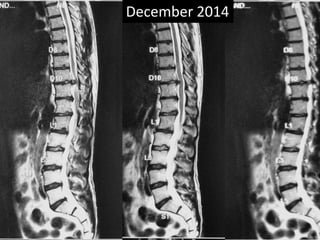
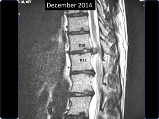
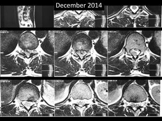
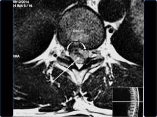
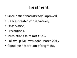
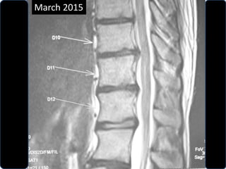
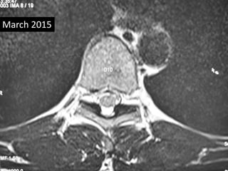
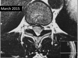
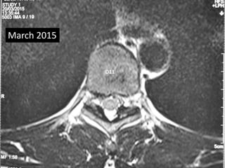
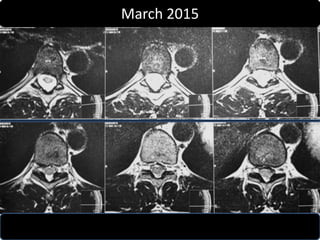
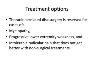
Rare case of Dorsal disc
D10 – D11
Vinod Naneria
Girish Yeotikar
Arjun Wadhwani
Choithram Hospital & Research Centre,
Indore, India
Case summary
• A 40 years old male, a regular visitor of gym.
• Complaint of sudden weakness & parasthesia
in whole right lower limb while doing weight
lifting in Gym.
• He was immediately made to lay down on a
bench.
• Gradual massage of the limb was done.
• Within 15 minutes he gradually regain the
control on the limb.
Case summary
• Out of fear – he attended the hospital for
check up.
• Clinically:
• Spine had stiffness, with limited flexion.
• Paraspinal rigidity was there.
• SLRT was negative.
• No obvious neuro-vascular deficit was
observed.
Case summary
• Hearing his abnormal history,
• Anxious but educated patient.
• A MRI was done.
• There was a disc extrusion at D10 – D11.
• The extruded fragment was on left side.
December 2014
December 2014
December 2014
Treatment
• Since patient had already improved,
• He was treated conservatively.
• Observation,
• Precautions,
• Instructions to report S.O.S.
• Follow up MRI was done March 2015
• Complete absorption of fragmant.
March 2015
March 2015
D10 – D11
March 2015
D11
March 2015
March 2015
Review Literature
• Neurosurgical Focus
The Pathophysiology of Thoracic Disc Disease
James Mcinerney, MD, and Perry A. Ball, MD,
Section of Neurosurgery, Dartmouth-
Hitchcock Medical Center, Lebanon, New
Hampshire.
Neurosurg Focus. 2000;9(4)
Review Literature
Incidence & Site:
• Thoracic disc herniation accounts for 0.15 - 4.4%
of all disc herniation.
• 75 % of all thoracic disc problems occur below T8,
with a peak of 26% at T11-12.
• The upper thoracic spine (T1-5) is the region least
often affected, with only 6% of all thoracic disc
herniation occurring here.
• To date, a total of 31 cases of T1-2 disc herniation
have been reported in the literature.
Review Literature
• Analysis of population studies suggests that
the overall incidence of thoracic disc
herniations is approximately 1 per 1,000,000
patient years.
• The majority of thoracic herniations have
been noted to be central or centrolateral, with
a minority of herniations truly lateral.
Review Literature
• Calcification is reported to occur in 30 to 70%
of cases of thoracic disc herniations,
• The cause of this phenomenon remains
unclear.
• Calcification is an important consideration,
however, because approximately 5 to 10% of
calcified discs are associated with intradural
extension.
Patho-physiology
• The decrease in matrix water is due to
decrease in the overall amount of
proteoglycans as well as a change in the ratio
of chondroitin sulfate to keratin sulfate.
• Keratin sulfate, with only one net negative
charge, tends to increase as compared with
chondroitin sulfate, which has two.
Patho-physiology
• Because KS attracts fewer small cations, the
osmotic gradient into the disc is decreased
and subsequently the overall water content is
reduced as well.
• With decreased water content, the disc loses
height and some ability to expand.
Patho-physiology
• As a result, more of the load is borne by the
annulus fibrosus. This, in turn, increases the
likelihood of injury to the annulus and the
overall rate of its degeneration.
• The disc's ability to expand does not begin to
decrease until the fourth decade.
Patho-physiology
• Thoracic disc prolapse peak during the fourth
and fifth decades of life.
• It is at this time that the intervertebral discs
experience a slight decrease in nuclear
expansion, causing increased stress on the
annulus fibrosus, and the decreased elasticity
of the annulus makes it more susceptible to
injury.
Patho-physiology
• An increase in annular tears coupled with a
persistently expansile nucleus then results in a
higher incidence of disc herniation.
• This would be especially true for active
individuals who place additional loads on the
spine.
Brown CW, Deffer PA Jr, Akmakjian J, et al:
The natural history of thoracic disc herniation.
Spine 17(Suppl):S97–S102, 1992• Brown, et al.,
• Retrospectively reviewed data obtained in 55
patients with 72 thoracic disc herniation.
• 15 (27%) of these patients eventually required
surgery, due to myelopathy.
• The vast majority of patients, however, did not
require surgery and have continued to perform
activities of daily living, including vigorous sports
activities.•
• There was no correlation between radiographic
depiction and the patient’s symptoms.
operative treatment of thoracic discs
• Posterior approach –
– Transpedicular,
– Transfacetal,
– Posterolateral approach modified
costotransversectomy,
• lateral extracavitary;
• Anterolateral approach - transthoracic;
• Thoracoscopic approach
Treatment options
• Thoracic herniated disc surgery is reserved for
cases of:
• Myelopathy,
• Progressive lower extremity weakness, and
• Intolerable radicular pain that does not get
better with non-surgical treatments.
References
• Adams MA, Hutton WC: Prolapsed intervertebral
disc. A hyperflexion injury. Spine 7:184-191, 1982
• Adams P, Muir H: Qualitative changes with age of
proteoglycans in human lumbar discs. Ann
Rheum Dis 35:289-296, 1976 Neurosurg. Focus /
Volume 9 / October, 2000 Pathophysiology of
thoracic disc disease 7
• Arce CA, Dohrmann GJ: Thoracic disc herniation.
Improved diagnosis with computed tomographic
scanning and a review of the literature. Surg
Neurol 23:356-361, 1985
References
• Awwad EE, Martin DS, Smith KR Jr, et al:
Asymptomatic versus symptomatic herniated
thoracic discs: their frequency and
characteristics as detected by computed
tomography after myelography. Neurosurgery
28:180-186, 1991
• Ball PA, Benzel EC: Pathology of disc
degeneration, in Menezes AH, Sonntag VKH
(eds): Principles of Spinal Surgery. New York:
McGraw-Hill, 1996, pp 507-516
References
• Benzel EC: Biomechanics of Spine Stabilization:
Principles and Clinical Practice. New York: McGraw-
Hill, 1995
• Brown CW, Deffer PA Jr, Akmakjian J, et al: The
natural history of thoracic disc herniation. Spine 17
(Suppl 6):S97-S102, 1992
• Compere EL, Cloward RB: Origin, anatomy,
physiology, and pathology of the intervertebral
disc. Instruct Lect Am Acad Orthop Surg 18:15-20,
1961
• DePalma AF, Rothman RH: The Intervetebral Disc.
Philadelphia: WB Saunders, 1970
References
• Fisher CM: Painful states: a neurological
commentary. Clin Neurosurg 31:32-53,
1983
• Hirsch C, et al: The anatomical basis for low
back pain. Studies on the presence of
sensory nerve endings in ligamentous,
capsular and intervertebral disc
structuresin the human lumbar spine. Acta
Orthop Scand 33:1-17, 1963
References
• Hitselberger WE, Witten RM: Abnormal
myelograms in asymptomatic patients. J
Neurosurg 28:204-206, 1968
• Holm S, Maroudas A, Urban JP, et al: Nutrition
of the intervertebral disc: solute transport
and metabolism. Connect Tissue Res 8:101-
119, 1981
• Kramer J, Schleberger R, Hedtmann A, et al:
Intervertebral Disk Diseases: Causes,
Diagnosis, Treatment, and Prophylaxis, ed 2.
Stuttgart: Thieme, 1990
References
• Moore K: The Developing Human: Clinically
Oriented Embryology, ed 4. Philadelphia: WB
Saunders, 1988, pp 334-340
• Nachemson A, Lewin T, Maroudas A, et al: In
vitro diffusion of dye through the end-plates and
annulus fibrosus of human intervertebral discs.
Acta Orthop Scand 41:589-607, 1970
• Smyth M, Wright V: Sciatica and the intrvertebral
disc: an experimental study. J Bone Joint Surg
(Am) 40:1401-1418, 1958
References
• Stillerman CB, Chen TC, Couldwell WT, et al:
Experience in the surgical management of 82
symptomatic herniated thoracic discs and
review of the literature. J Neurosurg 88:623-
633, 1998
• Stryer L: Biochemistry, ed 3. New York: WH
Freeman, 1988, pp 261-281
• White AA III, Panjabi MM: Clinical
Biomechanics of the Spine. Philadelphia:
Lippincott, 1990
DISCLAIMER
• Information contained and transmitted by this presentation is based
on personal experience and collection of cases at Choithram Hospital
& Research centre, Indore, India.
• It is intended for use only by the students of orthopaedic surgery.
• Views and opinion expressed in this presentation are personal.
• Depending upon the x-rays and clinical presentations viewers can
make their own opinion.
• For any confusion please contact the sole author for clarification.
• Every body is allowed to copy or download and use the material best
suited to him.
• We not responsible for any controversies arise out of this
presentation. For any correction or suggestion please contact
naneria@yahoo.com

Dorsal disc prolapse

  • 1.
    Rare case ofDorsal disc D10 – D11 Vinod Naneria Girish Yeotikar Arjun Wadhwani Choithram Hospital & Research Centre, Indore, India
  • 2.
    Case summary • A40 years old male, a regular visitor of gym. • Complaint of sudden weakness & parasthesia in whole right lower limb while doing weight lifting in Gym. • He was immediately made to lay down on a bench. • Gradual massage of the limb was done. • Within 15 minutes he gradually regain the control on the limb.
  • 3.
    Case summary • Outof fear – he attended the hospital for check up. • Clinically: • Spine had stiffness, with limited flexion. • Paraspinal rigidity was there. • SLRT was negative. • No obvious neuro-vascular deficit was observed.
  • 4.
    Case summary • Hearinghis abnormal history, • Anxious but educated patient. • A MRI was done. • There was a disc extrusion at D10 – D11. • The extruded fragment was on left side.
  • 5.
  • 6.
  • 7.
  • 10.
    Treatment • Since patienthad already improved, • He was treated conservatively. • Observation, • Precautions, • Instructions to report S.O.S. • Follow up MRI was done March 2015 • Complete absorption of fragmant.
  • 11.
  • 12.
  • 13.
  • 14.
  • 15.
  • 16.
    Review Literature • NeurosurgicalFocus The Pathophysiology of Thoracic Disc Disease James Mcinerney, MD, and Perry A. Ball, MD, Section of Neurosurgery, Dartmouth- Hitchcock Medical Center, Lebanon, New Hampshire. Neurosurg Focus. 2000;9(4)
  • 17.
    Review Literature Incidence &Site: • Thoracic disc herniation accounts for 0.15 - 4.4% of all disc herniation. • 75 % of all thoracic disc problems occur below T8, with a peak of 26% at T11-12. • The upper thoracic spine (T1-5) is the region least often affected, with only 6% of all thoracic disc herniation occurring here. • To date, a total of 31 cases of T1-2 disc herniation have been reported in the literature.
  • 18.
    Review Literature • Analysisof population studies suggests that the overall incidence of thoracic disc herniations is approximately 1 per 1,000,000 patient years. • The majority of thoracic herniations have been noted to be central or centrolateral, with a minority of herniations truly lateral.
  • 19.
    Review Literature • Calcificationis reported to occur in 30 to 70% of cases of thoracic disc herniations, • The cause of this phenomenon remains unclear. • Calcification is an important consideration, however, because approximately 5 to 10% of calcified discs are associated with intradural extension.
  • 20.
    Patho-physiology • The decreasein matrix water is due to decrease in the overall amount of proteoglycans as well as a change in the ratio of chondroitin sulfate to keratin sulfate. • Keratin sulfate, with only one net negative charge, tends to increase as compared with chondroitin sulfate, which has two.
  • 21.
    Patho-physiology • Because KSattracts fewer small cations, the osmotic gradient into the disc is decreased and subsequently the overall water content is reduced as well. • With decreased water content, the disc loses height and some ability to expand.
  • 22.
    Patho-physiology • As aresult, more of the load is borne by the annulus fibrosus. This, in turn, increases the likelihood of injury to the annulus and the overall rate of its degeneration. • The disc's ability to expand does not begin to decrease until the fourth decade.
  • 23.
    Patho-physiology • Thoracic discprolapse peak during the fourth and fifth decades of life. • It is at this time that the intervertebral discs experience a slight decrease in nuclear expansion, causing increased stress on the annulus fibrosus, and the decreased elasticity of the annulus makes it more susceptible to injury.
  • 24.
    Patho-physiology • An increasein annular tears coupled with a persistently expansile nucleus then results in a higher incidence of disc herniation. • This would be especially true for active individuals who place additional loads on the spine.
  • 25.
    Brown CW, DefferPA Jr, Akmakjian J, et al: The natural history of thoracic disc herniation. Spine 17(Suppl):S97–S102, 1992• Brown, et al., • Retrospectively reviewed data obtained in 55 patients with 72 thoracic disc herniation. • 15 (27%) of these patients eventually required surgery, due to myelopathy. • The vast majority of patients, however, did not require surgery and have continued to perform activities of daily living, including vigorous sports activities.• • There was no correlation between radiographic depiction and the patient’s symptoms.
  • 26.
    operative treatment ofthoracic discs • Posterior approach – – Transpedicular, – Transfacetal, – Posterolateral approach modified costotransversectomy, • lateral extracavitary; • Anterolateral approach - transthoracic; • Thoracoscopic approach
  • 27.
    Treatment options • Thoracicherniated disc surgery is reserved for cases of: • Myelopathy, • Progressive lower extremity weakness, and • Intolerable radicular pain that does not get better with non-surgical treatments.
  • 28.
    References • Adams MA,Hutton WC: Prolapsed intervertebral disc. A hyperflexion injury. Spine 7:184-191, 1982 • Adams P, Muir H: Qualitative changes with age of proteoglycans in human lumbar discs. Ann Rheum Dis 35:289-296, 1976 Neurosurg. Focus / Volume 9 / October, 2000 Pathophysiology of thoracic disc disease 7 • Arce CA, Dohrmann GJ: Thoracic disc herniation. Improved diagnosis with computed tomographic scanning and a review of the literature. Surg Neurol 23:356-361, 1985
  • 29.
    References • Awwad EE,Martin DS, Smith KR Jr, et al: Asymptomatic versus symptomatic herniated thoracic discs: their frequency and characteristics as detected by computed tomography after myelography. Neurosurgery 28:180-186, 1991 • Ball PA, Benzel EC: Pathology of disc degeneration, in Menezes AH, Sonntag VKH (eds): Principles of Spinal Surgery. New York: McGraw-Hill, 1996, pp 507-516
  • 30.
    References • Benzel EC:Biomechanics of Spine Stabilization: Principles and Clinical Practice. New York: McGraw- Hill, 1995 • Brown CW, Deffer PA Jr, Akmakjian J, et al: The natural history of thoracic disc herniation. Spine 17 (Suppl 6):S97-S102, 1992 • Compere EL, Cloward RB: Origin, anatomy, physiology, and pathology of the intervertebral disc. Instruct Lect Am Acad Orthop Surg 18:15-20, 1961 • DePalma AF, Rothman RH: The Intervetebral Disc. Philadelphia: WB Saunders, 1970
  • 31.
    References • Fisher CM:Painful states: a neurological commentary. Clin Neurosurg 31:32-53, 1983 • Hirsch C, et al: The anatomical basis for low back pain. Studies on the presence of sensory nerve endings in ligamentous, capsular and intervertebral disc structuresin the human lumbar spine. Acta Orthop Scand 33:1-17, 1963
  • 32.
    References • Hitselberger WE,Witten RM: Abnormal myelograms in asymptomatic patients. J Neurosurg 28:204-206, 1968 • Holm S, Maroudas A, Urban JP, et al: Nutrition of the intervertebral disc: solute transport and metabolism. Connect Tissue Res 8:101- 119, 1981 • Kramer J, Schleberger R, Hedtmann A, et al: Intervertebral Disk Diseases: Causes, Diagnosis, Treatment, and Prophylaxis, ed 2. Stuttgart: Thieme, 1990
  • 33.
    References • Moore K:The Developing Human: Clinically Oriented Embryology, ed 4. Philadelphia: WB Saunders, 1988, pp 334-340 • Nachemson A, Lewin T, Maroudas A, et al: In vitro diffusion of dye through the end-plates and annulus fibrosus of human intervertebral discs. Acta Orthop Scand 41:589-607, 1970 • Smyth M, Wright V: Sciatica and the intrvertebral disc: an experimental study. J Bone Joint Surg (Am) 40:1401-1418, 1958
  • 34.
    References • Stillerman CB,Chen TC, Couldwell WT, et al: Experience in the surgical management of 82 symptomatic herniated thoracic discs and review of the literature. J Neurosurg 88:623- 633, 1998 • Stryer L: Biochemistry, ed 3. New York: WH Freeman, 1988, pp 261-281 • White AA III, Panjabi MM: Clinical Biomechanics of the Spine. Philadelphia: Lippincott, 1990
  • 35.
    DISCLAIMER • Information containedand transmitted by this presentation is based on personal experience and collection of cases at Choithram Hospital & Research centre, Indore, India. • It is intended for use only by the students of orthopaedic surgery. • Views and opinion expressed in this presentation are personal. • Depending upon the x-rays and clinical presentations viewers can make their own opinion. • For any confusion please contact the sole author for clarification. • Every body is allowed to copy or download and use the material best suited to him. • We not responsible for any controversies arise out of this presentation. For any correction or suggestion please contact naneria@yahoo.com