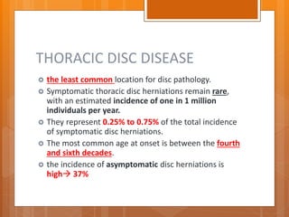
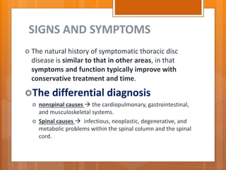
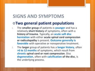
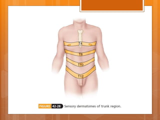
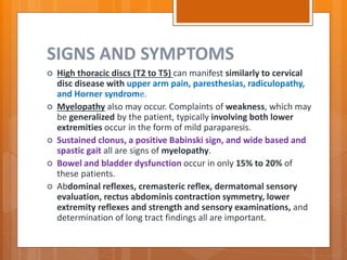
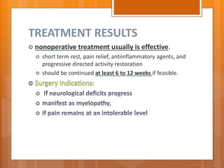
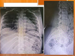
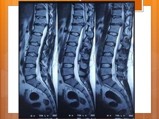
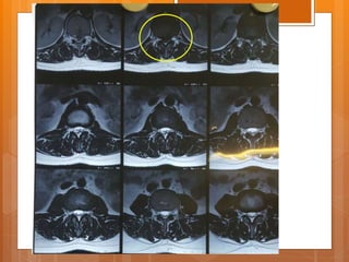
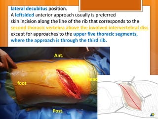
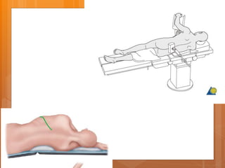
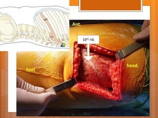
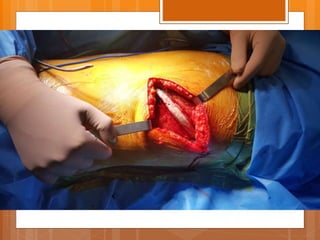
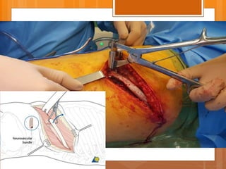
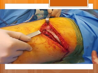
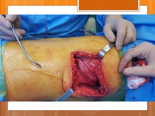
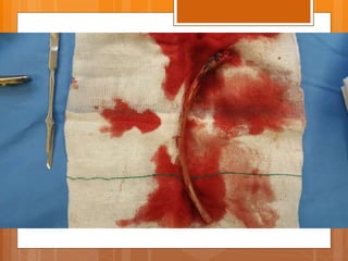
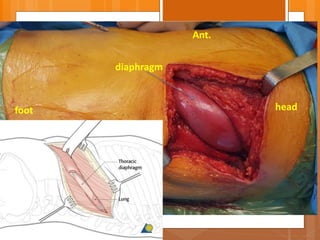
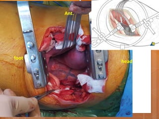
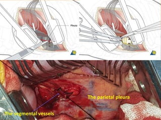
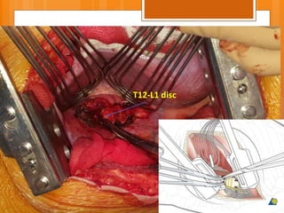
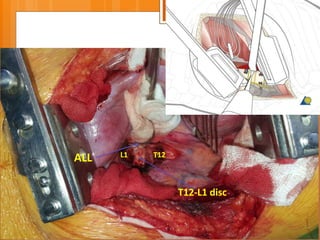
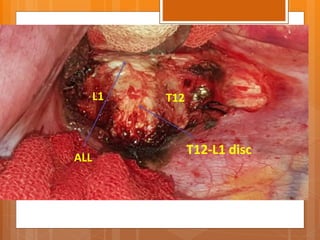
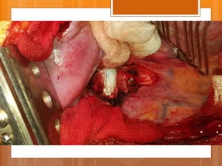
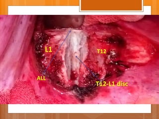
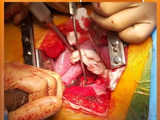
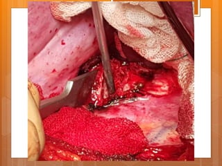
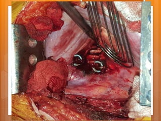
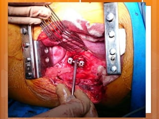
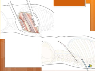
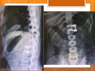
THORACIC
DISC DISEASE
m. dehnokhalaji
THORACIC DISC DISEASE
 the least common location for disc pathology.
 Symptomatic thoracic disc herniations remain rare,
with an estimated incidence of one in 1 million
individuals per year.
 They represent 0.25% to 0.75% of the total incidence
of symptomatic disc herniations.
 The most common age at onset is between the fourth
and sixth decades.
 the incidence of asymptomatic disc herniations is
high 37%
SIGNS AND SYMPTOMS
 The natural history of symptomatic thoracic disc
disease is similar to that in other areas, in that
symptoms and function typically improve with
conservative treatment and time.
The differential diagnosis
 nonspinal causes  the cardiopulmonary, gastrointestinal,
and musculoskeletal systems.
 Spinal causes  infectious, neoplastic, degenerative, and
metabolic problems within the spinal column and the spinal
cord.
SIGNS AND SYMPTOMS
Two general patient populations
1. The smaller group of patients is younger and has a
relatively short history of symptoms, often with a
history of trauma. Typically, an acute soft disc
herniation with either acute spinal cord compression
or radiculopathy is present. Outcome generally is
favorable with operative or nonoperative treatment.
2. The larger group of patients has a longer history, often
>6 to 12 months of symptoms, which result from
chronic spinal cord or root compression. Disc
degeneration, often with calcification of the disc, is
the underlying process.
SIGNS AND SYMPTOMS
 Pain the most common presenting
 Two patterns : , and along
the course of the intercostal nerve.
 The T10 dermatomal level is the most commonly
reported distribution, regardless of the level of
involvement.
 This is a band extending around the lower lateral thorax
and caudad to the level of the umbilicus.
 This radicular pattern is more common with upper
thoracic and lateral disc herniations.
 Associated sensory changes of paresthesias and
dysesthesia in a dermatomal distribution also occur .
SIGNS AND SYMPTOMS
 High thoracic discs (T2 to T5) can manifest similarly to cervical
disc disease with upper arm pain, paresthesias, radiculopathy,
and Horner syndrome.
 Myelopathy also may occur. Complaints of weakness, which may
be generalized by the patient, typically involving both lower
extremities occur in the form of mild paraparesis.
 Sustained clonus, a positive Babinski sign, and wide based and
spastic gait all are signs of myelopathy.
 Bowel and bladder dysfunction occur in only 15% to 20% of
these patients.
 Abdominal reflexes, cremasteric reflex, dermatomal sensory
evaluation, rectus abdominis contraction symmetry, lower
extremity reflexes and strength and sensory examinations, and
determination of long tract findings all are important.
TREATMENT RESULTS
 nonoperative treatment usually is effective.
 short term rest, pain relief, antiinflammatory agents, and
progressive directed activity restoration
 should be continued at least 6 to 12 weeks if feasible.



OPERATIVE TREATMENT
 Simple laminectomy has no role in the treatment of thoracic
disc herniations.
 Posterior approaches, including costotransversectomy,
transpedicular approach, transfacet pedicle sparing, transdural,
and lateral extracavitary approach, all have been used
successfully.
 Most more recent studies suggest that lateral rachiotomy
(modified costotransversectomy) or an anterior transthoracic
approach for discectomy produces considerably better results
with no evidence of worsening after the procedure.
 Anterior approaches via thoracotomy, a transsternal approach,
retropleural approach, or VATS also have been used
successfully.
THORACIC DISCECTOMY
ANTERIOR APPROACH
Case presentation
 Female
 21 y/o
 Back pain from 1.5 years ago
 Exacerbated after child delivery (9m ago)
 Pain radiated to right leg from 7m ago
P/E
 ↓L3 , L4 force
 right clonus
 ↓ achilles DTR
 QC DTR normal
 Babinski sign normal
 Intermittent Bladder dysfunction
 Sensory normal
head
foot
Ant.
lateral decubitus position.
A leftsided anterior approach usually is preferred
skin incision along the line of the rib that corresponds to the
second thoracic vertebra above the involved intervertebral disc
except for approaches to the upper five thoracic segments,
where the approach is through the third rib.
Post.
10th rib
head
foot
Ant.
headfoot
Ant.
diaphragm
head
foot
Ant.
The segmental vessels
The parietal pleura
T12-L1 disc
T12-L1 disc
ALL L1 T12
T12-L1 disc
ALL
L1 T12
T12-L1 disc
ALL
L1 T12
POSTOPERATIVE CARE
 Postoperative care is the same as for a thoracotomy.
 The patient is allowed to walk after the chest tubes are
removed.
 Extension in any position is prohibited.
 A brace or body cast that limits extension should be used if
the stability of the graft is questionable.
 The graft usually is stable without support if only one disc
space is removed.
 Postoperative care is the same as for anterior corpectomy
and fusion if more than one disc level is removed.
 If no fusion is done, the patient is mobilized as pain
permits without a brace.

Thoracic disc disease

  • 1.
  • 2.
    THORACIC DISC DISEASE the least common location for disc pathology.  Symptomatic thoracic disc herniations remain rare, with an estimated incidence of one in 1 million individuals per year.  They represent 0.25% to 0.75% of the total incidence of symptomatic disc herniations.  The most common age at onset is between the fourth and sixth decades.  the incidence of asymptomatic disc herniations is high 37%
  • 3.
    SIGNS AND SYMPTOMS The natural history of symptomatic thoracic disc disease is similar to that in other areas, in that symptoms and function typically improve with conservative treatment and time. The differential diagnosis  nonspinal causes  the cardiopulmonary, gastrointestinal, and musculoskeletal systems.  Spinal causes  infectious, neoplastic, degenerative, and metabolic problems within the spinal column and the spinal cord.
  • 4.
    SIGNS AND SYMPTOMS Twogeneral patient populations 1. The smaller group of patients is younger and has a relatively short history of symptoms, often with a history of trauma. Typically, an acute soft disc herniation with either acute spinal cord compression or radiculopathy is present. Outcome generally is favorable with operative or nonoperative treatment. 2. The larger group of patients has a longer history, often >6 to 12 months of symptoms, which result from chronic spinal cord or root compression. Disc degeneration, often with calcification of the disc, is the underlying process.
  • 5.
    SIGNS AND SYMPTOMS Pain the most common presenting  Two patterns : , and along the course of the intercostal nerve.  The T10 dermatomal level is the most commonly reported distribution, regardless of the level of involvement.  This is a band extending around the lower lateral thorax and caudad to the level of the umbilicus.  This radicular pattern is more common with upper thoracic and lateral disc herniations.  Associated sensory changes of paresthesias and dysesthesia in a dermatomal distribution also occur .
  • 7.
    SIGNS AND SYMPTOMS High thoracic discs (T2 to T5) can manifest similarly to cervical disc disease with upper arm pain, paresthesias, radiculopathy, and Horner syndrome.  Myelopathy also may occur. Complaints of weakness, which may be generalized by the patient, typically involving both lower extremities occur in the form of mild paraparesis.  Sustained clonus, a positive Babinski sign, and wide based and spastic gait all are signs of myelopathy.  Bowel and bladder dysfunction occur in only 15% to 20% of these patients.  Abdominal reflexes, cremasteric reflex, dermatomal sensory evaluation, rectus abdominis contraction symmetry, lower extremity reflexes and strength and sensory examinations, and determination of long tract findings all are important.
  • 8.
    TREATMENT RESULTS  nonoperativetreatment usually is effective.  short term rest, pain relief, antiinflammatory agents, and progressive directed activity restoration  should be continued at least 6 to 12 weeks if feasible.   
  • 9.
    OPERATIVE TREATMENT  Simplelaminectomy has no role in the treatment of thoracic disc herniations.  Posterior approaches, including costotransversectomy, transpedicular approach, transfacet pedicle sparing, transdural, and lateral extracavitary approach, all have been used successfully.  Most more recent studies suggest that lateral rachiotomy (modified costotransversectomy) or an anterior transthoracic approach for discectomy produces considerably better results with no evidence of worsening after the procedure.  Anterior approaches via thoracotomy, a transsternal approach, retropleural approach, or VATS also have been used successfully.
  • 10.
  • 11.
    Case presentation  Female 21 y/o  Back pain from 1.5 years ago  Exacerbated after child delivery (9m ago)  Pain radiated to right leg from 7m ago
  • 12.
    P/E  ↓L3 ,L4 force  right clonus  ↓ achilles DTR  QC DTR normal  Babinski sign normal  Intermittent Bladder dysfunction  Sensory normal
  • 16.
    head foot Ant. lateral decubitus position. Aleftsided anterior approach usually is preferred skin incision along the line of the rib that corresponds to the second thoracic vertebra above the involved intervertebral disc except for approaches to the upper five thoracic segments, where the approach is through the third rib. Post.
  • 18.
  • 24.
  • 25.
  • 26.
  • 27.
  • 28.
  • 29.
  • 31.
  • 38.
    POSTOPERATIVE CARE  Postoperativecare is the same as for a thoracotomy.  The patient is allowed to walk after the chest tubes are removed.  Extension in any position is prohibited.  A brace or body cast that limits extension should be used if the stability of the graft is questionable.  The graft usually is stable without support if only one disc space is removed.  Postoperative care is the same as for anterior corpectomy and fusion if more than one disc level is removed.  If no fusion is done, the patient is mobilized as pain permits without a brace.