Download to read offline

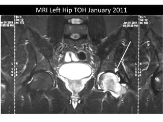
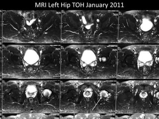
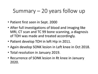

The document presents a detailed case study of a patient with transient osteoporosis of the hip (TOH) and spontaneous osteonecrosis of the knee (SONK) treated at Choithram Hospital & Research Centre in India. Over 20 years, the patient experienced multiple episodes of TOH and SONK with no history of trauma or co-morbidities, resulting in resolutions and recurrences of conditions. The information is intended for orthopedic surgery students and highlights personal experiences and case collections, with a disclaimer regarding content usage and potential controversies.


