Case record...Lumbar spondylosis
•
7 likes•6,833 views

Case record...Lumbar spondylosis http://yassermetwally.com http://yassermetwally.net

Recommended
More Related Content
What's hot
What's hot (20)
Similar to Case record...Lumbar spondylosis
Similar to Case record...Lumbar spondylosis (20)
More from Professor Yasser Metwally
More from Professor Yasser Metwally (20)
Recently uploaded
Recently uploaded (20)
Case record...Lumbar spondylosis
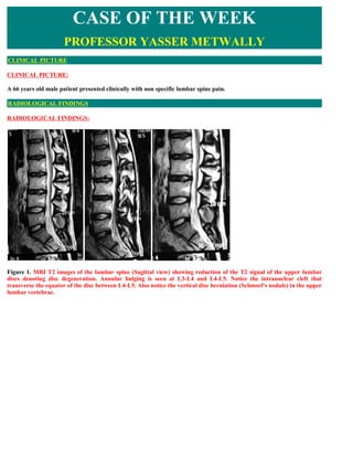
- 1. CASE OF THE WEEK PROFESSOR YASSER METWALLY CLINICAL PICTURE CLINICAL PICTURE: A 66 years old male patient presented clinically with non specific lumbar spine pain. RADIOLOGICAL FINDINGS RADIOLOGICAL FINDINGS: Figure 1. MRI T2 images of the lumbar spine (Sagittal view) showing reduction of the T2 signal of the upper lumbar discs denoting disc degeneration. Annular bulging is seen at L3-L4 and L4-L5. Notice the intranuclear cleft that transverse the equator of the disc between L4-L5. Also notice the vertical disc herniation (Schmorl's nodule) in the upper lumbar vertebrae.
- 2. Figure 2. MRI T1 images showing vertical disc herniation (Schmorl's nodule) in the upper lumbar vertebrae.
- 3. Figure 3. MRI T1 images showing vertical disc herniation (Schmorl's nodule) in the upper lumbar vertebrae
- 4. Figure 4. MRI T2 (A) and T1 (B) images images showing vertical disc herniation (Schmorl's nodule) in the upper lumbar vertebrae
- 5. Figure 5. Axial MRI T2 images showing the vertical disc herniation (Schmorl's nodule). The Schmorl's nodule appeared hyperintense (A) in the upper part of the vertebra and hypointense (B) in the lower part of the vertebra. The hypointensity in the lower part of the vertebra is surrounded by relatively hyperintense rings. The T2 hyperintensity is probably caused by inflammatory edema. Notice the annular bulge and the fact joint disease. Figure 6. MRI T2 images showing vertical disc herniation (Schmorl's nodule) in the upper lumbar vertebrae. Notice the
- 6. annular bulge and the facet joint disease. DIAGNOSIS: DIAGNOSIS: LUMBAR SPONDYLOSIS DISCUSSION DISCUSSION: Discogenic spine disease is the most common surgically treatable form of pain due to nerve root compression. Patients who present with reproducible radicular back and extremity pain that is unresponsive to conservative management can obtain excellent results with surgical excision of the offending herniated intervertebral disc. Careful consideration of a patient's clinical symptoms and signs and close correlation with the appropriate radiologic examination are mandatory. The spine surgeon and the radiologist must collaborate to determine the optimal imaging modality for an individual patient and his or her pathologic process. Radiographic imaging is a vital adjunct to the neurosurgical evaluation of the patient with potential discogenic disease of the spine. Magnetic resonance (MR) imaging has emerged as the imaging modality of choice for evaluation of lumbar disc disease. 1, 13,37 Technical advantages of MR imaging include superior soft-tissue contrast, direct multiplanar capability, and the lack of ionizing radiation. Computed tomography (CT) (enhanced, nonenhanced, and postmyelographic) is still widely used and also provides excellent images. The optimal combination of studies and individual study protocols for discogenic disease is quite variable and is under continuous investigation. Disc degeneration can often be attributable to the combined effects of biomechanical stress and age related changes. The cervical and lumbar discs are subject to more mechanical stress than thoracic discs. Structural support provided by the thoracic rib cage and adjacent musculature as well as the coronal orientation of the facet joints are factors that attenuate biomechanical stresses on the thoracic discs. Disc herniation most commonly occurs at the C5-6, C6-7, L4-5, and L5-S1 levels. 16,51 Symptoms referable to a specific spinal root level help define the optimal radiographic examination. Correlation of imaging results with the clinical characteristic of neurologic dysfunction from disc disease is of paramount importance in formulating an appropriate treatment plan.
- 7. Figure 1. Normal lumbar disc anatomy. Notice that it is difficult to differentiate between the nucleus pulposus and annulus fibrosus ID = Intervertebral disc S= Superior articular facet I = Inferior articular facet L = Ligamentum flavum G = Gray matter W= white matter F= Facet joint Figure 2. Normal lumbar disc anatomy. Notice that it is difficult to differentiate between the nucleus pulposus and annulus fibrosus 1. The intervertebral disc 2. Epidural fat 3. Superior articular facet
- 8. 4. Inferior articular facet 5. Spinal roots 6. Superior articular joint 7. Inferior articular facet 8. Tegmentum flavum Pathophysiology of disc degeneration Posterior elements of the lumbar spinal functional unit typically bear less weight than anterior elements in all positions. Anterior elements bear over 90% of forces transmitted through the lumbar spine in sitting; during standing, this portion decreases to approximately 80%. As the degenerative process progresses, relative anterior-to-posterior force transmission approaches parity. The spine functions best within a realm of static and dynamic stability. Bony architecture and associated specialized soft tissue structures, especially the intervertebral disc, provide static stability. Dynamic stability, however, is accomplished through a system of muscular and ligamentous supports acting in concert during various functional, occupational, and avocational activities. The overall mechanical effect of these structures maintains the histologic integrity of the tri-joint complex. Net shear and compressive forces must be maintained below respective critical minima to maintain tri-joint articulation integrity. Persistent, recurrent, nonmechanical, and/or excessive forces to the motion segment beyond minimal thresholds lead to microtrauma of the disc and facet joints, triggering and continuing the degenerative process. Degenerative cascade is the widely accepted pathophysiologic model describing the degenerative process as it affects the lumbar spine and individual motion segments. This process occurs in 3 phases that comprise a continuum with gradual transition, rather than 3 distinct clearly definable stages. Phase I The dysfunctional phase, or Phase I, is characterized histologically by circumferential tears or fissures in the outer annulus. Tears can be accompanied by endplate separation or failure, interrupting blood supply to the disc and impairing nutritional supply and waste removal. Such changes may be the result of repetitive microtrauma. Since the outer one third of the annular wall is innervated, tears or fissures in this area may be painful. Strong experimental evidence suggests that most episodes of LBP are a consequence of disc injury, rather than musculotendinous or ligamentous strain. Circumferential tears may coalesce to form radial tears. The nucleus pulposus may lose its normal water-imbibing abilities as a result of biochemical changes in aggregating proteoglycans. Studies suggest proteoglycan destruction may result from an imbalance between the matrix metalloproteinase-3 (MMP- 3) and tissue inhibitor of metalloproteinase-1 (TIMP-1). This imbalance results in diminished capacity for imbibing water, causing loss of nuclear hydrostatic pressure and leading to buckling of the annular lamellae. This phenomenon leads to increased focal segmental mobility and shear stress to the annular wall. Delamination and fissuring within the annulus can result. Annular delamination has been shown to occur as a separate and distinct event from annular fissures. Microfractures of collagen fibrils within the annulus have been demonstrated with electron microscopy. MRI at this stage may reveal desiccation, disc bulging without herniation, or a high-intensity zone (HIZ) within the annulus. Structural alteration of the facet joints following disc degeneration is acknowledged widely, but this expected pathologic alteration does not necessarily follow. Changes associated with zygapophyseal joints during the dysfunctional phase may include synovitis and hypomobility. The facet joints may serve as a pain generator.
- 9. Figure 3. Dessication of the nucleus pulposus associated with multiple annular tears (eg, radial, circumferential). Notice that the disc is bulging without actual herniation. Because of the existing disc degeneration, it is possible to differentiate between the nucleus pulposus and annulus fibrosus. 1. Annulus fibrosis 2. Circumferential annular tears 3- Posterior longitudinal ligament 4. The desiccated nucleus pulposus
- 10. Figure 4. Dessication of the nucleus pulposus associated with multiple annular tears (eg, radial, circumferential). Notice that the disc is bulging without actual herniation. Because of the existing disc degeneration, it is possible to differentiate between the nucleus pulposus and annulus fibrosus. Notice the existence of articular facet pathology and tegmental flavum hypertrophy. 1. Annulus fibrosis 2. Circumferential annular tears with infiltration by nuclear material 3,4 . The desiccated nucleus pulposus Phase II The unstable phase, or phase II, may result from progressive loss of mechanical integrity of the tri-joint complex. Disc- related changes include multiple annular tears (eg, radial, circumferential), internal disc disruption and resorption, or loss of disc-space height. Concurrent changes in the zygapophyseal joints include cartilage degeneration, capsular laxity, and subluxation. The biomechanical result of these alterations leads to segmental instability. Clinical syndromes of segmental instability, internal disc disruption syndrome, and herniated disc seem to fit within this phase.
- 11. Figure 5. Disc degeneration with multiple disc bulging without herniation G= Gray matter W= white matter PLL= Posterior longitudinal ligament DD= Disc degeneration B= Disc bulging Phase III The third and final phase, stabilization, is characterized by further disc resorption, disc-space narrowing, endplate destruction, disc fibrosis, and osteophyte formation. Discogenic pain from such discs may be of much lower incidence than pain from discs in phases I and II; however, great variation of phases can be expected within different discs in any given individual, since much variation exists between individuals of similar ages.
- 12. Figure 6. The desiccated nucleus (1) is unable to redistribute much of the vertical load radically, causing the annulus to bulge posteriorly (4), pushing the posterior spinal ligament (3), this can result in annular tear and herniation of the nucleus pulposus (2). Histologic Findings The lumbar intervertebral disc is composed of the nucleus pulposus and annulus fibrosus. The disc is related intimately as a functional unit to the cartilaginous endplate. The intervertebral disc contains water, collagen, and proteoglycans. The nucleus pulposus normally is well hydrated, containing approximately 85-90% water in the first decade and 70-80% water in the adult. Elongated fibrocytes are organized loosely, forming a gelatinous matrix. The nucleus has a higher content of proteoglycans than the disc annulus.
- 13. Figure 7. The desiccated nucleus (1) is unable to redistribute much of the vertical load radically, this can result in annular tear and herniation of the nucleus pulposus (2,4,7). Notice posterior annulus bulge (6). (3= the annulus, 5= the cauda roots) The annulus fibrosis contains 75% water in the first decade of life and 70-80% water in the adult. The peripheral annulus is composed primarily of type I collagen, lending tensile strength to the intervertebral disc. The inner annulus is composed primarily of type 2 collagen, which, in conjunction with the nucleus pulposus, provides compressive strength. Type 2 collagen may have greater water content than type 1 collagen. The collagenous lamellae are fewer, thinner, and more tightly packed posteriorly than anteriorly. The central depression of the vertebral endplate is covered by hyaline cartilage. With age-related degeneration, the volume of the nucleus pulposus diminishes with decreasing hydration and increasing fibrosis. Changes in water content are from alteration in the relative composition of proteoglycan, as well as decrease in the extent of aggregating proteoglycans. By age 30 years, in-growth of fibrous tissue into the nucleus results in an intranuclear cleft. Fibrocartilage, derived from cells in the annulus and endplate, gradually replaces mucoid material within the nucleus. Gradual loss of definition between nucleus and inner annular fibers occurs.
- 14. Figure 8. A normal nucleus has a sharply demarcated oval contour, and due to increased water content, the nucleus is hyperintense on the MRI T1 images surrounded by the hypointense annulus fibrosis. A firm fibrous band traverses the disc equator and blends imperceptibly with the anulus fibers, this band is seen as a hypointense band traversing the disc equator (L4,L5 disc in A). A degenerated disc is hypointense on The MRI T2 images due to reduced water contents. Degenerated discs are seen bulging in both A,B In the final stages of degeneration, the nucleus is replaced completely by fibrocartilage, indistinguishable from the fibrotic disc annulus. Specifically, the type 1 collagen content of the disc annulus increases, especially posteriorly, and type 2 collagen content diminishes. Cartilaginous metaplasia begins in the inner annular fibers with changes in the overall fiber direction from vertical to horizontal. Infolding of fibers of the outer annulus occurs early with myxoid degeneration of the outer annular fibers. Concentric and/or transverse tears in the annulus fibrosis are frequent findings. Peripheral tears are more frequent posterior or posterolateral where the annular lamellae are fewer. Development of a radial tear, particularly a tear extending to the disc nucleus, is one of the major hallmarks of disc degeneration. The degenerated intervertebral disc loses height and overall volume. Herniation of both nuclear material and annulus fibrosis may occur through the tear. With aging, the cartilage endplate may become thinner and eventually may calcify. In advanced disc degeneration, the cartilage endplate is calcified with fissuring and microfractures. At autopsy, 97% of adults aged 49 years or older demonstrate degenerative changes. For a structure to be considered a pain generator, it must meet the following 3 criteria: (1) it must have a nerve supply, (2) it must be susceptible to disease or injuries known to be painful, and (3) it must be capable of causing pain similar to that observed clinically. The superficial layers of the annulus fibrosis contain nerve fibers located in the posterior portion of the annulus, which are branches from the sinuvertebral nerves. The sinuvertebral nerves are branches of the ventral rami. They also contain fibers derived from the grey ramus. Small branches from the grey ramus communicans or sympathetic fibers innervate the anterior longitudinal ligament and lateral and anterior annulus. The grey ramus communicans joins the sinuvertebral nerve that reenters the intervertebral foramen and spinal canal to innervate the posterior annulus and the posterior longitudinal ligament. A dense nerve network on the posterior portion of the lumbar intervertebral disc has been demonstrated in rats, disappearing almost completely after total resection of bilateral sympathetic trunks at L2-L6. The authors concluded that, in rats, the posterior portion of the lumbar intervertebral disc and posterior longitudinal ligament are innervated by sympathetic nerves bilaterally and multisegmentally. A variety of free and complex nerve endings have been demonstrated in the outer one third to one half of the annulus. In addition to annular fissures or tears, Coppes et al observed more extensive disc innervation in the severely degenerated lumbar disc compared with the normal disc.
- 15. The nociceptive properties of at least some of these nerves have been suggested by substance P immunoreactivity, which provides further evidence for the existence of a morphologic substrate of discogenic pain. Nerve fibers were restricted to the outer or middle third of the annulus in control samples. In the patient population undergoing spinal fusion for chronic LBP, nerves extended into the inner third of the annulus fibrosis in 46% and into nucleus pulposus in 22% of patients. Their findings that isolated nerve fibers expressed substance P deep within diseased intervertebral discs and their association with pain suggests an important role for nerve in-growth into the intervertebral disc in the pathogenesis of chronic LBP. Weinstein et al 52 identified substance P, calcitonin gene-related peptide (CGRP), and vasoactive intestinal polypeptides (VIP) in the outer annular fibers of the disc in rats. These chemicals all are related to pain perception. Substance P, dopamine, and choline acetyltransferase immunoreactive nerve fibers are found in human longitudinal ligaments that have been removed surgically. These findings not only provide evidence to support the first criterion but also reveal changes associated with painful discs. LUMBAR DISC DISEASE Introduction Lumbar disc disease, a leading cause of correctable lower back pain, often manifests as posterior herniation with radicular symptoms from neural compression. The classical syndrome of lumbar disc herniation features stiffness in the back and pain radiating down to the thighs and feet associated with paresthesia, weakness, and reflex changes. 10, 16 Patients typically flex their backs to reduce the normal lumbar lordosis. Flexion of the low back may alleviate neural compression by widening the neuroforamina caused by posterior rotation of the superior facets. Nerve stretching by the straight leg test and other maneuvers can reproduce or exacerbate radicular pain. Nerve roots L4, L5, and S1 are most commonly affected. Physical impingement from disc herniation most often occurs in the quot;lateral gutterquot; of the spinal canal, adjacent to the posterior lateral border of a herniated disc fragment. For instance, at the L5-Sl disc level, the Sl nerve roots are poised in the antero- lateral margin of the thecal sac. They are quot;tetheredquot; at that location as they acquire an investment of dura and assume an oblique (and eventually transverse) orientation prior to exiting through the neural foramen more inferiorly. Nerve root impingement at each intervertebral disc level typically produces a distinctive and predictable pattern of radiating pain, sensory loss, weakness, and decreased reflexes. Intervertebral discs are composed of highly specialized connective tissues (glycosaminoglycans and collagen). 47 The central portion of the nucleus pulposus is comprised of a gelatinous mucoid material and is surrounded by the fibrocartilaginous anulus fibrosus. The nucleus pulposus and anulus fibrosus can be differentiated on MR images because of differing water content. With age, the water content of the disc decreases. This degenerative-senescent change is reflected by loss of signal on T2-weighted MR images and is often identified at levels that show disc bulging or frank herniation. The intervertebral disc can be classified as immature, transitional, adult, early degenerated, or severely degenerated. 47,50 Up to age 2, the nucleus has a sharply demarcated oval contour. The immature disc is also grossly translucent. By the second decade of life, quot;transitional discsquot; exhibit a fibrous structure oriented horizontally in the equator of the disc. The mature adult disc is characterized by a more homogeneous architecture and gross appearance with less distinction between the nucleus pulposus and anulus fibrosus. A firm fibrous band traverses the disc equator and blends imperceptibly with the anulus fibers. An early degenerated disc has a slightly narrowed disc height and reduced fibrocartilage in the nucleus. The severely degenerated disc has amorphous fibers and cystic spaces replacing normal fibrocartilage. The normal adult type of nucleus commonly has an anulus with small concentric or transverse tears but never a radial tear. The early degenerated disc may be associated with a peripheral radial tear of the anulus in up to 92% of cases. 48 The severely degenerated disc is typically associated with a complete disruption of the anulus. The normal anatomical/radiological picture of the intervertebral discs Functionally and pathologically, the intervertebral joints are an important aspect of the anatomy of the spine. These joints are amphiarthrodial, with only slightly movable articulations connected by fibrocartilage. Although only a slight amount of motion is possible at each joint, the spine has a considerable amount of mobility because of the number of joints present. The cervical disks are thicker anteriorly than posteriorly, and this shape contributes to the lordosis of the cervical spine. The major components of the intervertebral disk are the nucleus pulposus, the annulus fibrosus, and the cartilaginous endplates. These components are composed of proteoglycans, fibrocartilage, dense collagenous fibrous tissue, and hyaline cartilage. The cells present in the intervertebral disk include fibroblasts and chondrocytes. The ground substance, a gel-
- 16. like material, is most plentiful in the nucleus pulposus. After infancy the disk loses its vascularity. The nucleus pulposus, located centrally in the intervertebral disk, is composed of fibrocartilage that is predominantly type II collagen and proteoglycans that include hyaluronic acid and sulfated glycosaminoglycans. The disk absorbs and retains water because of the negative charge of the proteoglycans. In adults, the nucleus pulposus is hyperintense on T2- weighted imaging and has an indistinct boundary with the annulus fibrosus. The annulus fibrosus, which is a highly ordered laminated structure, is subdivided into an outer ring and an inner ring. The outer ring inserts onto the ring apophyses of adjacent vertebrae and the adjacent cartilaginous end-plates. The type of collagen present changes across the annulus. At the outer ring of the annulus the collagen is predominantly type I, and at the inner ring the collagen is predominantly type II. More collagen is present in the outer ring than in the inner ring. The proteoglycan content of the intervertebral disk varies inversely with the collagen content; the least amount of proteoglycan is present at the outer aspect of the annulus, and the greatest amount is in the nucleus pulposus. On T2-weighted images, the outer ring of the annulus fibrosus has a hypointense signal. On Tl-weighted images, the outer ring can appear isointense or slightly hypointense but has been described as hyperintense in cadaver specimens. The outer fibrous ring contains little ground substance that would produce hyperintense signal on T2-weighted images. The composition of the inner ring results in a hyperintense signal on T2-weighted images that is similar to that of the nucleus pulposus, and the two cannot be distinguished on MR imaging. Hyaline cartilage makes up the cranial and caudal aspects of the disk and covers the vertebral endplates. This cartilaginous endplate is attached to the osseous endplate by numerous collagenous fibers. The endplates appear hypointense on MR imaging and this appearance can be accentuated by chemical-shift artifact. In the vertebral endplates are numerous pores through Which vascular channels extend. Diffusion of gadolinium-containing chelates into the intervertebral disk has been shown to occur, presumably through these channels. The appearance of the intervertebral disk changes from infancy to adulthood, and the described MR imaging appearances of the intervertebral disks are for adults. The biochemical changes that occur with aging are related to the MR imaging characteristics within the intervertebral disks. These changes differ with the location of the disk in the spine and are also different in the annulus fibrosus and nucleus pulposus. In children and young adults, the nucleus pulposus is a semifluid gel with a water content of 80% or more, but the water content decreases markedly with aging. This decrease is slightly more marked in the cervical intervertebral disks than in thoracic or lumbar intervertebral disks. The collagen content increases and the glycosaminoglycan content of the intervertebral disks decreases with aging. The changes in collagen content and glycosaminoglycan content are less marked in the cervical nucleus pulposus where both remain roughly constant throughout On MR imaging, a transverse band of low signal intensity may be seen in the center of intervertebral disks. If the line is narrow and regular in appearance, it is probably caused by truncation artifact. An irregular thick line described in lumbar intervertebral disks has been found to correlate with a higher collagen concentration at the equator of the disks. The irregular thick line has been termed an intranuclear cleft. The intranuclear cleft is broader than the line caused by truncation. Table 1. The normal anatomical/radiological picture of the intervertebral discs Structure Comment Radiological picture Nucleus Located centrally in the intervertebral disk, is -In adults, the nucleus pulposus is hyperintense on T2- pulposus composed of fibrocartilage that is predominantly weighted imaging and has an indistinct boundary with the type II collagen and proteoglycans that include annulus fibrosus. The high T2 contrast of the nucleus hyaluronic acid and sulfated pulposus is due to its low specific gravity that results from glycosaminoglycans. The disk absorbs and increased water content. retains water because of the negative charge of the proteoglycans. In adults, the nucleus -A transverse band of low signal intensity, on T2-weighted pulposus is hyperintense on T2-weighted imaging imaging, may be seen in the center of intervertebral disks. and has an indistinct boundary with the annulus This irregular thick line has been termed an intranuclear fibrosus. cleft and has been found to correlate with a higher collagen concentration at the equator of the disks Annulus Is a highly ordered laminated structure, is On T2-weighted images, the outer ring of the annulus
- 17. fibrosus subdivided into an outer ring and an inner ring. fibrosus has a hypointense signal. On Tl-weighted images, the outer ring inserts onto the ring apophyses of the outer ring can appear isointense or slightly adjacent vertebrae and the adjacent hypointense but has been described as hyperintense in cartilaginous end-plates. The type of collagen cadaver specimens. The outer fibrous ring contains little present changes across the annulus. At the outer ground substance that would produce hyperintense signal ring of the annulus the collagen is predominantly on T2-weighted images. The composition of the inner ring type I, and at the inner ring the collagen is results in a hyperintense signal on T2-weighted images predominantly type II. More collagen is present that is similar to that of the nucleus pulposus, and the two in the outer ring than in the inner ring. The cannot be distinguished on MR imaging. proteoglycan content of the intervertebral disk varies inversely with the collagen content; the least amount of proteoglycan is present at the outer aspect of the annulus, and the greatest amount is in the nucleus pulposus. Figure 9. The internal disc structures with MRI correlation.
- 18. 1. Body of thoracic vertebra 2. Intervertebral disc 3. Spinal cord 4. Vertebral canal with spinal meninges 5. Spinous process of vertebra 6. Hyaline cartilage over articular surfaces of vertebral bodies 7. Anterior longitudinal ligament 8. Posterior longitudinal ligament Figure 10. A normal nucleus has a sharply demarcated oval contour, and due to increased water content, the nucleus is hyperintense on the MRI T2 images surrounded by the hypointense annulus fibrosus. A firm fibrous band traverses the disc equator and blends imperceptibly with the anulus fibers, this band is seen as a hypointense band traversing the disc equator.
- 19. Figure 11. Degenerated discs have reduced height, are hypointense on the MRI T2 images- due to reduced water contents- and might appear cystic (A) Figure 12. Dessication of the nucleus pulposus associated with multiple annular tears (eg, radial, circumferential). Notice that the disc is bulging without actual herniation. Because of the existing disc degeneration, it is possible to differentiate between the nucleus pulposus and annulus fibrosus. Notice the existence of articular facet pathology and tegmental flavum hypertrophy. 1,2. Circumferential annular tears with infiltration by nuclear material 3. Annulus fibrosus 4 . The desiccated nucleus pulposus Magnetic Resonance Imaging Protocol
- 20. Magnetic resonance (MR) imaging, with its excellent ability to provide soft-tissue contrast, can demonstrate signal changes indicative of both disc degeneration and disc herniation. In particular, the anatomic boundaries and interfaces that permit reliable identification of neural impingement caused by disc disease are often best delineated on MR images, provided that the appropriate sequence choices have been made. The set of instrument parameters that are quot;optimalquot; depend on several factors, including the field strength and software capabilities of the MR unit, variations in patient anatomy, and to a large extent, user preference. In general, sagittal images are acquired using both TI-weighted and T2- weighted techniques. A field of view and matrix size are chosen to provide ample coverage of the spine (from approximately T2 superiorly through Sl inferiorly) with good spatial resolution. The thickness of individual slices should not exceed 3 to 4 mm. We obtain Tl-weighted spin echo images in the sagittal plane using a TR of 500 to 600 msec and a TE of 16 msec. The image slab, or set of individual slices, spans the vertebral column from the lateral aspect of the left neural foramen to the lateral aspect of the opposite neural foramen on the right side. The low signal intensity of cerebrospinal fluid (CSF), high signal intensity of epidural fat, and intermediate signal intensity of disc material are characteristic features of these Tl-weighted images. Sagittal T2-weighted images can be acquired using either spin echo (SE) or gradient refocused echo (GRE) techniques. Using the SE method, a TR value of 1800 to 2500 msec is desirable with a single or dual echo image pair at each slice location (e.g., a single 80 msec echo, or a dual 30/80 msec echo pair). A multiplanar GRE sequence using a lowered flip angle is a suitable short acquisition time substitute for the SE method. Application of a presaturation zone to anatomy anterior to the vertebral column is a valuable adjunct that may help suppress artifacts from vascular pulsations and respiratory motion. Transaxial or oblique axial Tl- weighted SE images of the lumbar spine are also obtained. Our protocol calls for 4-mm thick axial Tl-weighted images from the pedicles of L3 through the L5-Sl disc space. Supplemental sections can be added more superiorly if needed for further evaluation of more cephalad abnormalities detected on the sagittal images. Presaturation zones are also applied anterior to the vertebral column to suppress signal from extraneous anatomy and eliminate artifacts from both vascular and respiratory pulsations. Three sets of images (two in the sagittal plane and one in the axial plane) are sufficient for basic screening purposes in most patients. Additional or modified sequences may occasionally be of value. For instance, patients who have had prior lumbar disc surgery or those with quot;atypicalquot; appearing disc herniations may benefit from gadolinium- enhanced Tl- weighted images. A quot;fat-saturationquot; technique that eliminates the signal of epidural fat may help clarify various tissue constituents or improve the visibility of important anatomic boundaries such as scar tissue versus recurrent or residual disc herniation adjacent to the thecal sac. 4 Development of even more advanced pulse sequence modifications as well as newer acquisition methods (such as quot;fast spin echoquot;) are under way. Availability of these newer technical adjuncts periodically redefine the optimal MR pulse sequences suitable for the evaluation of lumbar disc disease. Classification and Terminology A plethora of sometimes confusing terminology has been used for description of different degrees and types of disc herniation. The classification scheme defined by Macnab correlates well with MR images and clinical relevance and includes disc bulge, prolapse, extrusion, and sequestration. 25 Actual disc herniation implies prolapse, extrusion, or sequestration (free fragment). Bulging discs demonstrate normal native disc signal intensity, with a slight convexity extending beyond the adjacent vertebral disc margins. The classic teaching is that the anulus fibrosus and Sharpey's fibers remain intact. Yu et al, 48 examined cryotome sections of 149 bulging lumbar discs in 31 cadavers to investigate the association of radial tears of the anulus and intervertebral disc bulging, however. In all but one case of a maximum disc bulge more than 2.5 mm (adults), a radial tear or complete disruption of the anulus fibrosus was demonstrated. Their findings were also correlated with MR and CT images. Results indicated that small tears of the anulus that commonly accompany bulging disc margins may remain occult on CT or MR images, which refuted the concept that the anulus fibrosus is completely intact in bulging discs, although ruptured in only herniated discs.
- 21. Figure 13. A bulging disc at L4,L5 Figure 14. Disc herniation classification includes the following: A - Normal disc anatomy demonstrating nucleus pulposus (NP) and annular margin (AM) B - Disc protrusion with NP penetrating asymmetrically through annular fibers but confined within the AM C - Disc extrusion with NP extending beyond the AM D - Disc sequestration with nuclear fragment separated from extruded disc Table 2. Types of degenerative disc disease
- 22. Lumbar disk Comment disease Disk bulge Annular fibers intact Disk protrusion Localized bulging with damage of some annular fibers Disk extrusion Extended bulge with loss of annular fibers, but disk remains intact Disk Fragment of disk broken off from the nucleus pulposus sequestration Figure 15. Extruded disc herniation at L3-4. A, Left parasagittal Tl-weighted image shows disruption of the anulus fibrosus of L3-4 (open arrow). Note inferior extrusion of disc material into the left lateral recess beneath the posterior longitudinal ligament and behind the posterior body of L4, where it replaces the normal high-signal intensity of epidural fat (closed arrow). B, Axial Tl-weighted image shows the ovoid disc fragment interposed between the left pedicle of L4 with deformation of the thecal sac, which is displaced medially. Note lack of visibility of the descending L4 nerve root. C, Postgadolinium axial Tl-weighted image at the same location as that in B shows ring-like enhancement of granulation tissue around the perimeter of the herniated disc fragment. D, Postgadolinium axial fat saturation Tl-weighted image cancels signal intensity of yellow marrow and epidural fat, providing improved contrast between the disc fragment (large arrow) and laterally displaced L4 nerve root (small arrow). E, Fat saturation axial T2-weighted image shows high signal intensity of the extruded disc fragment, reflecting its high water content. Note high signal intensity of the thecal sac contents (CSF) interrupted by low signal intensity cauda equina. The sharp plane of demarcation between the disc fragment and thecal sac is caused by low signal intensity of the posterior longitudinal ligament and dura.
- 23. Prolapsed discs herniate posteriorly through an incomplete defect in the anulus fibrosus. Only the most peripheral (posterior) anulus fibers are intact and appear as low signal fibers on T2-weighted images. Herniated disc material is contiguous with the parent nucleus, connected by a high signal intensity isthmus on T2-weighted images. Figure 16. quot;Squeezed toothpaste sign.quot; A disc herniation at L4-5 with inferior extrusion (arrow) retains contiguity with the parent disc space through a narrow isthmus at the site of anulus disruption. Extruded discs herniate posteriorly through a complete defect in the anulus fibrosus. The parent nucleus and the extruded portion remain connected by a high signal intensity isthmus. The herniated disc retains high signal intensity on T2- weighted images and may lay anterior or lateral to the posterior longitudinal ligament. Classically, the extruded disc shows the quot;squeezed toothpaste signquot;. Lateral disc extrusion affects the adjacent nerve root, which has already exited the spinal canal (e.g., a lateral disc extrusion at L5-Sl would impinge on the exiting L5 nerve root). Lateral or foraminal disc herniations are usually well demonstrated on both axial and parasagittal images, where disc material can be shown protruding into the neural foramen. 13 Sequestered disc fragments also result from extrusion of nuclear material through a complete anulus fibrosus defect. The herniated disc material has lost continuity with the parent nucleus pulposus, however. This isolated fragment may be anterior or posterior to the posterior longitudinal ligament, superior or inferior to the parent disc, and may be extradural or (rarely) intradural. Table 3. Types of degenerative disc disease DISC PATHOLOGY COMMENT Bulging discs Demonstrate normal native disc signal intensity, with a slight convexity extending beyond the adjacent vertebral disc margins. The classic teaching is that the anulus fibrosus and Sharpey's fibers remain intact. Prolapsed discs Herniate posteriorly through an incomplete defect in the anulus fibrosus. Extruded discs Herniate posteriorly through a complete defect in the anulus fibrosus. The parent nucleus and (protruded, herniated) the extruded portion remain connected by a high signal intensity isthmus. The herniated disc retains high signal intensity on T2- weighted images and may lay anterior or lateral to the posterior longitudinal ligament. Classically, the extruded disc shows the quot;squeezed toothpaste signquot;
- 24. Sequestered disc Result from extrusion of nuclear material through a complete anulus fibrosus defect. The fragments herniated disc material has lost continuity with the parent nucleus pulposus na might migrates in the epidural spaces More than 90% of displaced disc components migrate into the right or left half of the anterior epidural space and rarely straddle the midline. Recently, Schellinger et al, 43 described the anatomy of the anterior epidural space. They evaluated lumbar spine MR imaging examinations in 300 patients and correlated their results to cadaver specimens. The anterior epidural space is a well defined space contained by the posterior longitudinal ligament and a thin translucent laterally attached membrane. There is a midline septum (septum posticum) that divides the anterior epidural space into two compartments. Compartmentalization of the anterior epidural space by the dural sac, posterior longitudinal ligament, and fibrous septae causes migrating discs to acquire a smooth and well defined posterior contour. With this model, the anatomy superior and inferior to the disc level is identical, and migration should occur in each direction with approximately equal frequency. Figure 17. quot;Squeezed toothpaste sign.quot; A disc herniation at L4-5 with inferior extrusion (arrow) retains contiguity with the parent disc space through a narrow isthmus at the site of anulus disruption. There is no consensus on the direction of predilection for longitudinal migration of herniated disc fragments. Fries et al, I2 observed higher frequency of superior migration (78%), possibly because of greater space availability superiorly. Dillon et al, 9 looked at 40 patients and found 50% inferior migration, however. Schellinger et al, 43 evaluated 47 patients with disc migration and found 42% with superior migration and 40% with inferior migration.
- 25. Figure 18. L5-Sl herniated disc with lateral extrusion into the right L5 foramen. Type 2 vertebral body marrow changes. A, Right parasagittal Tl -weighted images show disc material replaces epidural fat and impinges on the undersurface on the right L5 nerve root as it exits (arrows). The normal quot;keyholequot; appearance of high-signal epidural fat interrupted by the exiting nerve root is obliterated at this level. Also note type 2 subchondral vertebral body bone marrow changes as manifested by increased signal intensity. B, Right parasagittal T2-weighted images (same locations as A) reveal that the intraforaminal disc fragment retains high signal (curved arrows) and appears brighter than the exiting L5 nerve root (straight arrow). Note isointensity of type 2 subchondral vertebral body bone marrow changes at L5-Sl. C, Axial Tl - weighted image confirms the right lateral herniation (arrows). A bulging disc is less likely to cause sciatica than a herniated disc. Disc extrusions produce signs and symptoms indistinguishable from disc protrusions. On T2-weighted images, an extruded disc fragment usually lacks a boundary of low signal intensity because of complete disruption of anulus fibers at the apex of herniation .
- 26. Figure 19. Sequestered disc fragment with migration into right Sl foramen. A and B, Right parasagittal Tl-weighted image (A) and T2-weighted image (B) show a prominent ovoid focus of CSF-like signal occupying the right St foramen (open arrows), simulating a Tarlov cyst, swollen nerve root, or possibly a schwannoma. C and D, Pre-gadolinium (C) and postgadolinium (D) show lack of intrinsic enhancement within the ovoid Tl-weighted disc fragment that occupies the right lateral recess (arrows). E, Coronal Tl-weighted image obtained after gadolinium infusion shows the low signal intensity sequestered disc fragment wedged into the right Sl foramen (larger arrows). Note linear enhancement of right Sl spinal nerve extending cephalad in the thecal sac (small arrows). Magnetic Resonance imaging Signal Changes of Degenerative Disc Disease Although the pathophysiology of degenerative and normal aging changes of intervertebral discs are controversial, disc anatomy and degenerative disease correlate well with signal changes visible on MR imaging. 2,3,27,42,44,47-50 On Tl- weighted images, the adult disc (no MR imaging delineation between the anulus and the nucleus) has homogeneous and intermediate signal intensity framed by the low signal intensity of peripheral Sharpey's fibers. On T2-weighted images, the disc has a high signal intensity with a transverse central low signal from the fibrous plate in the nucleus pulposus. 47
- 27. Figure 20. MRI T2 images of the lumbar spine showing extruded disc herniation at L5,S1 (white arrow) with displacement of the thecal sac (open arrow) The early degenerated disc demonstrates diminished signal intensity on T2-weighted images. Scheibler et al, 42 described two specific findings of the early degenerated disc: infolding of the intervertebral disc and the appearance of a central low intensity within the disc on T2-weighted images. These signs are visible before disc height reduction and homogeneous signal loss. Concentric anulus fibrosus tears are not seen by standard MR imaging sequences, but transverse and radial tears may be manifested by a higher signal intensity in the periphery of the disc. 48 Gadolinium DTPA-enhanced Tl-weighted images may reveal enhancing granulation tissue adjacent to an anular tear. 36 The late degenerated disc shows loss of disc height and a reduction of signal intensity within the disc (on both Tl-weighted and T2- weighted images) as fibrous tissue replaces fibrocartilage. Vacuum phenomenon associated with degenerative disc disease appears as signal void on all sequences and may bloom or appear to enlarge in size on T2-weighted images obtained using GRE technique. Dense calcifications can produce a similar appearance. Thus, correlation with a plain film radiograph or CT scan may be needed. 15 In degenerative disease of the spine, the vacuum disc relates to the accumulation of gas (90% nitrogen) created by the deteriorating nucleus and anulus fibrosus. Gas collection may be seen intra-discal, intraosseous, or lying free in the spinal canal in relation to the posterior part of the cartilaginous end plate.
- 28. Figure 21. A, plain X ray, B, plain CT scan image of a lumbar vertebra showing the spinal vacuum phenomena demonstrated as a hypodense area. In degenerative disease of the spine, the vacuum disc relates to the accumulation of gas (90% nitrogen) created by the deteriorating nucleus and anulus fibrosus. Gas collection may be seen intra-discal, intraosseous, or lying free in the spinal canal in relation to the posterior part of the cartilaginous end plate. Early reports stated that herniated discs typically have a low signal on T2-weighted images. 30 This signal decrement was believed to reflect degeneration and lowered water content. Subsequent reports show that a significant percentage of extruded discs and the majority of sequestered discs demonstrate increased signal on T2-weighted images, however. 14, 25 Several mechanisms have been proposed to explain the apparent variability in the hydration status of extruded disc fragments. For instance, the surrounding physical milieu of an extruded fragment may allow imbibition of water and increased hydration compared with the desiccated environment of the parent disc space. In addition, the extruded fragment may be in a different physical environment, exposed to more water than the parent disc. The extruded fragment may represent complete extrusion of the nucleus pulposus (90% water content compared with the anulus, 75%), leaving the disc space devoid of hydrated tissue. 14 Sequestered discs have characteristic MR signals. 14,25 The isolated fragment typically manifests as a distinct and separate isointense or high signal focus in the ventral epidural space of T2-weighted images. The fragment often exhibits a rounded configuration when adjacent to its parent interspace and an oval shape superior or inferior to the interspace. The parent disc space may show low signal intensity on T2-weighted images, whereas the extruded fragment shows comparatively higher signal, reflecting increased hydration . Modic et al, 26 reported a spectrum of signal changes in the subchondral vertebral body bone marrow of patients with lumbar degenerative disc disease. Initially, two patterns of signal change adjacent to the end plates were described. Type 1 shows a decreased signal on Tl-weighted images and increased signal on T2-weighted images. Type 2 shows increased signal intensity on Tl-weighted images and isointense or slightly increased signal intensity on T2-weighted images. In either case, the intervening disc space atypically shows a decreased signal on T2-weighted images along with other signs of degenerative disc disease such as decreased disc height and osteophyte formation. These marrow changes can be distinguished from changes seen in vertebral osteomyelitis and discitis. Follow-up studies in most patients with type 1 marrow changes demonstrate a progression to type 2 changes. Some patients with type 1 changes may remain stable and may actually revert to normal, however. Type 1 endplate changes are characterized by decreased signal intensity in T1-weighted images and increased signal intensity on T2-weighted images. Disruption of the endplate with replacement of the hematopoietic elements within the adjacent marrow by fat result in type 2 changes. Type 2 endplate changes consequently are nearly isointense with fat, show increased signal intensity on T1-weighted images, and demonstrate isointensity or slightly diminished signal intensity on T2-weighted images. Type 1 changes appear to convert to type 2 changes over time. Extensive bony sclerosis with thickening of subchondral trabeculae results in type 2 endplate changes. The type 3 changes demonstrate decreased signal intensity on both T1-weighted and T2-weighted images.
- 29. Figure 22. Type 2 bone marrow changes Type 1 marrow changes are believed to reflect an acute reparative response to injury with marrow edema. Type 2 changes are related to conversion of red to yellow marrow. The amount of endplate sclerosis (woven bone) seen on plain film radiographs does not predict the MR marrow appearance. The marrow changes related to degenerative disc disease often differ from those produced by malignancy or vertebral osteomyelitis. Destructive features of infection include conspicuous involvement of the adjacent marrow spaces with dissolution of the intervening end plates. Enhancement of infected disc material on postgadolinium T1- weighted images, in addition to enhancement of adjoining vertebral bodies and epidural or paravertebral granulation tissue, may help differentiate discitis or osteomyelitis from reactive change. In contrast, destructive changes and multiplicity are hallmark features of neoplasia. Further study is needed to determine if marrow changes associated with degenerative disc disease themselves are associated with back pain and whether type 2 marrow changes indicate stability of the degenerative disease process. Occasionally, the vertebral marrow has a decreased signal on both Tl-weighted and T2-weighted images, a type 3 change, secondary to sclerotic marrow changes. 6,33 Table 4. MRI SIGNAL CHANGES OF THE BONE MARROW ADJACENT TO THE VERTEBRAL ENDPLATES OF DEGENERATED DISCS TYPE 1 REDUCED SIGNAL ON THE T1 IMAGES AND INCREASED SIGNAL ON THE T2 IMAGES CHANGES THIS IS DUE TO BONE MARROW OEDEMA TYPE 2 INCREASED SIGNAL INTENSITY ON THE T1 IMAGES AND SIGNAL ISOINTENSITY ON CHANGES THE T2 IMAGES ,THIS IS DUE TO CONVERSION OF RED MARROW TO YELLOW MARROW [INCREASED FAT CONTENT] TYPE 3 T1,T2 HYPOINTENSITY,THIS IS DUE TO BONE MARROW BONY SCLEROSIS CHANGES Schmorl's Nodes (Vertical disc herniation) Schmorl's nodes are considered to be vertical disc herniations through the cartilaginous vertebral body endplates. They can sometimes be seen radiographically, however they are more often seen on MRI, even when not visible on plain film x- ray. They may or may not be symptomatic, and their etiological significance for back pain is controversial. In a recent study in Spine by Hamanishi, et al.,53 Schmorl's nodes were observed on MRI in 19% of 400 patients with
- 30. back pain, and in only 9% of an asymptomatic control group. Plain film x-rays only revealed about 33% of the nodes identified on MRI. They also found a high incidence of nodes in the teenager group who had complaints of lower back pain and an increased level of participation in contact sports. The authors concluded that Schmorl's nodes are areas of quot;vertical disc herniationquot; through areas of weakness in the endplate. In younger patients, it seems to be more common because the annulus is strong and intact, and thus nuclear material herniates through the weaker endplate. As the annulus degenerates with time and age, transverse or posterolateral herniations are more common. In a more recent study published in the European Spine Journal by Takahashi, et al.,54 an analysis and correlation was made in symptomatic and asymptomatic patients with MRI evidence of Schmorl's nodes. There were five patients with pain and Schmorl's nodes, and 11 asymptomatic controls. Symptomatic Schmorl's nodes were classified by physical exam, radiographs, MRI, and lab tests. All other possible etiologies were reportedly ruled out. Patients with symptomatic Schmorl's nodes had pain on percussion, and manual compression of the vertebra was involved. Back pain was exacerbated by axial loading and extreme lumbar ROM. They found no differences in the two groups on plain film x-ray evaluation. However, on MRI, in symptomatic cases, the vertebral body bone marrow surrounding the node was seen as low-intensity on T1-weighted images, and high signal intensity on T2-weighted images. These changes were local to the area of the Schmorl's node. The signal changes on MRI are reflective of bone marrow edema and inflammation often seen in cases of fracture. The MRI findings in Takahashi's study were confirmed upon histological section in two cases where surgery was performed. Conservative care was delivered for three patients with symptomatic Schmorl's nodes. All three patients were asymptomatic after 3-4 months of conservative care. Symptomatic Schmorl's nodes represent a fresh fracture of the vertebral endplate, which allows vertical disc herniation and nuclear migration. This may cause diffuse lower back pain without associated radicular findings often seen in transverse type herniations. It must be emphasized that for a Schmorl's node to be considered symptomatic or active subsequent to trauma, an MRI should demonstrate the T1 and T2 signal changes described above. Figure 23. Schmorl's Nodes demonstrated as a hypodense poorly defined area on plain x Ray
- 31. Figure 24. MRI T2 images showing diffuse degeneration of the lumber discs, A Schmorl's Node is seen between L1, L2 vertebrae as a vertical disc herniations through the cartilaginous vertebral body endplates (Yelow arrows)
- 32. Figure 25. MRI T1 images showing diffuse degeneration of the lumber discs, A Schmorl's Node is seen between L1, L2 vertebrae as a vertical disc herniations through the cartilaginous vertebral body endplates (Yelow arrows)
- 33. Figure 26. MRI T2 images showing a Schmorl's Node Although Schmorl's nodes in the past have been considered clinically insignificant, clearly they may be an active symptomatic process and etiology of pain in some patients. Yochum 55 states that Schmorl's nodes may be caused by numerous factors: trauma; hyperparathyroidism; osteoporosis; Schuermann's disease; osteomalacia; infections; and neoplasm. Yochum, et al., and Walters, et al.,56 state that trauma in adolescent athletes may be responsible for symptomatic Schmorl's nodes. Yochum, et al. also describe a unique large Schmorl's node that can cause a quot;squared offquot; vertebral body. They represent vertical disc herniation through a pain-sensitive endplate. Recent studies have demonstrated that nucleus pulposis activates the release of inflammatory hormones and enzymes, such as leukotrenes, cytokines, PLA2, substance P, etc., and as such may be responsible for C-nociception or diffuse vertebrogenic pain seen in these types of cases. Lumbar Spine: Postoperative Magnetic Resonance Imaging Recurrent intractable back pain after spinal surgery (failed back surgery syndrome) may occur in up to 15% to 25% of surgically managed patients. 5,37 Postoperative symptoms are often related to epidural fibrosis or disc reherniation. Other rarer postsurgical causes of back pain include spinal stenosis, arachnoiditis, nerve injury, instability, infection, and wrong level surgery. 37 postoperative patients who have significant scar tissue formation without disc herniation invariably have a disappointing outcome with reoperation. Therefore, differentiation of scar from a recurrent or residual disc herniation is imperative. Magnetic resonance imaging has become the imaging modality of choice for evaluation of the postoperative spine. Typically, epidural fibrosis is poorly defined, distributed along the surgical tract, and has no significant mass effect. These features alone usually facilitate reliable differentiation of scar tissue from disc material. Tumefactive or focally prominent scar tissue can mimic a residual or recurrent disc herniation in location and configuration, however. Unenhanced MR images obtained using Tl- or T2-weighted technique may show signal differences between fibrosis and disc material. Signal intensity differences are often subtle or unreliable, however, requiring increased reliance on morphology, epidural location and extent, and topographic relationships. The use of gadolinium-enhanced MR images has evolved as a valuable adjunct for differentiation of scar tissue from disc herniation. 17, 35, 37, 38, 40 This MR contrast agent is injected intravenously after acquisition of quot;baselinequot;
- 34. pregadolinium MR images in the sagittal and axial planes. Gadolinium is most commonly administered in the form of gad-opentetate dimeglumine (Gd-DTPA). This intermediate molecular weight compound accumulates in a concentrated manner at sites of blood-brain barrier defects, amidst highly vascular tissue such as the neovasculature of certain tumors, and especially, the reactive inflammatory change associated with subacute scar tissue. Gadolinium is strongly paramagnetic, a property that causes surrounding water protons (in hydrated tissue) to gain signal more efficiently. Thus, Tl-weighted images obtained immediately after injection of GD-DTPA can be expected to show high signal intensity of enhancing scar tissue, whereas avascular disc material is shown with low signal intensity, virtually identical to its appearance on the baseline images. The strategy of obtaining pregadolinium and postgadolinium Tl-weighted images for differentiation of epidural fibrosis from disc herniation has emerged as an invaluable diagnostic aid for patients with presumed failed back surgery syndrome . Immediate imaging (less than 6 weeks) of the postoperative spine often appears identical to the preoperative images. 5,40 Although significant mass effect on the thecal sac is unusual, edema and anulus disruption do not resolve for 2 to 6 months. Immediate postoperative MR imaging may distinguish disc space infection, pseudomeningocele, or a significant hematoma but not a recurrent herniated disc from scar formation. 37, 40 Unfortunately, MR cannot distinguish between benign postoperative fluid collections and infection. In the immediate postoperative period, laminectomy defects are manifested by the loss of normal marrow signal, which is replaced by soft-tissue edema or iatrogenically placed fat. 40 Edematous tissue has a heterogeneous intermediate signal on Tl-weighted images and a hyperintense signal on T2-weighted images. Within 2 to 6 months after the operation, posterior soft-tissue scarring assumes a more homogeneous appearance. When microsurgical techniques have been used, the laminotomy site may be occult to MR imaging and manifests only indirectly as focal absence of the ligamentum flavum. Immediate postoperative MR imaging of the discectomy patient typically reveals a persistent anterior epidural quot;massquot; at the level of surgery. This defect is caused by a soft-tissue focus with T2- weighted image hyperintensity that usually persists for several months. Additionally, soft-tissue edema surrounds the nerve roots and thecal sac, essentially obliterating the normal epidural fat signal on Tl-weighted images. Increased signal on T2-weighted images is also apparent. After 2 to 3 months, this soft-tissue edema retracts (scar) but retains T2-weighted image hyperintensity. Thus, MR imaging within 2 months of surgery is seldom useful or definitive for differentiation of scar from disc material. Boden et al, 5 evaluated 15 patients after operation with clinically improved radicular symptoms. Tl- weighted gadolinium-enhanced MR images were obtained at 3 weeks, 3 months, and 6 months after surgery. At the level of surgery, the facet joints and paraspinal muscles enhanced on the initial studies demonstrated gradual resolution over time. The decompressed nerve root enhanced proximally toward the conus medullaris in 62% of the cases at 3 weeks, showing complete resolution at 6 months. Three weeks after the operation at the site of the original disc, lesions with mass effect and peripheral enhancement appeared identical to a herniated disc (38%) despite the asymptomatic clinical presentation. At 3 and even 6 months, 12% of the patients still had a radiographically identifiable lesion creating epidural mass effect with peripheral enhancement.
- 35. Figure 27. Pregadolinium axial Tl-weighted image shows a large extruded disc fragment (open arrow) in the ventral epidural space exerting mass effect on the right S2 and S3 nerve roots (straight arrows). The right Sl nerve root is unaffected (closed curved arrow). D, Post-gadolinium axial Tl-weighted image (same level as C) shows nonenhancing disc fragment (open arrow) bordered medially by enhancing granulation tissue (closed arrow). E and F, Pregadolinium (E) and postgadolinium (F) axial Tl-weighted images several months after surgery show ill-defined intermediate signal intensity postoperative change (scar tissue) occupying the surgical tract on the right side, extending from the laminotomy site to the posterolateral anulus margin. After gadolinium infusion, reactive fibrous scar tissue enhances homogeneously, delineating in the margins an inflamed right Sl nerve root (curved arrow) as well as a seroma within the laminectomy site posteriorly (straight arrow). Fusion masses have a variable appearance depending on their fatty marrow content. 37 Autografts tend to show a high signal on TI-weighted images secondary to marrow content. Allografts, however, show a decreased signal on Tl- and T2- weighted sequences. Because cortical bone is ill defined by MR imaging, CT, or tomography as well as flexion-extension, plain film views may be needed to help assess the integrity and stability of the fusion mass. Other postoperative abnormalities demonstrated by MR imaging include pseudo-meningocele-fluid collections , fat grafts, arachnoiditis, metal artifacts, and Gelfoam artifact. 37,40 A pseudomeningocele is caused by a dural tear. This fluid collection accumulates posteriorly, where it may be traced through the surgical tract to the thecal sac. Fluid contained within a pseudomeningocele usually displays signal intensity characteristics similar to CSF, although elevated protein content or blood products may increase signal intensity on Tl-weighted images. Fat grafts are sometimes placed at the laminectomy site in an attempt to diminish the amount of post- operative fibrosis. This tissue shows Tl-weighted image hyperintensity adjacent to the posterior lateral thecal sac within the surgical defect. Arachnoiditis manifests as central or peripheral (quot;empty sacquot;) clumping of the nerve roots. The affected spinal nerves within the thecal sac may enhance on postgadolinium images. 20 Microscopic metallic fragments, occult to plain film radiographs, can abrade from the surgical instruments at the time of surgery and be desposited in the foramina and recesses where resultant anatomic distortion may be observed on Tl- or T2-weighted images. Gel-foam, used for hemostasis, characteristically displays decreased signal on Tl- and T2-weighted images. The affinity of gadolinium for epidural scar tissue in patients evaluated more than several months after spine surgery is associated with conspicuous enhancement that serves to highlight the presence and delineate the extent of scar tissue as well as separate it from nonenhancing disc material . Hueftle et al, l7 evaluated 30 patients with failed back surgery syndrome, including 17 patients with either scar tissue or disc herniation confirmed by pathologic or surgical correlation. Early (less than 10 minutes after injection) postgadolinium enhanced Tl-weighted images of these 17 patients correctly
- 36. distinguished scar from disc in all cases. The typical MR imaging findings of scar issue include absence of mass effect, increased signal intensity on T2-weighted images, location unrelated to the disc space, and uniform enhancement on postgadolinium Tl-weighted images. Scar tissue can occasionally produce mass effect . Enhancement of tumefactive scar tissue on postgadolinium TI-weighted images is a most important discriminator. Distinctive enhancement characteristics can aid differentiation of a mass of scar tissue from postoperative disc herniation . The morphology and surface contour features (round or ovoid and smooth or polypoid) of a postoperative disc fragment are often characteristic, but it is the lack of contrast enhancement that lends specificity as the single most important MR imaging finding. A herniated disc, which is avascular, shows no intrinsic (internal) enhancement but may be quot;wrappedquot; by a shroud of enhancing vascular granulation tissue. The pattern of peripheral enhancement marginating the edges of a herniated disc fragment also may be observed in relation to disc herniations in patients who have not had prior surgery. This feature can be exploited in equivocal cases by generating improved contrast more accurately identifying the precise location and anatomic relationships of an extruded or migrated quot;freequot; fragment. This technique also can lend specificity by eliminating the likelihood of lesions that should enhance homogeneously such as schwannomas. Pathology That May Mimic Disc Herniation A wide variety of both degenerative and nondegenerative conditions such as postoperative granulation tissue, tumors (meningiomas, schwannoma, epidural metastases), infection (epidural abscess, discitis) as well as miscellaneous quot;massesquot; (conjoined nerve roots, synovial cysts, Tarlov cysts, epidural gas) can mimic the appearance of disc disease. Reliable differentiation of these possibilities is predicated on consideration of, clinical findings and detailed analysis of morphologic features and MR signal characteristics detailed in Table 5. Tumors Intraspinal tumors derived from nerve sheath elements (schwannoma) as well as arachnoidal tissue (meningioma) can simulate the appearance of a disc herniation. 24 The morphology of a schwannoma typically conforms to the vertical or oblique orientation of the involved nerve root. This geometry can be ascertained on inspection of the set of contiguous axial images or a sagittal view. The erosion of a neural foramen also may be an indirect clue favoring schwannoma. The presence of uniform enhancement of abnormal tissue in proximity to the posterior lateral disc margin or within the lateral gutter would corroborate the possibility of an enhancing tumor, whereas a disc fragment would not be expected to show homogeneous enhancement in the patient without prior surgery. Synovial (Ganglion) Cyst Synovial cysts derive from the facet joints and are most commonplace at the L4-5 level, where 75% of all synovial cysts are encountered. 18, 22, 23 Increased biomechanical stress at this level is believed to account for this peculiar anatomic predilection. Once a synovial cyst has protruded through the joint capsule and lost its connection with the parent joint space, it is referred to as a ganglion cyst. These appear as rounded or ovoid structures based along the dura adjacent to the facet joint, which may be visibly degenerated. Extruded or migrated disc fragments rarely extend posterior to the midcoronal plane of the thecal sac and are unlikely to insinuate posterolateral to the thecal sac within the epidural space, where synovial cysts are encountered. The signal intensity of a synovial cyst is variable and may reflect proteinaceous fluid (mildly hyperintense relative to CSF on all images) and blood products (which may yield a fluid-fluid level). Air may rarely accumulate within a synovial cyst because of,the vacuum joint phenomenon within the facet joint resulting in sublimation of nitrogen from the surrounding tissues. This gas then migrates into the synovial cyst where it accumulates in the nondependent portion. The peripheral margin or capsule of the synovial cyst may exhibit low signal intensity on T1- and T2-weighted images because of calcification or deposition of blood products. 18,23 Gadolinium enhancement may improve visibility of the location and contour of the synovial cyst, thus lending specificity to the diagnosis.
- 37. Figure 28. Synovial cyst arising from L4,L5 fact joint. A precontrast MRI T1 image, and B, post contrast MRI T1 image shows an ovoid mass with enhancement in the posterolateral epidural spaces. Sequestrated disc fragments rarely migrates to the lateral epidural spaces. C, MRI T2 image showing the synovial cyst as a high signal ovoid lesions with peripheral hypointensity caused by calcium or blood product. D, axial postcontrast MRI T1 image showing degeneration of the parent fact joint (open arrows), low signal intensity fluid within the cyst (starlight arrow) and enhanced medial margin of the cyst Conjoined Nerve Roots Conjoined nerve roots commonly impart asymmetry to the thecal sac contents. This is usually manifested as an asymmetric paucity of epidural fat in the anterolateral recess on a single axial image. An enlarged common proximal sleeve for the anterior and posterior nerve roots of two levels (e.g., L5 and Sl nerve roots) occupies more space in this compartment of the spinal canal, thus explaining the asymmetry. On a single slice adjacent to a posterior disc margin, the appearance of a conjoined nerve root can be very similar to a disc fragment. Careful inspection of the sequence of images in a cephalocaudad direction permits isolation of the individual components of the conjoined nerve root, however, which separate more caudally and exit through their respective foramina . Tl-weighted images following gadolinium infusion may help corroborate an impression of conjoined nerve roots by showing absence of abnormal enhancement in relation to the asymmetric quot;filling defect.quot;
- 38. Figure 29. Conjoined Nerve Roots at L5,S2 nerve roots sleeves (arrows) Table 5. SIGNAL CHARACTERISTICS OF INTERVERTEBRAL DISCS AND EXTRADURAL LESIONS Lesion TlWls T2WIs TlWls with Gadolinium Normal Disc Intermediate High Intermediate without enhancement Low with enhancing granulation Disc Protrusion/Extrusion Low Low-High tissue Disc Sequestration (Free Low with enhancing granulation Low Intermediate-High Fragment) tissue Granulation Tissue (Scar) Low High Uniform enhancement Meningioma, Nerve Sheath Low High Uniform enhancement Tumors High (may have regions of low Low with enhancing granulation Synovial Cysts Low signal secondary to calcification or tissue blood products) Tarlov Cyst Low High Low with no enhancement Uniform enhancement of granulation Epidural Abscess Low High tissue involving the disc and contiguous endplates Vacuum Phenomenon Signal Void Signal Void (Blooming) No enhancement Abbreviations: TlWl = Tl-weighted image, T2WI = T2-weighted image.
- 39. Tarlov Cyst A perineural or Tarlov cyst refers to asymmetric enlargement of the CSF-filled arachnoid-dural cuff of a proximal nerve root. 22 On the Tl-weighted images, a Tarlov cyst approximates signal intensity of a disc fragment. Heavily Tl-weighted images using short TR/TE technique show signal intensity closer to that of CSF compared with disc material, however. Isointensity of a Tarlov cyst with CSF is more readily appreciated on T2-weighted images where this normal variant can more easily be distinguished from a disc herniation. Infection An epidural abscess may occur in an isolated fashion from hematogenous dissemination, or by direct extension of infected material from discitis or osteomyelitis. 46 Pyogenic material or infected granulation tissue rarely accumulates in the ventral epidural space compared with the frequency of disc herniations. Signs of osteomyelitis or discitis on screening MR images would indicate a need to obtain supplemental gadolinium enhanced images, which may show characteristic features of infection, including homogeneous enhancement of granulation tissue that extends into the epidural space, whereas an avascular disc herniation would not show intrinsic enhancement . Epidural Gas Degenerative disc disease accompanied by disc height reduction, desiccation of disc material, and osteophyte formation also may feature the so called vacuum disc phenomenon. The correlative low density shown on plain films is often the result of gas accumulation (nitrogen) subsequent to the vacuum phenomenon. Rarely, this gas can migrate into the ventral epidural space. On MR imaging the appearance of low signal intensity at this location could erroneously suggest the presence of a desiccated or calcified disc fragment . Correlation with plain films or CT may be necessary in order to differentiate epidural gas or vacuum phenomenon from a typical disc herniation. Figure 30. Epidural gas simulating calcified disc herniation on MR image. Gas has accumulated from vacuum phenomenon in the parent disc space with subsequent migration into the epidural compartment adjacent to the left St nerve root. THORACIC DISC HERNIATION Thoracic disc herniation is often overlooked as a potential cause of thoracoabdominal complaints. Historically, even in patients with suspected thoracic disc herniation, documentation with traditional myelographic technique often remained an elusive goal. Both detection sensitivity and clarity of myelographic images are operator dependent, requiring considerable skill and experience. Postmyelographic CT scanning, however, provides excellent spatial detail and contrast resolution, permitting an accurate diagnosis, provided that the approximate level of involvement can be suspected clinically. More recently, MR imaging has emerged as the imaging modality of choice because of its multiplanar capability, superior image quality, and decreased dependence on operator skill. 31, 34, 39 The typical patient with thoracic disc herniation presents with midline back pain. Seventy-five percent of herniated thoracic discs occur at T7-8 through T12-LI, with the single most common levels T11 and T12. 8 Seventy-five percent of affected patients exhibit motor or sensory disturbances. Bladder or colon sphincter malfunction is present in up to 30% of patients at presentation. Unfortunately, the typical thoracic disc herniation patient is rare, and atypical quot;gastrointestinalquot; or quot;cardiacquot; symptoms may predominate. 31 Because thoracic disc herniation is such a rare entity, evaluation and assimilation of new imaging techniques occur at a slow pace. Ross et al, 39 reported the comparative results of MR imaging to CT myelography of 16 thoracic disc
- 40. herniations. MR imaging examinations consisted of three sequences: Tl-weighted sagittal, T2-weighted sagittal, and Tl- weighted axial images. CT myelography demonstrated all disc herniations as did MR imaging, provided all three image sequences were considered. Sagittal Tl-weighted images proved the single most useful sequence, identifying the abnormality in 13 of 16 herniations. Sagittal T2- weighted images revealed the pertinent findings with clarity in only 75% of patients. On Tl- weighted images, the herniated disc fragment was isointense or slightly hypointense relative to normal disc material and hypointense on T2-weighted images. Herniated fragments were contiguous with the parent disc and exerted mass effect on the thecal sac, often showing displacement or compression of neural elements. Calcification of a thoracic disc herniation is not uncommon and may result in a signal void on all MR imaging sequences. Calcification can diminish the conspicuousness of a disc herniation. In such cases, correlative CT scanning may be desirable for confirmation . Magnetic resonance imaging with GD-DTPA also has been evaluated. 32 Postcontrast TI-weighted images can reveal reactive enhancement of the posterior longitudinal ligament or dilated epidural veins adjacent to a herniated disc fragment. An inflamed posterior longitudinal ligament appears as a linear focus of enhancement dorsal to the disc herniation. Deflated epidural veins appear as triangular areas of enhancement above and below the herniation. The use of GD-DTPA may help in differentiating thoracic disc herniations from other categories of disease such as epidural metastases. Satisfactory MR imaging evaluation of the thoracic spine requires optimization of pulse sequence parameters and utilization of several technical adjuncts. 21, 34 Motion artifact from cardiac, respiratory, or vascular sources may degrade the MR image. Additional artifacts may derive from pulsatile CSF motion. Cardiac gating and the use of presaturation zones also can ameliorate the deleterious effects of physiologic motion. Despite somewhat unique physioanatomic conditions that require a tailored imaging protocol, MR imaging is the examination of choice for initial evaluation of suspected thoracic disc disease. Figure 31. Calcified disc extrusion in left T10 foramen. A, Left parasagittal Tl-weighted image shows an quot;emptyquot; foramen at the T10 level. The signal void within the normal-sized foramen (arrow) represents a calcified disc fragment, a determination that cannot be made on the basis of this image alone. Note the mild decrease in disc height at the T10-11 level. B, Axial Tl-weighted image localizes the calcified disc fragment within the left T10 foramen (arrow). C, Plain film lateral radiograph of the thoracic spine shows diminished disc height with calcification of the parent disc centrum (open curved arrow). Subtle high density is shown within the left T10 foramen (closed curved arrow) compared with normal appearing quot;lucentquot; foramina above and below this level. D, Axial CT scan confirms calcification of the disc fragment and parent disc centrum.
- 41. CERVICAL SPINE Cervical spine disc disease is a common cause of neck pain and upper extremity radicular symptoms in the adult population. Not uncommonly, symptoms are vague and do not conform to a classic or specific nerve root distribution. Careful radiographic assessment or evaluation with MR imaging is therefore essential in order to accurately diagnose and localize cervical disc disease. Cervical disc herniation may occur in an isolated fashion from trauma or in association with spondylosis. Other degenerative changes of spondylosis include desiccation of the nucleus pulposus, disc height reduction, and formation of peripheral osteophytes arising from the vertebral body end plate margins, leading to acquired stenosis of the lateral recesses or canal. Hypertrophic osteophyte formation at the posterior disc margin is usually the most important factor in the central spinal stenosis. Cervical disc herniation peaks in the third and fourth decades. Posterior herniation of a cervical disc fragment typically occurs at a paracentral location or laterally where the anulus fibrosus is weakest and the posterior longitudinal ligament comparatively thin. The most frequent level of herniation is C6-7 (60%), followed by C5- 6 (30%). 19, 41, 45 The most commonly used and definitive imaging techniques for evaluation of the cervical spine include myelography with postmyelography CT and MR imaging. In addition, intravenously contrast- enhanced CT is useful in screening patients with radicular symptoms. 19, 41 Figure 32. Acute nucleus pulposus herniation between C6-C7. This herniation resulted in radiculopathy without evidence of myelopathy in this 29 years old lady Postmyelographic CT scanning provides excellent cross-sectional evaluation of the bony elements of the spine. In addition, the spinal cord and proximal nerve roots are shown in relief, profiled by the high density of intrathecal contrast material. Compression of the spinal cord or nerve roots by bony structures is easily demonstrated. Even a small laterally placed lesion can be shown with clarity on postmyelographic CT images. Because this procedure is invasive and involves
- 42. ionizing radiation, however, MR imaging is often an attractive alternative. At many centers, MR imaging has gained favor as the most appropriate initial screening examination of degenerative cervical spine disease. Advantages include multiplanar capabilities and excellent soft-tissue contrast without ionizing radiation or need to instill intrathecal contrast material. Disadvantages include relatively poor osseus detail, long acquisition times, variable image quality related to equipment specification or sequence prescriptions, and patient claustrophobia. A variety of sequences, instrument parameters, and technical adjuncts have been developed for optimization of MR images of the cervical spine. 11 A basic screening study for evaluation of suspected cervical disc disease or spondylosis should include sagittal Tl- and T2-weighted images. Three- millimeter thick sagittal Tl-weighted images are typically obtained using a SE technique. Sagittal and axial T2-weighted images can be obtained using a GRE technique Combined with a low flip angle. As with the lumbar spine, the use of presaturation zones ventral to the spinal column helps reduce artifact from physiologic motion. Thin section images can be obtained using a 3-dimensional Fourier transformation acquisition technique for more detailed evaluation of the neural foramina if sections less than 2 mm in thickness are desired. Gadolinium-enhanced MR imaging seldom contributes additional information in patients who have had prior cervical spine surgery. 38 In patients with myelopathic symptoms, sagittal T2-weighted images may be desired using spin echo technique coupled with peripheral cardiac gating and flow compensation in order to reduce CSF pulsation artifacts and improve visibility of cord tissue. Figure 33. Disc degeneration with multiple disc bulging without herniation G= Gray matter W= white matter
- 43. PLL= Posterior longitudinal ligament DD= Disc degeneration B= Disc bulging Satisfactory preoperative evaluation of the cervical spine can be provided with MR imaging and plain film images or, alternatively, CT myelography. 19, 21, 28, 41, 45 In 1986, Modic et al, 28 showed a slight deficiency of MR compared with CT myelography for evaluation of extradural degenerative disease. According to Modic et al's data, MR imaging was able to accurately identify the level of disease but was nonspecific. Specifically, difficulty was encountered differentiating soft disc protrusion from spur formation. 28 In 1988, Brown et al, 7 confirmed these results but noted that with the addition of plain film radiographs (which easily demonstrate osteophytes), MR imaging can serve as the initial screening examination of patients with suspected degenerative disc disease of the cervical spine. For further evaluation, oblique MR imaging may help delineate neuroforaminal anatomy, but requires increased imaging, time. Contrast- enhanced MR imaging and CT myelography are viable alternatives. 29,41 Figure 34. Cervical Disc Herniation at C4 -C5 disc level Postoperative Cervical Spine Imaging Although multilevel cervical spine disease can be surgically treated with a posterior approach, most spine surgeons currently favor an anterior approach for optimal clinical results. Many surgeons perform intervertebral fusion after anterior discectomies. Bone grafts form rectangular areas of altered signal intensity within the disc space. 32 The actual signal intensity varies depending on graft site and marrow constituents. Allografts, comprised of cortical bone and negligible marrow, show decreased signal on all pulse sequences. The signal of autograft marrow constituents usually parallels the normal vertebral body marrow. Unfortunately, there is no systematic correlation of graft signal intensity with stability. The only definitive sign of graft fusion on MR images is a homogeneously high signal intensity from the contiguous vertebral bodies and graft site without the appearance of a discernible intervening disc. 32 Lack of this appearance does not imply nonunion. Bony stenosis occasionally occurs after cervical spine surgery. Stenosis may be caused by formation of uncovertebral osteophytes or overgrowth of the fusion mass. Most spurs consist of compact cortical bone and thus have low signal intensity, diminishing their visibility compared with spinal canal contents on Tl-weighted images. The presence of spurs can be appreciated directly on T2-weighted images owing to the high signal intensity of bordering CSF. T2-weighted images are also of fundamental importance for detection and assessment of cord impingement. Using currently available sequences, distinction between an osteophyte and cervical disc herniation is often imprecise. Specificity can be augmented by performing a noncontrast CT scan if clinically indicated. SUMMARY
- 44. SUMMARY Magnetic resonance imaging has gained favor as the imaging modality of choice for evaluation of disc disease that affects the lumbar and thoracic spinal segments. This new noninvasive modality also competes favorably with myelography and CT for evaluation of cervical spine disc disease. An algorithmic approach to use of various imaging modalities for evaluation of suspected disc disease is provided in. Important advantages of MR imaging relate to its multiplanar capability and unprecedented soft-tissue contrast for simultaneous evaluation of the thecal sac and spinal canal contents. These advantages often translate to more accurate and specific diagnoses related to degenerative disc disease. Addendum A new version of this PDF file (with a new case) is uploaded in my web site every week (every Saturday and remains available till Friday.) To download the current version follow the link quot;http://pdf.yassermetwally.com/case.pdfquot;. You can also download the current version from my web site at quot;http://yassermetwally.comquot;. To download the software version of the publication (crow.exe) follow the link: http://neurology.yassermetwally.com/crow.zip The case is also presented as a short case in PDF format, to download the short case follow the link: http://pdf.yassermetwally.com/short.pdf At the end of each year, all the publications are compiled on a single CD-ROM, please contact the author to know more details. Screen resolution is better set at 1024*768 pixel screen area for optimum display. For an archive of the previously reported cases go to www.yassermetwally.net, then under pages in the right panel, scroll down and click on the text entry quot;downloadable case records in PDF formatquot; Also to view a list of the previously published case records follow the following link (http://wordpress.com/tag/case-record/) or click on it if it appears as a link in your PDF reader REFERENCES References 1. Bates D, Ruggieri P: Imaging modalities for evaluation of the spine. Radiol Clin North Am 29:675, 1991 2. Bernick S, Walker J, Paule W: Age changes to the anulus fibrosus in human intervertebral discs. Spine 16:520-524, 1991 3. Blumberg M, Ostrum B, Ostrum D: Changes in MR signal intensity of the intervertebral disk. Radiology 179:584-585, 1991 4. Bobman S, Atlas S, Listerud J, et al: Postoperative lumbar spine: Contrast-enhanced chemical shift MR imaging. Radiology 179:557-562, 1991 5. Boden S, Davis D, Dina T, et al: Contrast-enhanced MR imaging performed after successful lumbar disk surgery: prospective study. Radiology 182:59-64, 1992 6. Borne J, Daniels D: Guidelines for differentiating vertebral marrow abnormalities on MRI. MRI decisions 5:2-18, 1991 7. Brown B, Schwartz R, Frank E, et al: Preoperative evaluation of cervical radiculopathy and myelopathy by surface- coil MR imaging. AJNR 9:859-866, 1988 8. Chambers A: Thoracic disk herniation. Semin Roentgenol 23:111-117, 1988 9. Dillon W, Kaseff L, Knackstedt V, et al: Computed tomography and differential diagnosis of the extruded lumbar
- 45. disk. J Comput Tomogr 7:969-975, 1983 10. Eighazawi A: Clinical syndromes and differential diagnosis of spinal disorders. Radiol Clin North Am 29:651, 1991 11. Enzman D, Rubin J: Cervical spine: MR imaging with a partial flip angle, gradient-refocused pulse sequence. Radiology 166:467-472, 1988 12. Fries J, Abodeely D, Vijungco J, et al: Computed tomography of herniated and extruded nucleus pulposus. J Comput Tomogr 6:874-887, 1982 13. Gaskill M, Lukin R, Wiot J: Lumbar disc disease and stenosis. Radiol Clin North Am 29:753, 1991 14. Glickstein M, Burke D, Kressel H: Magnetic resonance demonstration of hyperintense herniated discs and extruded disc fragments. Skeletal Radiol 18:527- 530, 1989 15. Grenier N, Grossman R, Schiebler M, et al: Degenerative lumbar disk disease: Pitfalls and usefulness of MR imaging in detection of vacuum phenomenon. Radiology 164:861-865, 1987 16. Heiss J, Tew J: Diskogenic diseases of the spine: Clinical aspects. Semin Roentgenol 23:93-99, 1988 17. Hueftle M, Modic M, Ross J, et al: Lumbar spine: Postoperative MR imaging with GD-DTPA. Radiology 167:817- 824, 1988 18. Jackson D, Atlas S, Mani J, et al: Intraspinal synovial cysts: MR imaging. Radiology 170:527-530, 1989 19. jahnke R, Hart B: Cervical stenosis, spondylosis, and herniated disc disease. Radiol Clin North Am 29:777, 1991 20. Johnson C, Sze G: Benign lumbar arachnoiditis: MR imaging with gadopentetate dimeglumine. AJR Am j Roentgenol 155:873-880, 1990 21. Karnaze M, Gado M, Sartor K, et al: Comparison of MR and CT myelography in imaging the cervical and thoracic spine. AJNR 8:983-989, 1987 22. Kochan J, Quencer R: Imaging of cystic and cavitary lesions of the spinal cord and canal. Radiol Clinic North Am 29:903, 1991 23. Liu S, Williams K, Drayer B, et al: Synovial cysts of the lumbosacral spine: AJNR 10:1239-1242, 1990 24. Masaryk T: Neoplastic disease of the spine. Radiol Clin North Am 29:829, 1991 25. Masaryk T, Ross J, Modic M, et al: High-resolution MR imaging of sequestered lumbar intervertebral disks. AJR Am J Roentgenol 150:1155-1162, 1988 26. Modic M, Steinberg P, Ross J, et al: Degenerative disk disease: Assessment of changes in vertebral body marrow with MR imaging. Radiology 166:193-199, 1988 27. Modic M, Herfkens R: Intervertebral disk: Normal age-related changes in MR signal intensity. Radiology 177:332- 334, 1990 28. Modic M, Masaryk T, Mulopulos G, et al: Cervical radiculopathy: Prospective evaluation with surface coil MR imaging, CT with metrizamide, and metrizamide myelography. Radiology 161:753-759, 1986 29. Modic M, Masaryk T, Ross J, et al: Cervical radiculopathy: Value of oblique MR imaging. Radiology 163:227-231, 1987 30. Modic M, Pavlicek W, Weinstein M, et al: Magnetic resonance imaging of intervertebral disk disease. Radiology 152:103-111, 1984 31. Otani K, Yoshida M, Fujii E, et al: Thoracic disc herniation, surgical treatment in 23 patients. Spine 13:1262-1267, 1988
- 46. 32. Parizel P, Rodesch G, Baleriaux D, et al: GD-DTPA- enhanced MR in thoracic disc herniations. Neuroradiology 31:75-79, 1989 33. de Roos A, Kressel H, Spritzer C, et al: MR imaging of marrow changes adjacent to end plates in degenerative lumbar disk disease. AJR Am J Roentgenol 149:531-534, 1987 34. Rosenbloom S: Thoracic disc disease and stenosis. Radiol Clin North Am 29:765, 1991 35. Ross J, Masaryk T, Schrader M, et al: MR imaging of the postoperative lumbar spine: Assessment with gadopentetate dimeglumine. AJNR 11:771-776, 1990 36. Ross J, Modic M, Masaryk T: Tears of the anulus fibrosus: Assessment with Gd-DTPA-enhanced MR imaging. AJNR 10: 1251-1254, 1989 37. Ross J, Modic M, Masaryk T, et al: The postoperative lumbar spine. Semin Roentgenol 23:125-136, 1988 38. Ross J, Modic M, Masaryk T, et al: Assessment of extradural degenerative disease with GD-DTPA- enhanced MR imaging: Correlation with surgical and pathologic findings. AJNR 10:1243-1249, 1989 39. Ross J, Perez-Reyes N, Masaryk T, et al: Thoracic disk herniation: MR imaging. Radiology 165:511-515, 1987 40. Ross J: Magnetic resonance assessment of the postoperative spine. Radiol Clin North Am 29:793, 1991 Russell E: Cervical disk disease. Radiology 177:313- 325, 1990 41. Scheibler M, Camerino V, Fallon M, et al: In vivo and ex vivo magnetic resonance imaging evaluation of early disc degeneration with histopathologic correlation. Spine 16:635-640, 1991 42.Schellinger D, Manz H, Vidic B, et al: Disk fragment migration. Radiology 175:831-836, 1990 43.Sether L, Shiwei Y, Haughton V, et al: Intervertebral disk: Normal age-related changes in MR signal intensity. Radiology 177:385-388, 1990 44. Simon J, Lukin R: Diskogenic disease of the cervical spine. Semin Roentgenol 23:118-124, 1988 45. Smith A, Blaser S: Infectious and inflammatory processes of the spine. Radiol Clin North Am 29:809, 1991 46. Yu S, Haughton V, Rosenbaum A: Magnetic resonance imaging and anatomy of the spine. Radiol Clin North Am 29:691, 1991 47. Yu S, Haughton V, Sether L, et al: Anulus fibrosus in bulging intervertebral disks. Radiology 169:761-763, 1988 48. Yu S, Haughton V, Lynch K, et al: Fibrous structure in the intervertebral disk: Correlation of MR appearance with anatomic sections. AJNR 10:1105-1110, 1989 49. Yu S, Haughton V, Sether L, et al: Criteria for classifying normal and degenerated lumbar intervertebral disks. Radiology 170:523-526, 1989 50.Yu S. Haughton V, Lych K, et al :Fibrous structure in the intervertebral disc,correlation of MRI appearance with anatomical section. AJNR 10 : 1105-1110,1989 51. Metwally, MYM :Value of CT scan in the evaluation of spinal cord lesions. MD thesis,ain shams university,cairo egypt [department of neurology],1991 52. Weinstein J, Claverie W, Gibson S: The pain of discography. Spine 1988 Dec; 13(12): 1344-8. 53. Hamanashi C, Kawabati T, Yosii T. Schmorl's nodes on MRI. Their incidence and clinical relevance. Spine 19(4) p.450-453 1994. 54. Takahashi T, Miyazaki T, Ohnari H, Takino T. Schmorl's nodes and low back pain. Eur Spine J 1995, 4:56-59.
- 47. 55. Yochum TR, Wylie J, Green RL. Schmorl's node phenomenon. JNMS 2(1) 19-22,1994. 56. Walters G, Coumas JM. MRI of acute symptomatic Schmorl's nodes formation. Pediat Emerg Care 1991 7:294-296. 57. Metwally, MYM: Textbook of neuroimaging, A CD-ROM publication, (Metwally, MYM editor) WEB-CD agency for electronic publication, version 10.1a January 2009 58. Metwally, MYM: Textbook of neuroimaging, A CD-ROM publication, (Metwally, MYM editor) WEB-CD agency for electronic publication, version 10.1a January 2009