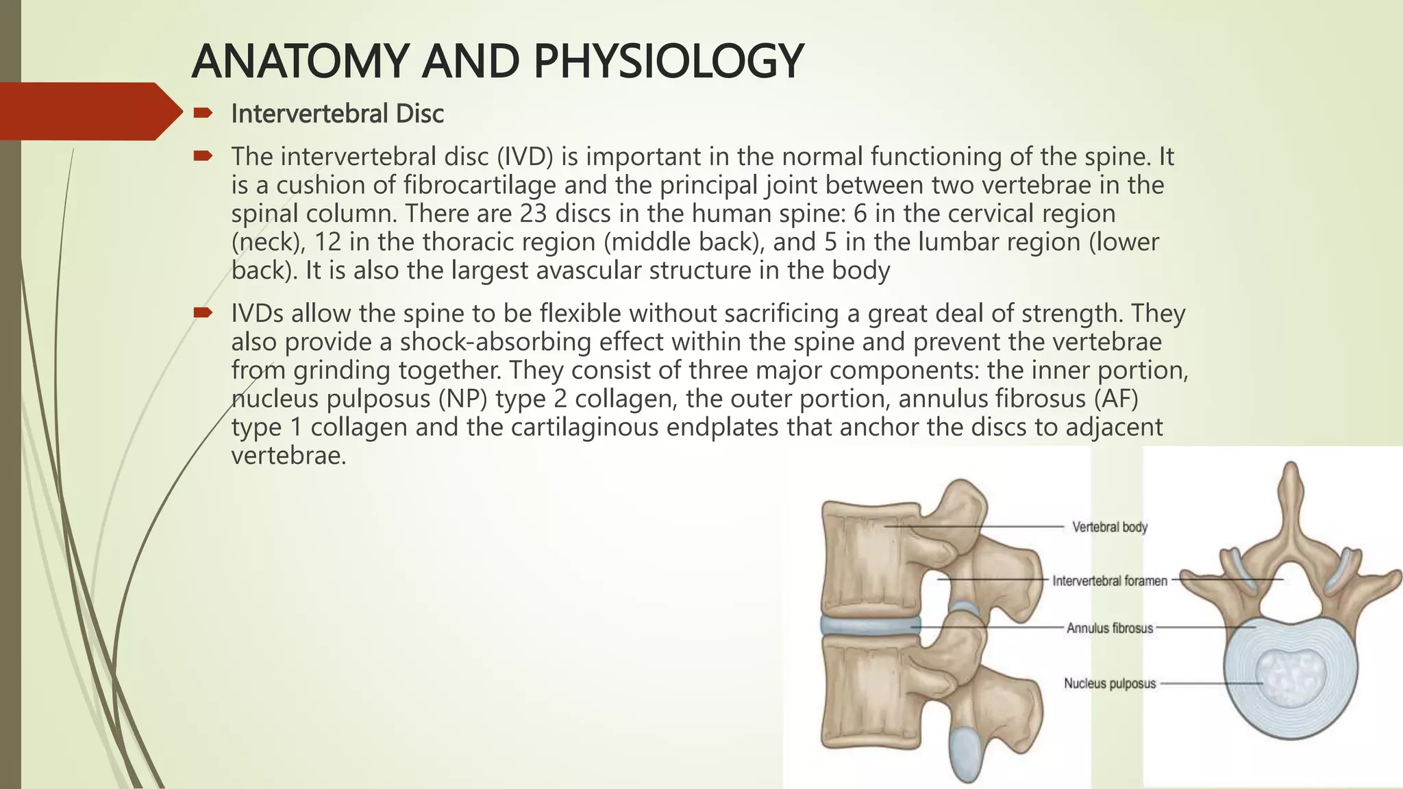
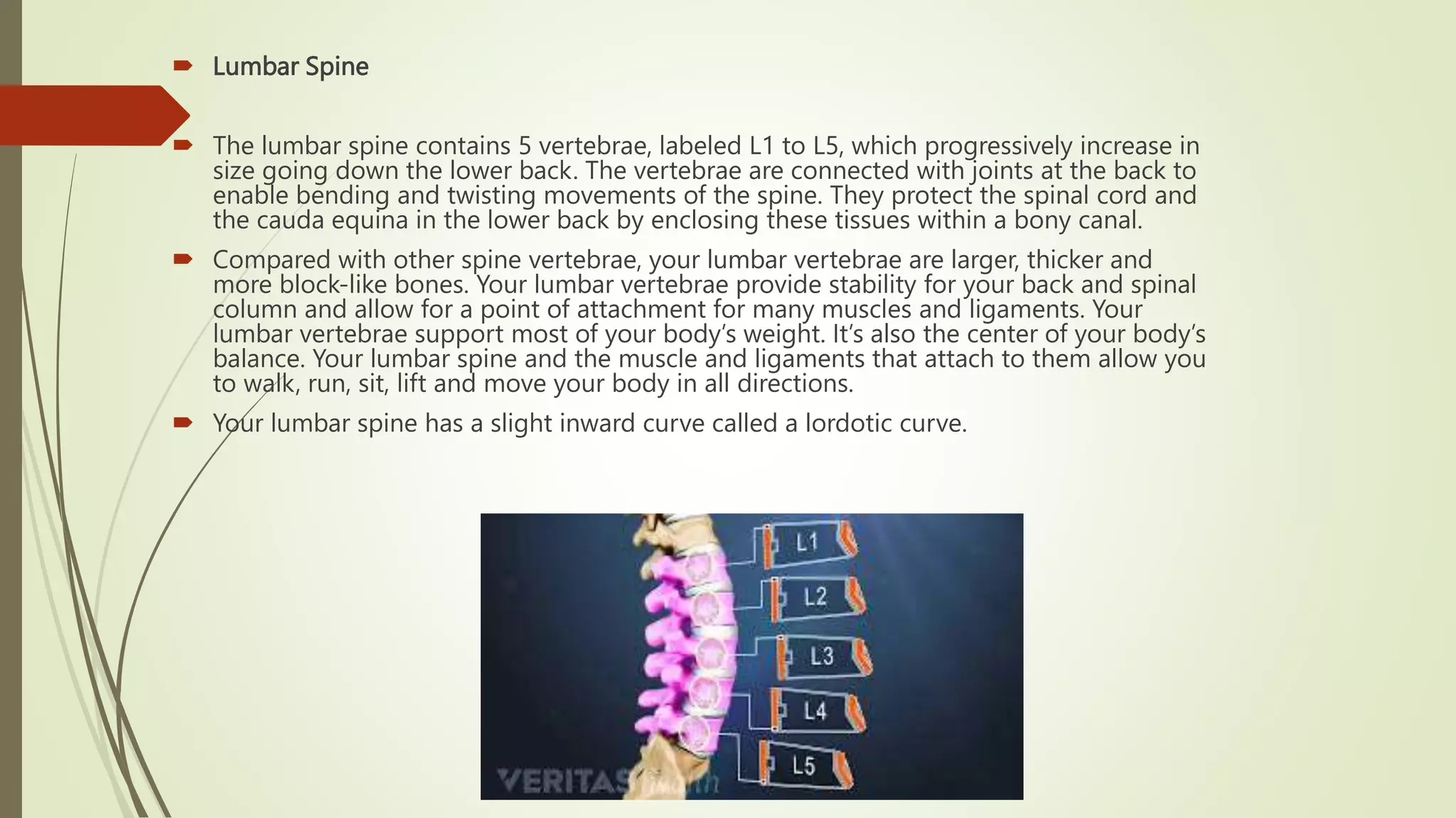
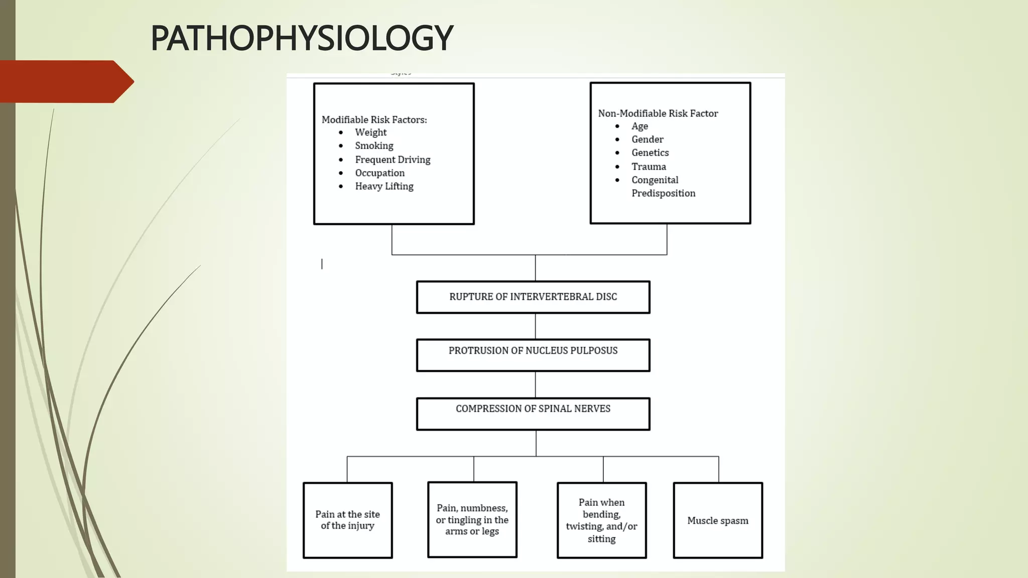
Herniated Nucleus Pulposus
(HNP)
INTRODUCTION
 Nucleus pulposus herniation is the most common cause of sciatic pain and one of
the most common indications for spine surgery worldwide. This condition presents
as a displacement of the nucleus pulposus beyond the intervertebral disc space.
 Nucleus pulposus herniation results from a failure in the annulus fibrosis integrity,
making the content of the nucleus to protrude into the neural canal, the
intervertebral foramen (foraminal) or lateral to the foramen (extraforaminal).
OBJECTIVES
By the end of this case analysis students will be able to:
 Understand what Herniated Nucleus Pulposus.
 Explain the causes & predisposing factors of Herniated Nucleus Pulposus.
 Retain the etiology of Herniated Nucleus Pulposus.
 Identify and understand the particular assessment and the clinical manifestation regarding
Herniated Nucleus Pulposus.
 Understand the general medical and surgical management, pharmacologic management,
and as well as the nursing responsibilities for patients with Herniated Nucleus Pulposus.
 Discuss the general specific Pathophysiology of Herniated Nucleus Pulposus.
 Identify the Nursing Care plan that the patient needs regarding with Herniated Nucleus
Pulposus.
CLINICAL MANIFESTATION
 Herniated discs most commonly affect the lower back, although they can also affect
the neck. The signs and symptoms vary depending on the location of the disc and
whether it is pushing on a nerve. Herniated discs are most commonly found on one
side of the body. Arm or leg pain are common symptoms of a herniated nucleus
pulposus. If the herniated disk is in the lower back, pain in the buttocks, thigh, and calf
may be experienced as well, a condition known as Sciatica.
DIAGNOSTIC TEST
 X- RAY
 Getting X-rays helps rule out other causes of back or neck pain.
 Magnetic Resonance Imaging (MRI)
 The most common and accurate imaging test for a suspected herniated disk is an MRI.
 Computed Tomography
 The most common and accurate imaging test for a suspected herniated disk is an MRI.
 Myelogram
 Involves an injection of dye into your spine using X-ray guidance for a CT scan. The dye can
reveal a narrowing of the spinal canal (spinal stenosis) and location of your herniated disk.
 Electromyogram (EMG)
 This test involves placing small needles into various muscles and evaluate the function of your
nerves. An EMG helps determine which nerve a herniated disk affects.
ANATOMY AND PHYSIOLOGY
 Intervertebral Disc
 The intervertebral disc (IVD) is important in the normal functioning of the spine. It
is a cushion of fibrocartilage and the principal joint between two vertebrae in the
spinal column. There are 23 discs in the human spine: 6 in the cervical region
(neck), 12 in the thoracic region (middle back), and 5 in the lumbar region (lower
back). It is also the largest avascular structure in the body
 IVDs allow the spine to be flexible without sacrificing a great deal of strength. They
also provide a shock-absorbing effect within the spine and prevent the vertebrae
from grinding together. They consist of three major components: the inner portion,
nucleus pulposus (NP) type 2 collagen, the outer portion, annulus fibrosus (AF)
type 1 collagen and the cartilaginous endplates that anchor the discs to adjacent
vertebrae.
 Lumbar Spine
 The lumbar spine contains 5 vertebrae, labeled L1 to L5, which progressively increase in
size going down the lower back. The vertebrae are connected with joints at the back to
enable bending and twisting movements of the spine. They protect the spinal cord and
the cauda equina in the lower back by enclosing these tissues within a bony canal.
 Compared with other spine vertebrae, your lumbar vertebrae are larger, thicker and
more block-like bones. Your lumbar vertebrae provide stability for your back and spinal
column and allow for a point of attachment for many muscles and ligaments. Your
lumbar vertebrae support most of your body’s weight. It’s also the center of your body’s
balance. Your lumbar spine and the muscle and ligaments that attach to them allow you
to walk, run, sit, lift and move your body in all directions.
 Your lumbar spine has a slight inward curve called a lordotic curve.
PATHOPHYSIOLOGY
Herniated Nucleus Pulposus (HNP).pptx

Herniated Nucleus Pulposus (HNP).pptx

  • 1.
  • 2.
    INTRODUCTION  Nucleus pulposusherniation is the most common cause of sciatic pain and one of the most common indications for spine surgery worldwide. This condition presents as a displacement of the nucleus pulposus beyond the intervertebral disc space.  Nucleus pulposus herniation results from a failure in the annulus fibrosis integrity, making the content of the nucleus to protrude into the neural canal, the intervertebral foramen (foraminal) or lateral to the foramen (extraforaminal).
  • 3.
    OBJECTIVES By the endof this case analysis students will be able to:  Understand what Herniated Nucleus Pulposus.  Explain the causes & predisposing factors of Herniated Nucleus Pulposus.  Retain the etiology of Herniated Nucleus Pulposus.  Identify and understand the particular assessment and the clinical manifestation regarding Herniated Nucleus Pulposus.  Understand the general medical and surgical management, pharmacologic management, and as well as the nursing responsibilities for patients with Herniated Nucleus Pulposus.  Discuss the general specific Pathophysiology of Herniated Nucleus Pulposus.  Identify the Nursing Care plan that the patient needs regarding with Herniated Nucleus Pulposus.
  • 4.
    CLINICAL MANIFESTATION  Herniateddiscs most commonly affect the lower back, although they can also affect the neck. The signs and symptoms vary depending on the location of the disc and whether it is pushing on a nerve. Herniated discs are most commonly found on one side of the body. Arm or leg pain are common symptoms of a herniated nucleus pulposus. If the herniated disk is in the lower back, pain in the buttocks, thigh, and calf may be experienced as well, a condition known as Sciatica.
  • 5.
    DIAGNOSTIC TEST  X-RAY  Getting X-rays helps rule out other causes of back or neck pain.  Magnetic Resonance Imaging (MRI)  The most common and accurate imaging test for a suspected herniated disk is an MRI.  Computed Tomography  The most common and accurate imaging test for a suspected herniated disk is an MRI.  Myelogram  Involves an injection of dye into your spine using X-ray guidance for a CT scan. The dye can reveal a narrowing of the spinal canal (spinal stenosis) and location of your herniated disk.  Electromyogram (EMG)  This test involves placing small needles into various muscles and evaluate the function of your nerves. An EMG helps determine which nerve a herniated disk affects.
  • 6.
    ANATOMY AND PHYSIOLOGY Intervertebral Disc  The intervertebral disc (IVD) is important in the normal functioning of the spine. It is a cushion of fibrocartilage and the principal joint between two vertebrae in the spinal column. There are 23 discs in the human spine: 6 in the cervical region (neck), 12 in the thoracic region (middle back), and 5 in the lumbar region (lower back). It is also the largest avascular structure in the body  IVDs allow the spine to be flexible without sacrificing a great deal of strength. They also provide a shock-absorbing effect within the spine and prevent the vertebrae from grinding together. They consist of three major components: the inner portion, nucleus pulposus (NP) type 2 collagen, the outer portion, annulus fibrosus (AF) type 1 collagen and the cartilaginous endplates that anchor the discs to adjacent vertebrae.
  • 7.
     Lumbar Spine The lumbar spine contains 5 vertebrae, labeled L1 to L5, which progressively increase in size going down the lower back. The vertebrae are connected with joints at the back to enable bending and twisting movements of the spine. They protect the spinal cord and the cauda equina in the lower back by enclosing these tissues within a bony canal.  Compared with other spine vertebrae, your lumbar vertebrae are larger, thicker and more block-like bones. Your lumbar vertebrae provide stability for your back and spinal column and allow for a point of attachment for many muscles and ligaments. Your lumbar vertebrae support most of your body’s weight. It’s also the center of your body’s balance. Your lumbar spine and the muscle and ligaments that attach to them allow you to walk, run, sit, lift and move your body in all directions.  Your lumbar spine has a slight inward curve called a lordotic curve.
  • 8.