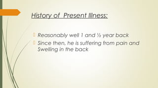
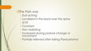
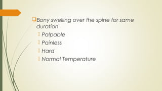
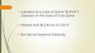
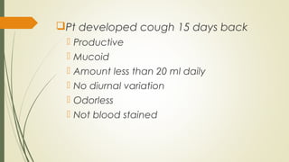
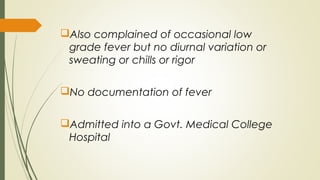
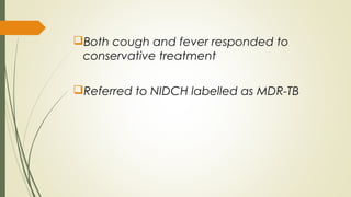
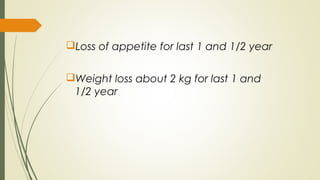
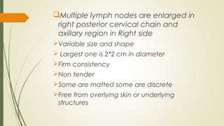
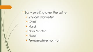
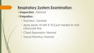
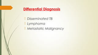
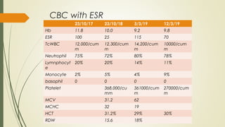
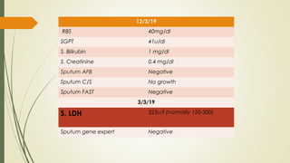
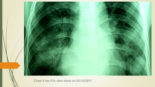
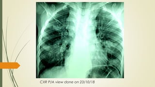
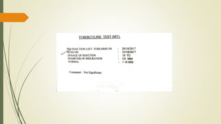
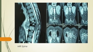
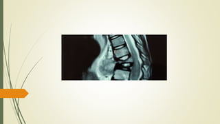
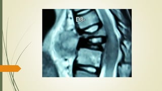
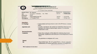
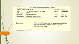
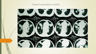
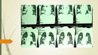
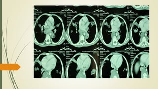
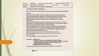
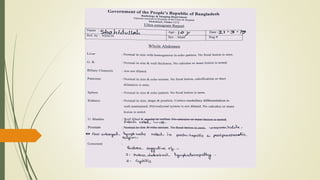
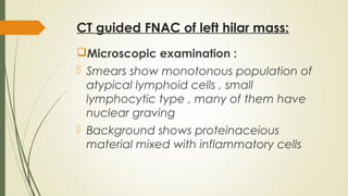
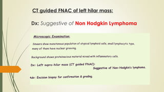
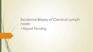
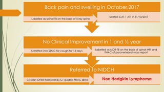
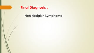
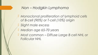
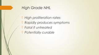
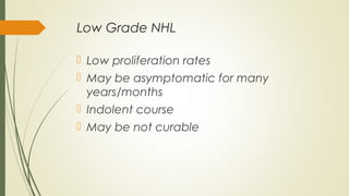
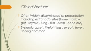
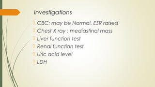
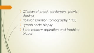
Central Case Presentation
Diseases With Access to All
Ages
May Not Always Present
Classically !!!
A 10 Year Old Boy With
Back Pain and Swelling
Dr Tanveer Kamal Fahim
Resident
Pulmonology , Phase B
Medicine Unit 4
NIDCH
Particulars of the patient
Master Shahid Ullah
Shahed
10 years
Student of class 6
Bikrampur , Munsiganj
Admitted in NIDCH on
11/3/19
Chief Complaints:
 Pain in the back for last 1 and ½ years
 Swelling in the back for same duration
 Cough for 15 days
History of Present Illness:
 Reasonably well 1 and ½ year back
 Since then, he is suffering from pain and
Swelling in the back
The Pain was
 Dull aching
 Localized in the back over the spine
 3/10
 Constant
 Non radiating
 Increased during posture change or
movement
 Partially relieved after taking Paracetamol
Bony swelling over the spine for same
duration
 Palpable
 Painless
 Hard
 Normal Temperature
 Labelled as a case of Spinal TB (Pott”s
Disease) on the basis of X ray Spine
 Started Anti TB CAT-on 31/10/17
 But did not improve Clinically
Pt developed cough 15 days back
 Productive
 Mucoid
 Amount less than 20 ml daily
 No diurnal variation
 Odorless
 Not blood stained
Also complained of occasional low
grade fever but no diurnal variation or
sweating or chills or rigor
No documentation of fever
Admitted into a Govt. Medical College
Hospital
Both cough and fever responded to
conservative treatment
Referred to NIDCH labelled as MDR-TB
Loss of appetite for last 1 and 1/2 year
Weight loss about 2 kg for last 1 and
1/2 year
History of Past Illness:
 Nothing significant
Family History :
 Lives with his parents and one sister
 All are in good health
Personal History
 Student of class 6
Socio-economic History
 Low-middle class family
 Monthly house hold income about 30
thousand taka
Treatment History
CAT - since 31/10/17
Antibiotics , can’t mention names
Paracetamol
Antiulcerant
Immunization History : Immunized
according to EPI schedule
Allergic History : Nothing significant
General Examination
 Ill – looking
 Mildly Anaemic
 Pulse : 90 beats/minute
 Temperature : 98 °F
 Respiratory rate :18 breaths/minute
Multiple lymph nodes are enlarged in
right posterior cervical chain and
axillary region in Right side
Variable size and shape
 Largest one is 2*2 cm in diameter
Firm consistency
Non tender
Some are matted some are discrete
Free from overlying skin or underlying
structures
Bony swelling over the spine
 2*2 cm diameter
 Oval
 Hard
 Non tender
 Fixed
 Temperature normal
Systemic Examination
Respiratory System Examination
Inspection : Normal
Palpation :
 Trachea : Central
 Apex beat: At left 5th
ICS just medial to mid
clavicular line
 Chest Expansion: Normal
 Vocal Fremitus: Normal
Percussion : Resonant
Auscultation :
 Breath sound : Vesicular
 Added sound : Absent
 Vocal resonance : normal
 Neurological System : No abnormality
detected
 Cardiovascular system : No abnormality
detected
 Gastro-Intestinal System : No abnormality
detected
What are the possibilities ??
Differential Diagnosis
 Disseminated TB
 Lymphoma
 Metastatic Malignancy
Investigations
CBC with ESR
23/10/17 23/10/18 3/3/19 12/3/19
Hb 11.8 10.0 9.2 9.8
ESR 100 25 115 70
TcWBC 12,000/cum
m
12,300/cum
m
14,200/cum
m
10000/cum
m
Neutrophil 75% 72% 80% 78%
Lymnphocyt
e
20% 20% 14% 11%
Monocyte 2% 5% 4% 9%
basophil 0 0 0 0
Platelet 368,000/cu
mm
361000/cum
m
270000/cum
m
MCV 31.2 62
MCHC 32 19
HCT 31.2% 29% 30%
RDW 15.6 18%
12/3/19
RBS 40mg/dl
SGPT 41u/dl
S. Bilirubin 1 mg/dl
S. Creatinine 0.4 mg/dl
Sputum AFB Negative
Sputum C/S No growth
Sputum FAST Negative
3/3/19
S. LDH 325u/l (normally 120-300)
Sputum gene expert Negative
Chest X ray P/A view done on 25/10/2017
CXR P/A view done on 23/10/18
CXR P/A View on 23/2/19
MRI Spine
Chest CT scan done on 15/3/19
CT guided FNAC of left hilar mass:
Microscopic examination :
 Smears show monotonous population of
atypical lymphoid cells , small
lymphocytic type , many of them have
nuclear graving
 Background shows proteinaceious
material mixed with inflammatory cells
CT guided FNAC of left hilar mass:
Dx: Suggestive of Non Hodgkin Lymphoma
Excisional Biopsy of Cervical Lymph
node:
Report Pending
Final Diagnosis :
Non Hodgkin Lymphoma
Non – Hodgkin Lymphoma
 Monoclonal proliferation of lymphoid cells
of B-cell (90%) or T-cell (10%) origin
 Slight male excess
 Median age 65-70 years
 Most common – Diffuse Large B cell NHL or
Follicular NHL
High Grade NHL
 High proliferation rates
 Rapidly produces symptoms
 Fatal if untreated
 Potentially curable
Low Grade NHL
 Low proliferation rates
 May be asymptomatic for many
years/months
 Indolent course
 May be not curable
Clinical Features
 Often Widely disseminated at presentation,
including extranodal sites (bone marrow ,
gut , thyroid , lung , skin , brain , bone etc)
 Systemic upset : Weight loss , sweat , fever ,
itching common
 Lymphadenopathy
 hepatosplenomegaly
 Compression syndrome may occur - cord
compression , SVCO , ascities , gut
obstruction etc
Investigations
 CBC: may be Normal. ESR raised
 Chest X ray : mediastinal mass
 Liver function test
 Renal function test
 Uric acid level
 LDH
 CT scan of chest , abdomen , pelvis :
staging
 Positron Emission Tomography ( PET)
 Lymph node biopsy
 Bone marrow aspiration and Trephine
biopsy
 Immune phenotyping of surface
Antigen
 Cytogenetic analysis
 Immunoglobulin determination
Treatment
Low Grade NHL
 Asymptomatic patient : may not need
treatment – “ Watch and Waiting”
 Indication of treatment:
 Systemic symptoms
 Lymphadenopathy
 Marrow failure
 compression syndrome
Treatment options :
Chemotherapy:
Rituximub combined with
Cyclophosphamide,Prednisolone,Vincristine(R-
CVP) or
Cyclophosphamide,Prednisolone,Vincristine,D
oxorubicine (R-CHOP)or Bendamustine
Kinase inhibitor : Idelasib
HSCT
Radiotherapy : Rarely, stage 1 disease
High Grade NHL
 Chemotherapy : R-CHOP
(Cyclophosphamide,Prednisolone,Vincristi
ne,Doxorubicine) is first line therapy
 Radiotherapy: Stage 1 disease
 HSCT
Prognosis
 Low Grade NHL : Median survival -12 years
 Diffuse Large B cell NHL (High Grade)
treated with R-CHOP , 50% patient have
disease free survival of 5 years
 Patients under 65 years , HSCT improves
survival
Take Home message :
 Tuberculosis like presentation may not
always be Tuberculosis
 Bone erosion or collapse may not always
be bone tumor or pott’s Disease
 Routine follow-up and regular physical
examination is very important as
pathological findings may appear at late
Lets Pray For the Departed Souls of New Zealand Attack
Thank You All !!!

A 10 year old boy with back pain

  • 1.
    Central Case Presentation DiseasesWith Access to All Ages May Not Always Present Classically !!!
  • 2.
    A 10 YearOld Boy With Back Pain and Swelling Dr Tanveer Kamal Fahim Resident Pulmonology , Phase B Medicine Unit 4 NIDCH
  • 3.
    Particulars of thepatient Master Shahid Ullah Shahed 10 years Student of class 6 Bikrampur , Munsiganj Admitted in NIDCH on 11/3/19
  • 4.
    Chief Complaints:  Painin the back for last 1 and ½ years  Swelling in the back for same duration  Cough for 15 days
  • 5.
    History of PresentIllness:  Reasonably well 1 and ½ year back  Since then, he is suffering from pain and Swelling in the back
  • 6.
    The Pain was Dull aching  Localized in the back over the spine  3/10  Constant  Non radiating  Increased during posture change or movement  Partially relieved after taking Paracetamol
  • 7.
    Bony swelling overthe spine for same duration  Palpable  Painless  Hard  Normal Temperature
  • 8.
     Labelled asa case of Spinal TB (Pott”s Disease) on the basis of X ray Spine  Started Anti TB CAT-on 31/10/17  But did not improve Clinically
  • 9.
    Pt developed cough15 days back  Productive  Mucoid  Amount less than 20 ml daily  No diurnal variation  Odorless  Not blood stained
  • 10.
    Also complained ofoccasional low grade fever but no diurnal variation or sweating or chills or rigor No documentation of fever Admitted into a Govt. Medical College Hospital
  • 11.
    Both cough andfever responded to conservative treatment Referred to NIDCH labelled as MDR-TB
  • 12.
    Loss of appetitefor last 1 and 1/2 year Weight loss about 2 kg for last 1 and 1/2 year
  • 14.
    History of PastIllness:  Nothing significant
  • 15.
    Family History : Lives with his parents and one sister  All are in good health
  • 16.
  • 17.
    Socio-economic History  Low-middleclass family  Monthly house hold income about 30 thousand taka
  • 18.
    Treatment History CAT -since 31/10/17 Antibiotics , can’t mention names Paracetamol Antiulcerant
  • 19.
    Immunization History :Immunized according to EPI schedule Allergic History : Nothing significant
  • 20.
  • 21.
     Ill –looking  Mildly Anaemic  Pulse : 90 beats/minute  Temperature : 98 °F  Respiratory rate :18 breaths/minute
  • 22.
    Multiple lymph nodesare enlarged in right posterior cervical chain and axillary region in Right side Variable size and shape  Largest one is 2*2 cm in diameter Firm consistency Non tender Some are matted some are discrete Free from overlying skin or underlying structures
  • 23.
    Bony swelling overthe spine  2*2 cm diameter  Oval  Hard  Non tender  Fixed  Temperature normal
  • 24.
  • 25.
    Respiratory System Examination Inspection: Normal Palpation :  Trachea : Central  Apex beat: At left 5th ICS just medial to mid clavicular line  Chest Expansion: Normal  Vocal Fremitus: Normal
  • 26.
    Percussion : Resonant Auscultation:  Breath sound : Vesicular  Added sound : Absent  Vocal resonance : normal
  • 27.
     Neurological System: No abnormality detected  Cardiovascular system : No abnormality detected  Gastro-Intestinal System : No abnormality detected
  • 28.
    What are thepossibilities ??
  • 29.
    Differential Diagnosis  DisseminatedTB  Lymphoma  Metastatic Malignancy
  • 30.
  • 31.
    CBC with ESR 23/10/1723/10/18 3/3/19 12/3/19 Hb 11.8 10.0 9.2 9.8 ESR 100 25 115 70 TcWBC 12,000/cum m 12,300/cum m 14,200/cum m 10000/cum m Neutrophil 75% 72% 80% 78% Lymnphocyt e 20% 20% 14% 11% Monocyte 2% 5% 4% 9% basophil 0 0 0 0 Platelet 368,000/cu mm 361000/cum m 270000/cum m MCV 31.2 62 MCHC 32 19 HCT 31.2% 29% 30% RDW 15.6 18%
  • 32.
    12/3/19 RBS 40mg/dl SGPT 41u/dl S.Bilirubin 1 mg/dl S. Creatinine 0.4 mg/dl Sputum AFB Negative Sputum C/S No growth Sputum FAST Negative 3/3/19 S. LDH 325u/l (normally 120-300) Sputum gene expert Negative
  • 33.
    Chest X rayP/A view done on 25/10/2017
  • 34.
    CXR P/A viewdone on 23/10/18
  • 35.
    CXR P/A Viewon 23/2/19
  • 37.
  • 46.
    Chest CT scandone on 15/3/19
  • 51.
    CT guided FNACof left hilar mass: Microscopic examination :  Smears show monotonous population of atypical lymphoid cells , small lymphocytic type , many of them have nuclear graving  Background shows proteinaceious material mixed with inflammatory cells
  • 52.
    CT guided FNACof left hilar mass: Dx: Suggestive of Non Hodgkin Lymphoma
  • 53.
    Excisional Biopsy ofCervical Lymph node: Report Pending
  • 55.
    Final Diagnosis : NonHodgkin Lymphoma
  • 56.
    Non – HodgkinLymphoma  Monoclonal proliferation of lymphoid cells of B-cell (90%) or T-cell (10%) origin  Slight male excess  Median age 65-70 years  Most common – Diffuse Large B cell NHL or Follicular NHL
  • 57.
    High Grade NHL High proliferation rates  Rapidly produces symptoms  Fatal if untreated  Potentially curable
  • 58.
    Low Grade NHL Low proliferation rates  May be asymptomatic for many years/months  Indolent course  May be not curable
  • 59.
    Clinical Features  OftenWidely disseminated at presentation, including extranodal sites (bone marrow , gut , thyroid , lung , skin , brain , bone etc)  Systemic upset : Weight loss , sweat , fever , itching common
  • 60.
     Lymphadenopathy  hepatosplenomegaly Compression syndrome may occur - cord compression , SVCO , ascities , gut obstruction etc
  • 61.
    Investigations  CBC: maybe Normal. ESR raised  Chest X ray : mediastinal mass  Liver function test  Renal function test  Uric acid level  LDH
  • 62.
     CT scanof chest , abdomen , pelvis : staging  Positron Emission Tomography ( PET)  Lymph node biopsy  Bone marrow aspiration and Trephine biopsy
  • 63.
     Immune phenotypingof surface Antigen  Cytogenetic analysis  Immunoglobulin determination
  • 64.
    Treatment Low Grade NHL Asymptomatic patient : may not need treatment – “ Watch and Waiting”  Indication of treatment:  Systemic symptoms  Lymphadenopathy  Marrow failure  compression syndrome
  • 65.
    Treatment options : Chemotherapy: Rituximubcombined with Cyclophosphamide,Prednisolone,Vincristine(R- CVP) or Cyclophosphamide,Prednisolone,Vincristine,D oxorubicine (R-CHOP)or Bendamustine Kinase inhibitor : Idelasib HSCT Radiotherapy : Rarely, stage 1 disease
  • 66.
    High Grade NHL Chemotherapy : R-CHOP (Cyclophosphamide,Prednisolone,Vincristi ne,Doxorubicine) is first line therapy  Radiotherapy: Stage 1 disease  HSCT
  • 67.
    Prognosis  Low GradeNHL : Median survival -12 years  Diffuse Large B cell NHL (High Grade) treated with R-CHOP , 50% patient have disease free survival of 5 years  Patients under 65 years , HSCT improves survival
  • 68.
    Take Home message:  Tuberculosis like presentation may not always be Tuberculosis  Bone erosion or collapse may not always be bone tumor or pott’s Disease  Routine follow-up and regular physical examination is very important as pathological findings may appear at late
  • 69.
    Lets Pray Forthe Departed Souls of New Zealand Attack
  • 70.