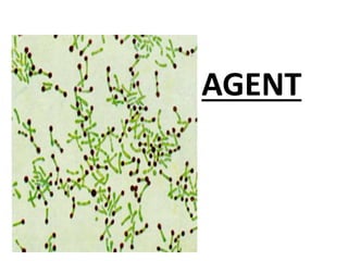
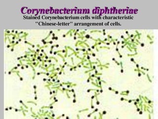
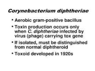
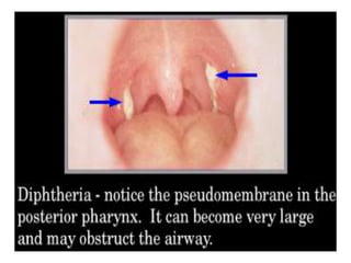
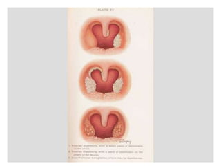
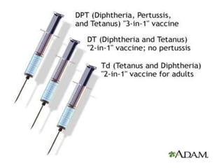
p
EPIDEMIOLOGY OF
DIPHTHERIA
DR. MAHESWARI JAIKUMAR.
maheswarijaikumar2103@gmail.com
DIPHTHERIA
• Is an acute infectious disease
caused by toxigenic strains of
Cornybacterium diphtheriae.
• The bacilli multiply locally,
usually in the throat, and
elaborate a powerful exotoxin
which is responsible for the
following pathology.
• The formation of grayish or
yellowish membrane (false
membrane) commonly over the
tonsils, pharynx, with well
defined edges and the
membrane cannot be wiped
away.
• Marked congestion, edema or
local tissue destruction.
• Enlargement of the regional
lymph nodes.
• Signs and symptoms of toxemia.
EPIDEMIOLOGICAL
DETERMINANTS
AGENT
• The causative agent, C diphtheriae
is a gram-positive, non motile
organism.
• It has no invasive power, but
produces a powerful exotoxin
which can affect the heart leading
to myocarditis.
• Four types of Diphtheria bacilli
are differentiated.
• gravis, mitis, belfanti and
intermedius, all pathogenic to
man.
SOURCE OF INFECTION
• The source of infection may be a
case or a carrier.
CASE
• Cases range from subclinical
infection to frank clinical cases.
• Mild or silent infections may
exhibit no more than a mere
running nose or sore throat.
These cases pose more threat in
the spread than the active cases.
CARRIER
• Carriers are common sources of
infection.
• Carriers may be temporary or
chronic nasal or throat carriers.
INFECTIVE MATERIAL
• Nasopharyngeal secretions,
discharges from skin lesions,
contaminated fomites and
possibly infected dust.
INFECTIVITY
• The period of infectivity may
vary from 14 to 28 days from the
onset of the disease.
• But carriers may remain infective
for much longer periods.
• A case or a carrier may be
considered non communicable,
when at least 2 cultures properly
obtained from nose and throat,
24 hours apart are negative for
diphtheria bacilli.
HOST FACTORS
• AGE : Diphtheria particularly
affects children (1-5 yrs).
• GENDER : Both genders are
affected.
IMMUNITY
• Infants born of immune mothers
are relatively immune during the
first few months of life.
• Children in developing countries
seem to acquire active immunity
through active infection.
ENVIRONMENTAL
FACTORS
• Case of diphtheria occur in all
seasons, although winter months
favour its spread.
MODE OF TRANSMISSION
• The disease is spread mainly by
droplet infection.
• It can also be transmitted
directly to susceptible persons
from infected cutaneous lesions.
• Transmission by objects (cups,
thermometers, toys, pencils)
contaminated by the
nasopharyngeal secretions of the
patients is possible, but only for
short periods.
PORTAL OF ENTRY
• RESPIRATORY ROUTE : Commonly
the portal of entry is the respiratory
tract.
• NON RESPIRATORY ROUTE : The
portal of entry sometimes may be
the skin where cuts, wounds and
ulcers not properly attended to,
may get infected with diphtheria
bacilli.(umbilical cord)
INCUBATION PERIOD
• 2 To 6 days.
• Occasionally longer.
CLINICAL FEATURE
• Respiratory tract forms of
diphtheria consists of
pharygotonsillar, larygotonsillar,
nasal and combinations thereof.
• Patients with pharyngotonsillar
diphtheria usually have a sore
throat, difficulty in swallowing
and low grade fever at
presentation.
• Examination of the throat may
show only mild erythema,
localized exudate or a
pseudomembrane.
PSEUDOMEMBRANE
• The membrane may be a
localized patch of the posterior
pharynx or tonsil, may cover the
entire tonsil, or less frequently,
may spread to cover the soft
palates and the posterior portion
of the pharynx.
• In the early stage the pseudo-
membrane may be whitish and
may wipe off easily.
• The membrane may extend to
become thick, blue-white to
grey-black and adherent.
• Attempts to remove the
membrane may result in
bleeding.
• A minimal area of mucosal
erythema surrounds the
membrane.
• Patients with severe disease may
have marked edema of the sub
mandibular area and the
antererior portion of the neck,
along with lymphadenopathy,
giving a characteristic “bull-
necked” appearance.
BULL NECK APPEARANCE
• Laryngeotracheal diphtheria is
most often preceded by
pharygotonsillar disease, usually
is associated with fever,
hoarseness and croupy cough at
presentation.
• If the infection extends into the
bronchial tree, it is the most
severe of disease.
• Initially it may be clinically
indistinguishable from viral
croup or epiglottitis.
• Prostration and dyspnoea soon
follow because of the
obstruction caused by the
membrane.
• This obstruction may even cause
suffocation if not promptly
relieved by intubation or
tracheostomy.
• The diphtheria bacilli within the
membrane continue to produce
toxin actively.
• This is absorbed and results in
distant toxic damage,
particularly paranchymatous
degeneration, fatty infiltration
and necrosis in heart muscle,
liver, kidneys and adrenals and
some time accompanied by gross
hemorrhage.
• Irregularities of the cardiac
rhythm indicate damage to the
heart.
• Later there may be difficulties
with vision, speech, swallowing,
or movement of the arms or
legs.
• The toxin also produces nerve
damage, resulting in paralysis of
the soft palate, eye muscles or
extremities.
• Patients who survive
complications recover
completely.
• Nasal diphtheria, the mildest
form of respiratory diphtheria,
usually is localized to the septum
or turbinates of one side of the
nose.
• Occasionally a membrane mat
extend into pharynx.
• Non respiratory mucosal surface
i.e., the conjunctivae and
genitals may also be sites of
infection.
• Cutaneous form of diphtheria is
common in tropical areas.
• Cutaneous diphtheria appears as
a secondary infection of a
previous skin abrasion or
infection.
• The presenting lesion, often an
ulcer, may be surrounded by
erythema and covered with a
membrane.
• Patients generally seek
treatment because of the
chronicity of the skin lesion.
CONTROL OF
DIPHTHERIA
(Cases & Controls)
EARLY DETECTION
• An active search for cases and
carriers should start immediately
amongst family and school
contacts.
• Carriers can be detected only by
culture method.
• Swabs should be taken from
both the nose and throat and
examined by culture methods for
diphtheria bacilli.
• Test should be made for the
virulence of the organism.
ISOLATION
• All cases, suspected cases and
carriers should be promptly
isolated, preferably in a hospital,
for at least 14 days or until
proved free of infection.
• At least 2 consecutive nose and
throat swabs , taken 24 hrs apart,
should be negative before
terminating isolation.
TREATMENT (CASES)
• When diphtheria is suspected,
diphtheria antitoxin should be given
without delay, IM or IV , in doses
ranging from 20,000 to 100,000 units or
more, depending on the severity of the
case following a test dose of 0.2ml
subcutaneously to detect sensitization
to horse serum.
• For mild early pharyngeal of
laryngeal disease the dose is
20,000-40,000 units.
• For moderate nasopharyngeal
disease, 40,000-60,000 units is
administered.
• For severe, extensive or late (3
days or more) disease, 80,000-
100,000 units.
• In addition to anti toxin every case
should be treated with penicillin or
erythromycin for 5 to 6 days to
clear the throat of C. diphtheriae
(this decreases toxin production)
CARRIERS
• The carriers should be treated
with 10 day course of oral
erythromycin, which is the most
effective drug for the treatment
of carriers.
• The immunity status should be
upgraded as follows.
CONTACTS
• Contacts merits special
attention. They should be throat
swabbed and their immunity
status determined.
• Non immunized close contact
should receive prophylactic
penicillin or erythromycin.
(1000-2000 units of diphtheria
antitoxin and actively immunized
against diphtheria).
• Contacts should be under
medical surveillance and
examined daily for evidence of
diphtheria for at least a week
after exposure.
COMMUNITY
• Effective control measure is by
active immunization with
diphtheria toxoid of all infants as
early in life as possible (national
immunization schedule).
• Subsequent booster doses every
10 years there after.
• Immunization does not prevent
the carrier state.
DIPHTHERIA IMMUNIZATION
DPT VACCINE
• It is a combined vaccine.
• The preparation of choice is DPT,
because it can be immunized
simultaneously against three
diseases, viz., Diphtheria,
pertussis, and tetanus.
• There are two types of vaccine :
plain and adsorbed.
• Adsorption is usually out on a
mineral carrier like aluminum
phosphate or hydroxide.
• Adsorption increases
immunological effectiveness
of the vaccine.
• WHO recommends the use of
adsorbed vaccines.
DPT STORAGE
• DPT/DT vaccine should not be
frozen.
• They should be stored in a
refrigerator between 2 to 8
degree Celsius.
• The vaccine should be used
before the date of expiry
indicated on the vial.
• The vaccine will lose it’s potency
if it is kept in room temperature
over a long period of time.
OPTIMUM AGE
• Young infants respond well to
immunization with potent
vaccines and toxoids, even in the
presence of low to moderate
levels of maternal antibodies.
• The Expanded programme of
Immunization has recommended
that DPT vaccine can be safely
and effectively administered as
early as 6 weeks after birth.
NUMBER OF DOSES
• Three doses of DPT each of
which is usually 0.5ml, should be
considered optimal for primary
immunization.
INTERVAL BETWEEN DOSES
• The current recommendation is
to allow an interval of 4 weeks
between 3 doses, with a booster
injection at one and a half years
or two years, followed by
another booster (DT only)at the
age of 5 to 6 years.
MODE OF ADMINISTRATION
• All vaccines containing mineral
carriers or adjuvants should be
injected deep intramuscular.
• This applies to DPT also.
• DPT is given in the upper and
outer quadrant of the gluteal
region.
• For children under one year of
age, DPT should be administered
in (lateral aspect) of the thigh.
(1984 Global Advisory Group).
• The most severe complications
that could follow DPT
immunization are encephalitis,
encephalopathy, prolonged
convulsions, infantile spasms
and Reye’s syndrome.
CONTRAINDICATIONS
• Minor conditions such as cough,
cold, mild fever are not
contraindications.
• Seriously ill children are not
vaccinated.
• DPT should not be repeated if a
severe reaction occurred after a
previous dose.
• Such reactions include collapse,
shock like state, persistent
screaming episodes,
temperature above 40 degree
Celsius, convulsions and other
neurological symptoms.
• DT only is recommended in such
case. (2 doses with 4 weeks
apart with a booster dose 6
months to one year).
• Children who have received DPT
earlier, should receive only DT as
booster at 5-6 years or at school
entry.
ANTISERA
Diphtheria
antitoxin prepared
in horse serum is
still the mainstay
of passive
prophylaxis and
also for treatment
of diphtheria.
THANK YOU

Ddd.pdf

  • 1.
  • 2.
    EPIDEMIOLOGY OF DIPHTHERIA DR. MAHESWARIJAIKUMAR. maheswarijaikumar2103@gmail.com
  • 3.
    DIPHTHERIA • Is anacute infectious disease caused by toxigenic strains of Cornybacterium diphtheriae.
  • 4.
    • The bacillimultiply locally, usually in the throat, and elaborate a powerful exotoxin which is responsible for the following pathology.
  • 5.
    • The formationof grayish or yellowish membrane (false membrane) commonly over the tonsils, pharynx, with well defined edges and the membrane cannot be wiped away.
  • 6.
    • Marked congestion,edema or local tissue destruction. • Enlargement of the regional lymph nodes. • Signs and symptoms of toxemia.
  • 7.
  • 8.
  • 11.
    • The causativeagent, C diphtheriae is a gram-positive, non motile organism. • It has no invasive power, but produces a powerful exotoxin which can affect the heart leading to myocarditis.
  • 12.
    • Four typesof Diphtheria bacilli are differentiated. • gravis, mitis, belfanti and intermedius, all pathogenic to man.
  • 13.
    SOURCE OF INFECTION •The source of infection may be a case or a carrier.
  • 14.
    CASE • Cases rangefrom subclinical infection to frank clinical cases. • Mild or silent infections may exhibit no more than a mere running nose or sore throat. These cases pose more threat in the spread than the active cases.
  • 15.
    CARRIER • Carriers arecommon sources of infection. • Carriers may be temporary or chronic nasal or throat carriers.
  • 16.
    INFECTIVE MATERIAL • Nasopharyngealsecretions, discharges from skin lesions, contaminated fomites and possibly infected dust.
  • 17.
    INFECTIVITY • The periodof infectivity may vary from 14 to 28 days from the onset of the disease. • But carriers may remain infective for much longer periods.
  • 18.
    • A caseor a carrier may be considered non communicable, when at least 2 cultures properly obtained from nose and throat, 24 hours apart are negative for diphtheria bacilli.
  • 19.
    HOST FACTORS • AGE: Diphtheria particularly affects children (1-5 yrs). • GENDER : Both genders are affected.
  • 20.
    IMMUNITY • Infants bornof immune mothers are relatively immune during the first few months of life. • Children in developing countries seem to acquire active immunity through active infection.
  • 21.
  • 22.
    • Case ofdiphtheria occur in all seasons, although winter months favour its spread.
  • 23.
    MODE OF TRANSMISSION •The disease is spread mainly by droplet infection. • It can also be transmitted directly to susceptible persons from infected cutaneous lesions.
  • 24.
    • Transmission byobjects (cups, thermometers, toys, pencils) contaminated by the nasopharyngeal secretions of the patients is possible, but only for short periods.
  • 25.
    PORTAL OF ENTRY •RESPIRATORY ROUTE : Commonly the portal of entry is the respiratory tract. • NON RESPIRATORY ROUTE : The portal of entry sometimes may be the skin where cuts, wounds and ulcers not properly attended to, may get infected with diphtheria bacilli.(umbilical cord)
  • 26.
    INCUBATION PERIOD • 2To 6 days. • Occasionally longer.
  • 27.
    CLINICAL FEATURE • Respiratorytract forms of diphtheria consists of pharygotonsillar, larygotonsillar, nasal and combinations thereof.
  • 28.
    • Patients withpharyngotonsillar diphtheria usually have a sore throat, difficulty in swallowing and low grade fever at presentation.
  • 29.
    • Examination ofthe throat may show only mild erythema, localized exudate or a pseudomembrane.
  • 30.
  • 31.
    • The membranemay be a localized patch of the posterior pharynx or tonsil, may cover the entire tonsil, or less frequently, may spread to cover the soft palates and the posterior portion of the pharynx.
  • 34.
    • In theearly stage the pseudo- membrane may be whitish and may wipe off easily. • The membrane may extend to become thick, blue-white to grey-black and adherent.
  • 35.
    • Attempts toremove the membrane may result in bleeding. • A minimal area of mucosal erythema surrounds the membrane.
  • 36.
    • Patients withsevere disease may have marked edema of the sub mandibular area and the antererior portion of the neck, along with lymphadenopathy, giving a characteristic “bull- necked” appearance.
  • 37.
  • 38.
    • Laryngeotracheal diphtheriais most often preceded by pharygotonsillar disease, usually is associated with fever, hoarseness and croupy cough at presentation.
  • 39.
    • If theinfection extends into the bronchial tree, it is the most severe of disease. • Initially it may be clinically indistinguishable from viral croup or epiglottitis.
  • 40.
    • Prostration anddyspnoea soon follow because of the obstruction caused by the membrane. • This obstruction may even cause suffocation if not promptly relieved by intubation or tracheostomy.
  • 41.
    • The diphtheriabacilli within the membrane continue to produce toxin actively.
  • 42.
    • This isabsorbed and results in distant toxic damage, particularly paranchymatous degeneration, fatty infiltration and necrosis in heart muscle, liver, kidneys and adrenals and some time accompanied by gross hemorrhage.
  • 43.
    • Irregularities ofthe cardiac rhythm indicate damage to the heart. • Later there may be difficulties with vision, speech, swallowing, or movement of the arms or legs.
  • 44.
    • The toxinalso produces nerve damage, resulting in paralysis of the soft palate, eye muscles or extremities. • Patients who survive complications recover completely.
  • 45.
    • Nasal diphtheria,the mildest form of respiratory diphtheria, usually is localized to the septum or turbinates of one side of the nose. • Occasionally a membrane mat extend into pharynx.
  • 46.
    • Non respiratorymucosal surface i.e., the conjunctivae and genitals may also be sites of infection. • Cutaneous form of diphtheria is common in tropical areas.
  • 47.
    • Cutaneous diphtheriaappears as a secondary infection of a previous skin abrasion or infection. • The presenting lesion, often an ulcer, may be surrounded by erythema and covered with a membrane.
  • 48.
    • Patients generallyseek treatment because of the chronicity of the skin lesion.
  • 49.
  • 50.
    EARLY DETECTION • Anactive search for cases and carriers should start immediately amongst family and school contacts. • Carriers can be detected only by culture method.
  • 51.
    • Swabs shouldbe taken from both the nose and throat and examined by culture methods for diphtheria bacilli. • Test should be made for the virulence of the organism.
  • 52.
    ISOLATION • All cases,suspected cases and carriers should be promptly isolated, preferably in a hospital, for at least 14 days or until proved free of infection.
  • 53.
    • At least2 consecutive nose and throat swabs , taken 24 hrs apart, should be negative before terminating isolation.
  • 54.
    TREATMENT (CASES) • Whendiphtheria is suspected, diphtheria antitoxin should be given without delay, IM or IV , in doses ranging from 20,000 to 100,000 units or more, depending on the severity of the case following a test dose of 0.2ml subcutaneously to detect sensitization to horse serum.
  • 55.
    • For mildearly pharyngeal of laryngeal disease the dose is 20,000-40,000 units. • For moderate nasopharyngeal disease, 40,000-60,000 units is administered.
  • 56.
    • For severe,extensive or late (3 days or more) disease, 80,000- 100,000 units. • In addition to anti toxin every case should be treated with penicillin or erythromycin for 5 to 6 days to clear the throat of C. diphtheriae (this decreases toxin production)
  • 57.
    CARRIERS • The carriersshould be treated with 10 day course of oral erythromycin, which is the most effective drug for the treatment of carriers. • The immunity status should be upgraded as follows.
  • 58.
    CONTACTS • Contacts meritsspecial attention. They should be throat swabbed and their immunity status determined.
  • 59.
    • Non immunizedclose contact should receive prophylactic penicillin or erythromycin. (1000-2000 units of diphtheria antitoxin and actively immunized against diphtheria).
  • 60.
    • Contacts shouldbe under medical surveillance and examined daily for evidence of diphtheria for at least a week after exposure.
  • 61.
    COMMUNITY • Effective controlmeasure is by active immunization with diphtheria toxoid of all infants as early in life as possible (national immunization schedule).
  • 62.
    • Subsequent boosterdoses every 10 years there after. • Immunization does not prevent the carrier state.
  • 63.
  • 64.
    DPT VACCINE • Itis a combined vaccine. • The preparation of choice is DPT, because it can be immunized simultaneously against three diseases, viz., Diphtheria, pertussis, and tetanus.
  • 66.
    • There aretwo types of vaccine : plain and adsorbed. • Adsorption is usually out on a mineral carrier like aluminum phosphate or hydroxide.
  • 67.
    • Adsorption increases immunologicaleffectiveness of the vaccine. • WHO recommends the use of adsorbed vaccines.
  • 68.
    DPT STORAGE • DPT/DTvaccine should not be frozen. • They should be stored in a refrigerator between 2 to 8 degree Celsius.
  • 69.
    • The vaccineshould be used before the date of expiry indicated on the vial. • The vaccine will lose it’s potency if it is kept in room temperature over a long period of time.
  • 70.
    OPTIMUM AGE • Younginfants respond well to immunization with potent vaccines and toxoids, even in the presence of low to moderate levels of maternal antibodies.
  • 71.
    • The Expandedprogramme of Immunization has recommended that DPT vaccine can be safely and effectively administered as early as 6 weeks after birth.
  • 72.
    NUMBER OF DOSES •Three doses of DPT each of which is usually 0.5ml, should be considered optimal for primary immunization.
  • 73.
    INTERVAL BETWEEN DOSES •The current recommendation is to allow an interval of 4 weeks between 3 doses, with a booster injection at one and a half years or two years, followed by another booster (DT only)at the age of 5 to 6 years.
  • 74.
    MODE OF ADMINISTRATION •All vaccines containing mineral carriers or adjuvants should be injected deep intramuscular. • This applies to DPT also.
  • 75.
    • DPT isgiven in the upper and outer quadrant of the gluteal region. • For children under one year of age, DPT should be administered in (lateral aspect) of the thigh. (1984 Global Advisory Group).
  • 76.
    • The mostsevere complications that could follow DPT immunization are encephalitis, encephalopathy, prolonged convulsions, infantile spasms and Reye’s syndrome.
  • 77.
    CONTRAINDICATIONS • Minor conditionssuch as cough, cold, mild fever are not contraindications. • Seriously ill children are not vaccinated.
  • 78.
    • DPT shouldnot be repeated if a severe reaction occurred after a previous dose.
  • 79.
    • Such reactionsinclude collapse, shock like state, persistent screaming episodes, temperature above 40 degree Celsius, convulsions and other neurological symptoms.
  • 80.
    • DT onlyis recommended in such case. (2 doses with 4 weeks apart with a booster dose 6 months to one year). • Children who have received DPT earlier, should receive only DT as booster at 5-6 years or at school entry.
  • 82.
    ANTISERA Diphtheria antitoxin prepared in horseserum is still the mainstay of passive prophylaxis and also for treatment of diphtheria.
  • 84.