EPIDEMIOLOGY OF DIPTHERIA
•Download as PPTX, PDF•
45 likes•11,844 views

EPIDEMIOLOGY OF DIPTHERIA

Recommended
More Related Content
What's hot
What's hot (20)
Similar to EPIDEMIOLOGY OF DIPTHERIA
Similar to EPIDEMIOLOGY OF DIPTHERIA (20)
More from MAHESWARI JAIKUMAR
More from MAHESWARI JAIKUMAR (20)
Recently uploaded
Recently uploaded (20)
EPIDEMIOLOGY OF DIPTHERIA
- 1. p
- 2. EPIDEMIOLOGY OF DIPHTHERIA DR. MAHESWARI JAIKUMAR. maheswarijaikumar2103@gmail.com
- 3. DIPHTHERIA • Is an acute infectious disease caused by toxigenic strains of Cornybacterium diphtheriae.
- 4. • The bacilli multiply locally, usually in the throat, and elaborate a powerful exotoxin which is responsible for the following pathology.
- 5. • The formation of grayish or yellowish membrane (false membrane) commonly over the tonsils, pharynx, with well defined edges and the membrane cannot be wiped away.
- 6. • Marked congestion, edema or local tissue destruction. • Enlargement of the regional lymph nodes. • Signs and symptoms of toxemia.
- 8. AGENT
- 11. • The causative agent, C diphtheriae is a gram-positive, non motile organism. • It has no invasive power, but produces a powerful exotoxin which can affect the heart leading to myocarditis.
- 12. • Four types of Diphtheria bacilli are differentiated. • gravis, mitis, belfanti and intermedius, all pathogenic to man.
- 13. SOURCE OF INFECTION • The source of infection may be a case or a carrier.
- 14. CASE • Cases range from subclinical infection to frank clinical cases. • Mild or silent infections may exhibit no more than a mere running nose or sore throat. These cases pose more threat in the spread than the active cases.
- 15. CARRIER • Carriers are common sources of infection. • Carriers may be temporary or chronic nasal or throat carriers.
- 16. INFECTIVE MATERIAL • Nasopharyngeal secretions, discharges from skin lesions, contaminated fomites and possibly infected dust.
- 17. INFECTIVITY • The period of infectivity may vary from 14 to 28 days from the onset of the disease. • But carriers may remain infective for much longer periods.
- 18. • A case or a carrier may be considered non communicable, when at least 2 cultures properly obtained from nose and throat, 24 hours apart are negative for diphtheria bacilli.
- 19. HOST FACTORS • AGE : Diphtheria particularly affects children (1-5 yrs). • GENDER : Both genders are affected.
- 20. IMMUNITY • Infants born of immune mothers are relatively immune during the first few months of life. • Children in developing countries seem to acquire active immunity through active infection.
- 22. • Case of diphtheria occur in all seasons, although winter months favour its spread.
- 23. MODE OF TRANSMISSION • The disease is spread mainly by droplet infection. • It can also be transmitted directly to susceptible persons from infected cutaneous lesions.
- 24. • Transmission by objects (cups, thermometers, toys, pencils) contaminated by the nasopharyngeal secretions of the patients is possible, but only for short periods.
- 25. PORTAL OF ENTRY • RESPIRATORY ROUTE : Commonly the portal of entry is the respiratory tract. • NON RESPIRATORY ROUTE : The portal of entry sometimes may be the skin where cuts, wounds and ulcers not properly attended to, may get infected with diphtheria bacilli.(umbilical cord)
- 26. INCUBATION PERIOD • 2 To 6 days. • Occasionally longer.
- 27. CLINICAL FEATURE • Respiratory tract forms of diphtheria consists of pharygotonsillar, larygotonsillar, nasal and combinations thereof.
- 28. • Patients with pharyngotonsillar diphtheria usually have a sore throat, difficulty in swallowing and low grade fever at presentation.
- 29. • Examination of the throat may show only mild erythema, localized exudate or a pseudomembrane.
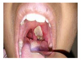
- 30. PSEUDOMEMBRANE
- 31. • The membrane may be a localized patch of the posterior pharynx or tonsil, may cover the entire tonsil, or less frequently, may spread to cover the soft palates and the posterior portion of the pharynx.
- 34. • In the early stage the pseudo- membrane may be whitish and may wipe off easily. • The membrane may extend to become thick, blue-white to grey-black and adherent.
- 35. • Attempts to remove the membrane may result in bleeding. • A minimal area of mucosal erythema surrounds the membrane.
- 36. • Patients with severe disease may have marked edema of the sub mandibular area and the antererior portion of the neck, along with lymphadenopathy, giving a characteristic “bull- necked” appearance.
- 38. • Laryngeotracheal diphtheria is most often preceded by pharygotonsillar disease, usually is associated with fever, hoarseness and croupy cough at presentation.
- 39. • If the infection extends into the bronchial tree, it is the most severe of disease. • Initially it may be clinically indistinguishable from viral croup or epiglottitis.
- 40. • Prostration and dyspnoea soon follow because of the obstruction caused by the membrane. • This obstruction may even cause suffocation if not promptly relieved by intubation or tracheostomy.
- 41. • The diphtheria bacilli within the membrane continue to produce toxin actively.
- 42. • This is absorbed and results in distant toxic damage, particularly paranchymatous degeneration, fatty infiltration and necrosis in heart muscle, liver, kidneys and adrenals and some time accompanied by gross hemorrhage.
- 43. • Irregularities of the cardiac rhythm indicate damage to the heart. • Later there may be difficulties with vision, speech, swallowing, or movement of the arms or legs.
- 44. • The toxin also produces nerve damage, resulting in paralysis of the soft palate, eye muscles or extremities. • Patients who survive complications recover completely.
- 45. • Nasal diphtheria, the mildest form of respiratory diphtheria, usually is localized to the septum or turbinates of one side of the nose. • Occasionally a membrane mat extend into pharynx.
- 46. • Non respiratory mucosal surface i.e., the conjunctivae and genitals may also be sites of infection. • Cutaneous form of diphtheria is common in tropical areas.
- 47. • Cutaneous diphtheria appears as a secondary infection of a previous skin abrasion or infection. • The presenting lesion, often an ulcer, may be surrounded by erythema and covered with a membrane.
- 48. • Patients generally seek treatment because of the chronicity of the skin lesion.
- 49. CONTROL OF DIPHTHERIA (Cases & Controls)
- 50. EARLY DETECTION • An active search for cases and carriers should start immediately amongst family and school contacts. • Carriers can be detected only by culture method.
- 51. • Swabs should be taken from both the nose and throat and examined by culture methods for diphtheria bacilli. • Test should be made for the virulence of the organism.
- 52. ISOLATION • All cases, suspected cases and carriers should be promptly isolated, preferably in a hospital, for at least 14 days or until proved free of infection.
- 53. • At least 2 consecutive nose and throat swabs , taken 24 hrs apart, should be negative before terminating isolation.
- 54. TREATMENT (CASES) • When diphtheria is suspected, diphtheria antitoxin should be given without delay, IM or IV , in doses ranging from 20,000 to 100,000 units or more, depending on the severity of the case following a test dose of 0.2ml subcutaneously to detect sensitization to horse serum.
- 55. • For mild early pharyngeal of laryngeal disease the dose is 20,000-40,000 units. • For moderate nasopharyngeal disease, 40,000-60,000 units is administered.
- 56. • For severe, extensive or late (3 days or more) disease, 80,000- 100,000 units. • In addition to anti toxin every case should be treated with penicillin or erythromycin for 5 to 6 days to clear the throat of C. diphtheriae (this decreases toxin production)
- 57. CARRIERS • The carriers should be treated with 10 day course of oral erythromycin, which is the most effective drug for the treatment of carriers. • The immunity status should be upgraded as follows.
- 58. CONTACTS • Contacts merits special attention. They should be throat swabbed and their immunity status determined.
- 59. • Non immunized close contact should receive prophylactic penicillin or erythromycin. (1000-2000 units of diphtheria antitoxin and actively immunized against diphtheria).
- 60. • Contacts should be under medical surveillance and examined daily for evidence of diphtheria for at least a week after exposure.
- 61. COMMUNITY • Effective control measure is by active immunization with diphtheria toxoid of all infants as early in life as possible (national immunization schedule).
- 62. • Subsequent booster doses every 10 years there after. • Immunization does not prevent the carrier state.
- 64. DPT VACCINE • It is a combined vaccine. • The preparation of choice is DPT, because it can be immunized simultaneously against three diseases, viz., Diphtheria, pertussis, and tetanus.
- 66. • There are two types of vaccine : plain and adsorbed. • Adsorption is usually out on a mineral carrier like aluminum phosphate or hydroxide.
- 67. • Adsorption increases immunological effectiveness of the vaccine. • WHO recommends the use of adsorbed vaccines.
- 68. DPT STORAGE • DPT/DT vaccine should not be frozen. • They should be stored in a refrigerator between 2 to 8 degree Celsius.
- 69. • The vaccine should be used before the date of expiry indicated on the vial. • The vaccine will lose it’s potency if it is kept in room temperature over a long period of time.
- 70. OPTIMUM AGE • Young infants respond well to immunization with potent vaccines and toxoids, even in the presence of low to moderate levels of maternal antibodies.
- 71. • The Expanded programme of Immunization has recommended that DPT vaccine can be safely and effectively administered as early as 6 weeks after birth.
- 72. NUMBER OF DOSES • Three doses of DPT each of which is usually 0.5ml, should be considered optimal for primary immunization.
- 73. INTERVAL BETWEEN DOSES • The current recommendation is to allow an interval of 4 weeks between 3 doses, with a booster injection at one and a half years or two years, followed by another booster (DT only)at the age of 5 to 6 years.
- 74. MODE OF ADMINISTRATION • All vaccines containing mineral carriers or adjuvants should be injected deep intramuscular. • This applies to DPT also.
- 75. • DPT is given in the upper and outer quadrant of the gluteal region. • For children under one year of age, DPT should be administered in (lateral aspect) of the thigh. (1984 Global Advisory Group).
- 76. • The most severe complications that could follow DPT immunization are encephalitis, encephalopathy, prolonged convulsions, infantile spasms and Reye’s syndrome.
- 77. CONTRAINDICATIONS • Minor conditions such as cough, cold, mild fever are not contraindications. • Seriously ill children are not vaccinated.
- 78. • DPT should not be repeated if a severe reaction occurred after a previous dose.
- 79. • Such reactions include collapse, shock like state, persistent screaming episodes, temperature above 40 degree Celsius, convulsions and other neurological symptoms.
- 80. • DT only is recommended in such case. (2 doses with 4 weeks apart with a booster dose 6 months to one year). • Children who have received DPT earlier, should receive only DT as booster at 5-6 years or at school entry.
- 82. ANTISERA Diphtheria antitoxin prepared in horse serum is still the mainstay of passive prophylaxis and also for treatment of diphtheria.
- 84. THANK YOU