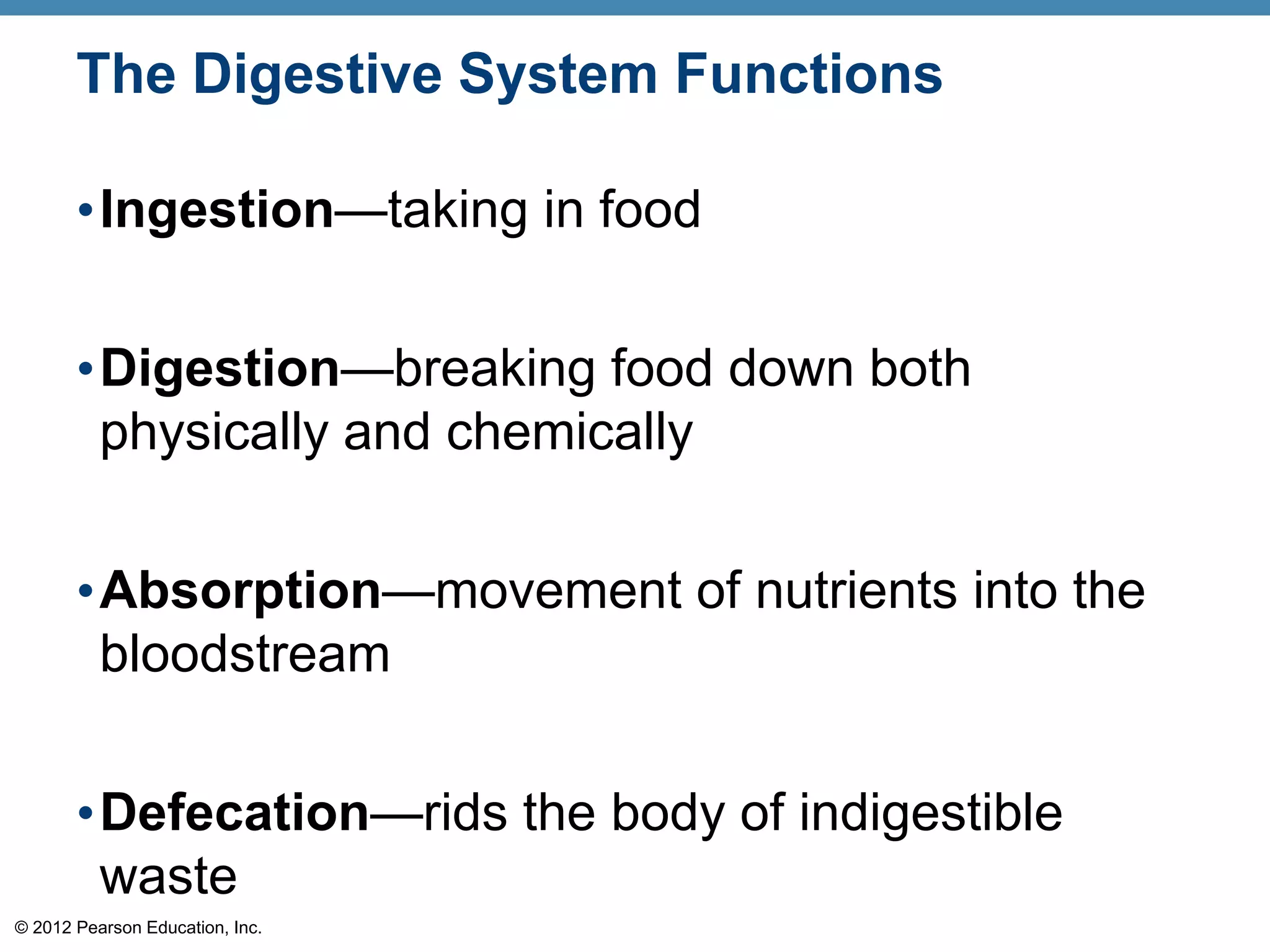
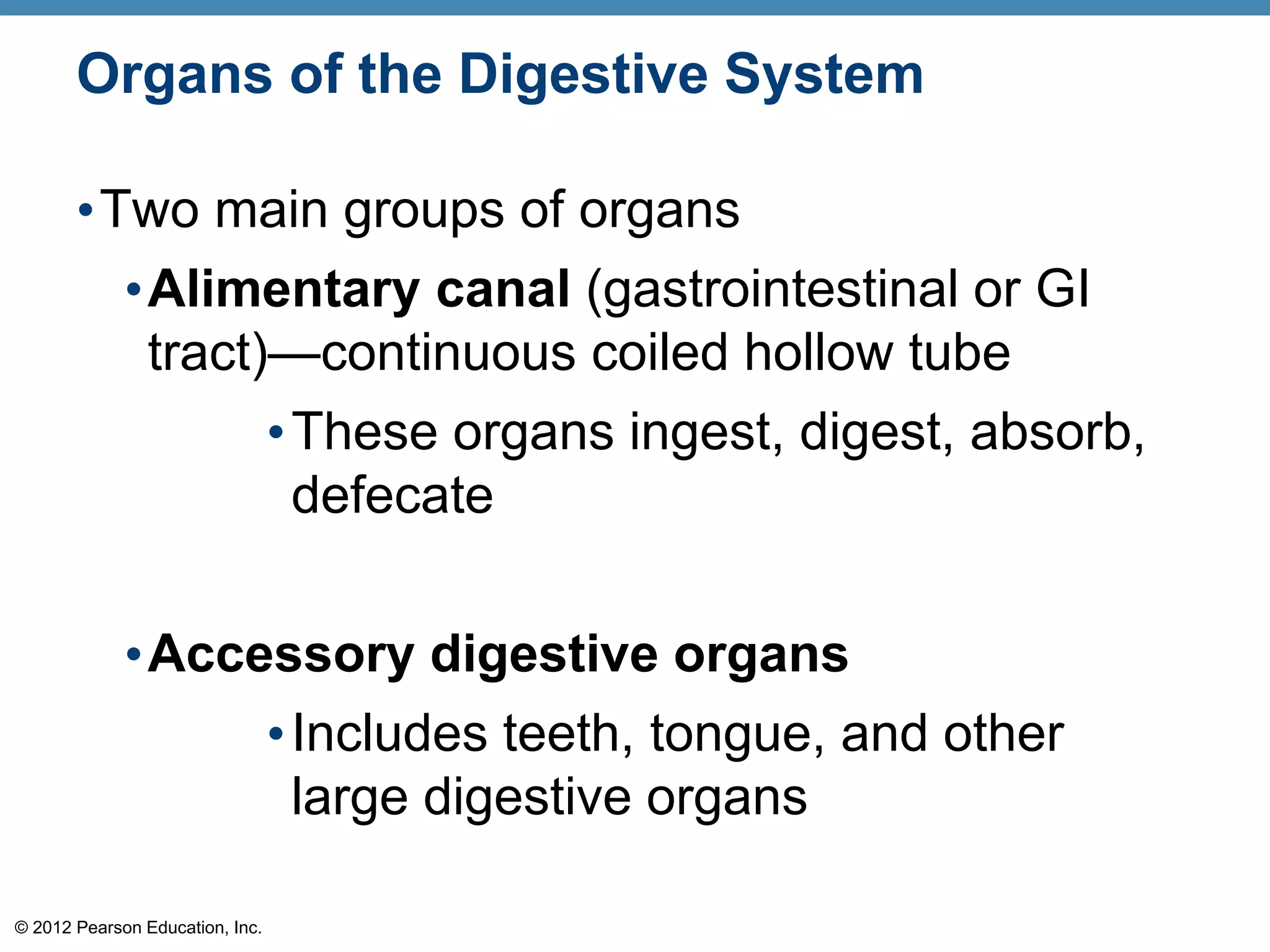
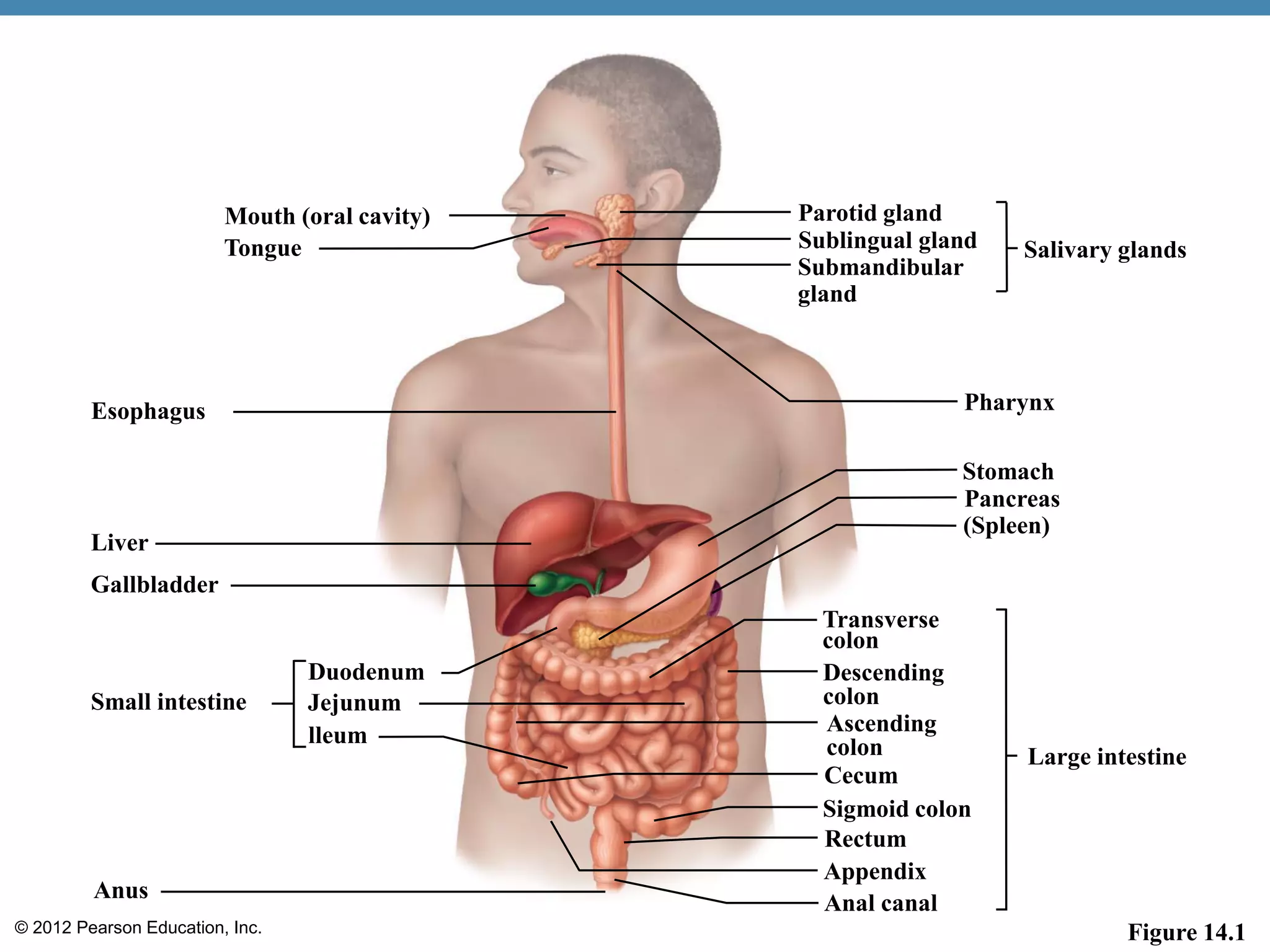
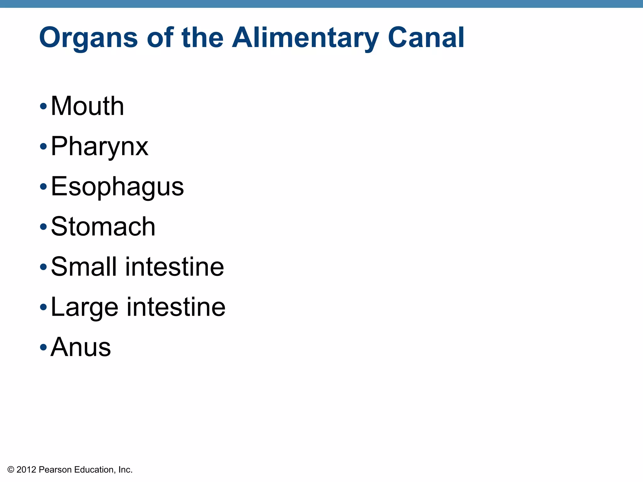
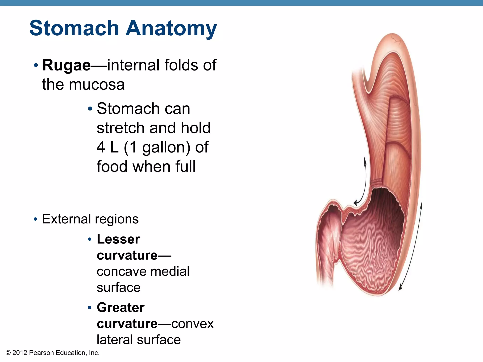
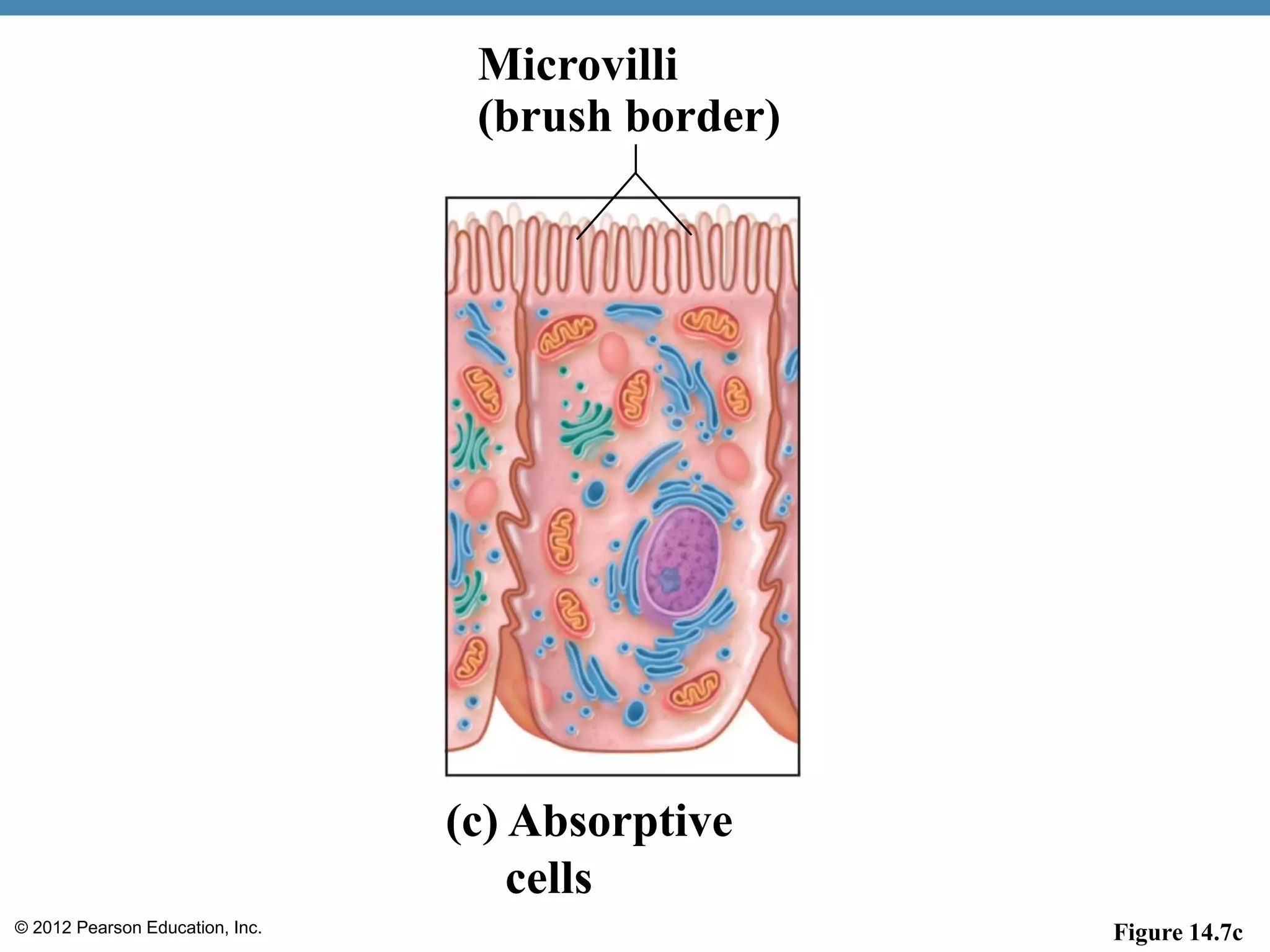
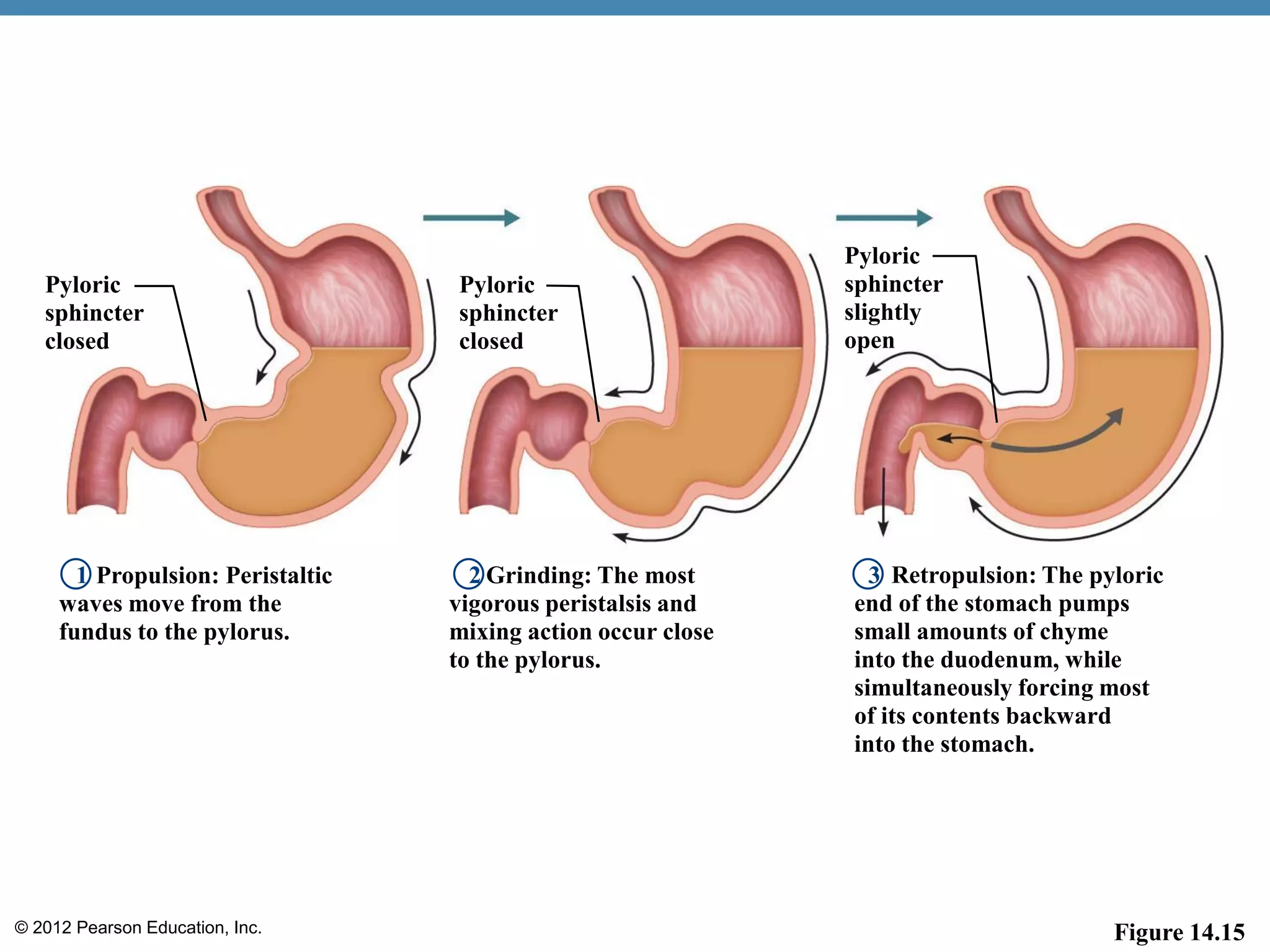
© 2012 Pearson Education, Inc.
The Digestive System Functions
•Ingestion—taking in food
•Digestion—breaking food down both
physically and chemically
•Absorption—movement of nutrients into the
bloodstream
•Defecation—rids the body of indigestible
waste
© 2012 Pearson Education, Inc.
Organs of the Digestive System
•Two main groups of organs
•Alimentary canal (gastrointestinal or GI
tract)—continuous coiled hollow tube
•These organs ingest, digest, absorb,
defecate
•Accessory digestive organs
•Includes teeth, tongue, and other
large digestive organs
© 2012 Pearson Education, Inc.
Mouth (oral cavity)
Tongue
Esophagus
Liver
Gallbladder
Small intestine
Duodenum
Jejunum
lleum
Anus
Parotid gland
Salivary glandsSublingual gland
Submandibular
gland
Pharynx
Stomach
Pancreas
(Spleen)
Large intestine
Descending
colon
Cecum
Sigmoid colon
Rectum
Appendix
Anal canal
Transverse
colon
Ascending
colon
Figure 14.1
© 2012 Pearson Education, Inc.
Organs of the Alimentary Canal
•Mouth
•Pharynx
•Esophagus
•Stomach
•Small intestine
•Large intestine
•Anus
© 2012 Pearson Education, Inc.
Mouth (Oral Cavity) Anatomy
•Lips (labia)—protect the anterior opening
•Cheeks—form the lateral walls
•Hard palate—forms the anterior roof
•Soft palate—forms the posterior roof
•Uvula—fleshy projection of the soft palate
© 2012 Pearson Education, Inc. Figure 14.2b
Hard palate
Soft palate
Uvula
Palatine tonsil
Oropharynx
Tongue
Upper lip
Gingivae
(gums)
(b)
© 2012 Pearson Education, Inc.
Mouth (Oral Cavity) Anatomy
• Vestibule—space
between lips externally
and teeth and gums
internally
• Oral cavity proper—area
contained by the teeth
• Tongue—attached at
hyoid bone and styloid
processes of the skull,
and by the lingual
frenulum to the floor of the
mouth
© 2012 Pearson Education, Inc. Figure 14.2a
(a)
Esophagus
Laryngopharynx
Epiglottis
Oropharynx
Lingual tonsil
Palatine tonsil
Uvula
Soft palate
Nasopharynx
Hard
palate
Oral
cavity
Lips (labia)
Vestibule
Lingual
frenulum
Tongue
Hyoid bone
Trachea
© 2012 Pearson Education, Inc.
Mouth Physiology
•Mastication (chewing) of food
•Mixing masticated food with saliva
•Initiation of swallowing by the tongue
•Allows for the sense of taste
© 2012 Pearson Education, Inc.
Pharynx Anatomy
•Nasopharynx—not part of the digestive
system
•Oropharynx—posterior to oral cavity
•Laryngopharynx—below the oropharynx and
connected to the esophagus
© 2012 Pearson Education, Inc.
Pharynx Physiology
•Serves as a passageway for air and food
•Food is propelled to the esophagus by two
muscle layers
•Longitudinal inner layer
•Circular outer layer
•Food movement is by alternating contractions
of the muscle layers (peristalsis)
© 2012 Pearson Education, Inc.
Esophagus Anatomy and Physiology
• Anatomy
• About 10 inches long
• Runs from pharynx to stomach through the
diaphragm
• Physiology
• Conducts food by peristalsis (slow rhythmic
squeezing)
• Passageway for food only (respiratory system
branches off after the pharynx)
© 2012 Pearson Education, Inc.
Stomach Anatomy
•Located on the left side of the abdominal
cavity
•Food enters at the cardioesophageal
sphincter
•Food empties into the small intestine at the
pyloric sphincter
© 2012 Pearson Education, Inc.
Stomach Anatomy
•Regions of the stomach
•Cardiac region—near the heart
•Fundus—expanded portion lateral to the
cardiac region
•Body—midportion
•Pylorus—funnel-shaped terminal end
© 2012 Pearson Education, Inc.
Stomach Anatomy
• Rugae—internal folds of
the mucosa
• Stomach can
stretch and hold
4 L (1 gallon) of
food when full
• External regions
• Lesser
curvature—
concave medial
surface
• Greater
curvature—convex
lateral surface
© 2012 Pearson Education, Inc. Figure 14.4a
Pyloric
antrum
Esophagus
Fundus
Serosa
Body
Rugae
of
mucosa
Greater
curvature
Pyloric
Sphincter
(valve)
(a)
Pylorus
Lesser
curvature
Cardioesophageal
sphincter
Muscularis
externa
• Circular layer
• Oblique layer
• Longitudinal layer
Duodenum
© 2012 Pearson Education, Inc. Figure 14.4b
Fundus
Rugae
of
mucosa
Body
(b) Pyloric
sphincter
Pyloric
antrum
© 2012 Pearson Education, Inc.
Stomach Physiology
•Temporary storage tank for food
•Site of food breakdown
•Chemical breakdown of protein begins
•Delivers chyme (processed food) to the small
intestine
© 2012 Pearson Education, Inc.
Structure of the Stomach Mucosa
•Mucosa is simple columnar epithelium
•Mucous neck cells—produce a sticky alkaline
mucus
•Gastric glands—situated in gastric pits and
secrete gastric juice
•Chief cells—produce protein-digesting enzymes
(pepsinogens)
•Parietal cells—produce hydrochloric acid
•Enteroendocrine cells—produce gastrin
© 2012 Pearson Education, Inc.
Pyloric
sphincter
Gastric pits
Surface
epithelium
Mucous
neck cells
Parietal cells
Gastric
glands
Chief cells
GastricpitGastricgland
(c)
Figure 14.4c
© 2012 Pearson Education, Inc.
Small Intestine
•The body’s major digestive organ
•Site of nutrient absorption into the blood
•Muscular tube extending from the pyloric
sphincter to the ileocecal valve
•Suspended from the posterior abdominal wall
by the mesentery
© 2012 Pearson Education, Inc.
Subdivisions of the Small Intestine
•Duodenum
•Attached to the stomach
•Curves around the head of the pancreas
•Jejunum
•Attaches anteriorly to the duodenum
•Ileum
•Extends from jejunum to large intestine
© 2012 Pearson Education, Inc.
Chemical Digestion in the Small Intestine
•Chemical digestion begins in the small
intestine
•Enzymes are produced by
•Intestinal cells
•Pancreas
•Pancreatic ducts carry enzymes to the small
intestine
•Bile, formed by the liver, enters via the bile
duct
© 2012 Pearson Education, Inc.
Small Intestine Anatomy
•Three structural modifications that increase
surface area
•Microvilli—tiny projections of the
plasma membrane (create a brush
border appearance)
•Villi—fingerlike structures formed by the
mucosa
•Circular folds (plicae circulares)—deep
folds of mucosa and submucosa
© 2012 Pearson Education, Inc.
Blood vessels
serving the small
intestine
Lumen
Circular folds
(plicae circulares)
Muscle
layers
Villi
(a) Small intestine
Figure 14.7a
© 2012 Pearson Education, Inc.
Absorptive
cells
Lacteal
Blood
capillaries
(b) Villi
Lymphoid
tissue
Intestinal
crypt
Muscularis
mucosae
Villus
Venule
Lymphatic vessel
Submucosa
Figure 14.7b
© 2012 Pearson Education, Inc. Figure 14.7c
(c) Absorptive
cells
Microvilli
(brush border)
© 2012 Pearson Education, Inc.
Large Intestine
•Larger in diameter, but shorter in length, than
the small intestine
•Extends from the ileocecal valve to the anus
•Subdivisions:
•Cecum
•Appendix
•Colon
•Rectum
•Anal canal
© 2012 Pearson Education, Inc.
Large Intestine Anatomy
•Cecum—saclike first part of the large intestine
•Appendix
•Accumulation of lymphatic tissue that
sometimes becomes inflamed (appendicitis)
•Hangs from the cecum
© 2012 Pearson Education, Inc.
Right colic
(hepatic) flexure
Transverse colon
Haustrum
Ascending colon
IIeum (cut)
IIeocecal valve
Cecum
Appendix Rectum
Alan canal External anal sphincter
Sigmoid colon
Teniae coli
Cut edge of
mesentery
Descending colon
Left colic
(splenic) flexure
Transverse
mesocolon
Figure 14.8
© 2012 Pearson Education, Inc.
Large Intestine Anatomy
•Colon
•Ascending colon—travels up right side of
abdomen
•Transverse colon—travels across the
abdominal cavity
•Descending colon—travels down the left
side
•Sigmoid colon—S-shaped region; enters
the pelvis
•Rectum and anus also are located in the
pelvis
© 2012 Pearson Education, Inc.
Large Intestine Anatomy
•Anus—opening of the large intestine
•External anal sphincter—formed by
skeletal muscle and under voluntary control
•Internal involuntary sphincter—formed by
smooth muscle
•These sphincters are normally closed except
during defecation
© 2012 Pearson Education, Inc.
Large Intestine Anatomy
• No villi present
• Goblet cells produce alkaline mucus which
lubricates the passage of feces
• Muscularis externa layer is reduced to three bands
of muscle called teniae coli
• These bands cause the wall to pucker into haustra
(pocketlike sacs)
© 2012 Pearson Education, Inc.
Accessory Digestive Organs
•Teeth
•Salivary glands
•Pancreas
•Liver
•Gallbladder
© 2012 Pearson Education, Inc.
Teeth
•Function is to masticate (chew) food
•Humans have two sets of teeth
•Deciduous (baby or “milk”) teeth
•A baby has 20 teeth by age two
•First teeth to appear are the lower
central incisors
© 2012 Pearson Education, Inc.
Teeth
•Permanent teeth
•Replace deciduous teeth between the
ages of 6 and 12
•A full set is 32 teeth, but some people
do not have wisdom teeth (third molars)
•If they do emerge, the wisdom teeth
appear between ages of 17 and 25
© 2012 Pearson Education, Inc.
Classification of Teeth
•Incisors—cutting
•Canines (eyeteeth)—tearing or piercing
•Premolars (bicuspids)—grinding
•Molars—grinding
© 2012 Pearson Education, Inc.
Incisors
Central
(6–8 mo)
Lateral
(8–10 mo)
Canine (eyetooth)
(16–20 mo)
Molars
First molar
(10–15 mo)
Second molar
(about 2 yr)
Incisors
Central (7 yr)
Lateral (8 yr)
Canine (eyetooth)
(11 yr)
Premolars
(bicuspids)
First premolar
(11 yr)
Second premolar
(12–13 yr)
Molars
First molar
(6–7 yr)
Second molar
(12–13 yr)
Third molar
(wisdom tooth)
(17–25 yr)
Deciduous
(milk) teeth
Permanent
teeth
Figure 14.9
© 2012 Pearson Education, Inc.
Regions of a Tooth
•Crown—exposed part
•Enamel—hardest substance in the body
•Dentin—found deep to the enamel and
forms the bulk of the tooth
•Pulp cavity—contains connective tissue,
blood vessels, and nerve fibers
•Root canal—where the pulp cavity extends
into the root
© 2012 Pearson Education, Inc.
Enamel
Dentin
Pulp cavity
Gum
(gingiva)
Periodontal
membrane
Bone
Cement
Root canal
Blood
vessels and
nerves in
pulp
Crown
Neck
Root
Figure 14.10
© 2012 Pearson Education, Inc.
Salivary Glands
•Three pairs of salivary glands empty
secretions into the mouth
•Parotid glands
•Found anterior to the ears
•Submandibular glands
•Sublingual glands
•Both submandibular and sublingual
glands empty saliva into the floor of
the mouth through small ducts
© 2012 Pearson Education, Inc.
Mouth (oral cavity)
Tongue
Esophagus
Liver
Gallbladder
Small intestine
Duodenum
Jejunum
lleum
Anus
Parotid gland
Salivary glandsSublingual gland
Submandibular
gland
Pharynx
Stomach
Pancreas
(Spleen)
Large intestine
Descending
colon
Cecum
Sigmoid colon
Rectum
Appendix
Anal canal
Transverse
colon
Ascending
colon
Figure 14.1
© 2012 Pearson Education, Inc.
Saliva
•Mixture of mucus and serous fluids
•Helps to form a food bolus
•Contains salivary amylase to begin starch
digestion
•Dissolves chemicals so they can be tasted
© 2012 Pearson Education, Inc.
Pancreas
•Found posterior to the parietal peritoneum
•Its location is retroperitoneal
•Extends across the abdomen from spleen to
duodenum
© 2012 Pearson Education, Inc.
Pancreas
• Produces a wide spectrum of digestive enzymes
that break down all categories of food
• Enzymes are secreted into the duodenum
• Alkaline fluid introduced with enzymes neutralizes
acidic chyme coming from stomach
• Hormones produced by the pancreas
• Insulin
• Glucagon
© 2012 Pearson Education, Inc.
Gallbladder
Duodenal
papilla
Hepatopancreatic
ampulla and sphincter Duodenum
Right and left
hepatic ducts
from liver
Cystic duct
Common hepatic duct
Bile duct and sphincter
Accessory pancreatic duct
Pancreas
Jejunum
Main pancreatic duct and sphincter
Figure 14.6
© 2012 Pearson Education, Inc.
Liver
• Largest gland in the body
• Located on the right side of the body under the
diaphragm
© 2012 Pearson Education, Inc.
Bile
•Produced by cells in the liver
•Composition is
•Bile salts
•Bile pigments (mostly bilirubin from the
breakdown of hemoglobin)
•Cholesterol
•Phospholipids
•Electrolytes
© 2012 Pearson Education, Inc.
Bile
•Function—
•emulsify fats by physically breaking large
fat globules into smaller ones
© 2012 Pearson Education, Inc.
Gallbladder
• Sac found in hollow fossa of liver
• When no digestion is occurring, bile backs up the
cystic duct for storage in the gallbladder
• When digestion of fatty food is occurring, bile is
introduced into the duodenum from the gallbladder
• Gallstones are crystallized cholesterol which can
cause blockages
© 2012 Pearson Education, Inc.
Functions of the Digestive System
• Ingestion—placing food into the mouth
• Propulsion—moving foods from one region of the
digestive system to another
• Peristalsis—alternating waves of contraction
and relaxation that squeezes food along the GI
tract
• Segmentation—moving materials back and
forth to aid with mixing in the small intestine
© 2012 Pearson Education, Inc.
Ingestion
Mechanical
digestion
Small
intestine
Large
intestine
Food
Pharynx
Esophagus
Propulsion
• Swallowing
(oropharynx)
Stomach
Absorption
Lymph
vessel
Blood
vessel
Mainly H2O
Feces
Anus
Chemical
digestion
Defecation
• Chewing (mouth)
• Churning (stomach)
• Segmentation
(small intestine)
• Peristalsis
(esophagus,
stomach,
small intestine,
large intestine)
Figure 14.11
© 2012 Pearson Education, Inc. Figure 14.12a-b
© 2012 Pearson Education, Inc.
Functions of the Digestive System
•Food breakdown as mechanical digestion
•Examples:
• Mixing food in the mouth by the tongue
• Churning food in the stomach
• Segmentation in the small intestine
•Mechanical digestion prepares food for further
degradation by enzymes
© 2012 Pearson Education, Inc.
Functions of the Digestive System
• Food breakdown as chemical digestion
• Enzymes break down food molecules into their
building blocks
• Each major food group uses different enzymes
• Carbohydrates  simple sugars
• Proteins  amino acids
• Fats  fatty acids, alcohols
© 2012 Pearson Education, Inc. Figure 14.13 (1 of 3)
© 2012 Pearson Education, Inc. Figure 14.13 (2 of 3)
© 2012 Pearson Education, Inc. Figure 14.13 (3 of 3)
© 2012 Pearson Education, Inc.
Functions of the Digestive System
• Absorption
• End products of digestion are absorbed in the
blood or lymph
• Food must enter mucosal cells and then into
blood or lymph capillaries
• Defecation
• Elimination of indigestible substances from the
GI tract in the form of feces
© 2012 Pearson Education, Inc.
Control of Digestive Activity
•Mostly controlled by reflexes via the
parasympathetic division
•Chemical and mechanical receptors are
located in organ walls that trigger reflexes
© 2012 Pearson Education, Inc.
Control of Digestive Activity
•Stimuli include
•Stretch of the organ
•pH of the contents
•Presence of breakdown products
•Reflexes include
•Activation or inhibition of glandular
secretions
•Smooth muscle activity
© 2012 Pearson Education, Inc.
Digestive Activities of the Mouth
•Mechanical breakdown
•Food is physically broken down by
chewing
•Chemical digestion
•Food is mixed with saliva
•Starch is broken down into maltose by
salivary amylase
© 2012 Pearson Education, Inc.
Activities of the Pharynx and Esophagus
•These organs have no digestive function
•Serve as passageways to the stomach
© 2012 Pearson Education, Inc.
Deglutition (Swallowing)
•Buccal phase
•Voluntary
•Occurs in the mouth
•Food is formed into a bolus
•The bolus is forced into the pharynx by the
tongue
© 2012 Pearson Education, Inc.
Deglutition (Swallowing)
• Pharyngeal-esophageal phase
• Involuntary transport of the bolus
• All passageways except to the stomach are
blocked
• Tongue blocks off the mouth
• Soft palate (uvula) blocks the nasopharynx
• Epiglottis blocks the larynx
© 2012 Pearson Education, Inc.
Deglutition (Swallowing)
•Pharyngeal-esophogeal phase (continued)
•Peristalsis moves the bolus toward the
stomach
•The cardioesophageal sphincter is
opened when food presses against it
© 2012 Pearson Education, Inc.
Bolus of food
Tongue
Pharynx
Epiglottis
up
Glottis (lumen)
of larynx
(a) Upper esophageal
sphincter contracted
Upper
esophageal
sphincter
EsophagusTrachea
Figure 14.14a
© 2012 Pearson Education, Inc. Figure 14.14b
Larynx up
Esophagus
(b) Upper esophageal
sphincter relaxed
Uvula
Bolus
Epiglottis
down
© 2012 Pearson Education, Inc.
Bolus
(c) Upper esophageal
sphincter contracted
Figure 14.14c
© 2012 Pearson Education, Inc. Figure 14.14d
Relaxed
muscles
Cardioesophageal
sphincter open
(d) Cardioesophageal
sphincter relaxed
© 2012 Pearson Education, Inc.
Food Breakdown in the Stomach
•Gastric juice is regulated by neural and
hormonal factors
•Presence of food or rising pH causes the
release of the hormone gastrin
•Gastrin causes stomach glands to produce
•Protein-digesting enzymes
•Mucus
•Hydrochloric acid
© 2012 Pearson Education, Inc.
Food Breakdown in the Stomach
•Hydrochloric acid makes the stomach
contents very acidic
•Acidic pH
•Activates pepsinogen to pepsin for
protein digestion
•Provides a hostile environment for
microorganisms
© 2012 Pearson Education, Inc.
Digestion and Absorption in the Stomach
•Protein digestion enzymes
•Pepsin—an active protein-digesting
enzyme
•Rennin—digests milk protein in infants,
not adults
•Alcohol and aspirin are the only items
absorbed in the stomach
© 2012 Pearson Education, Inc.
Propulsion in the Stomach
•Food must first be well mixed
•Rippling peristalsis occurs in the lower
stomach
•Propulsion
•Grinding
•Retropulsion
•The pylorus meters out chyme into the small
intestine (3 mL at a time)
•The stomach empties in 4–6 hours
© 2012 Pearson Education, Inc.
Pyloric
sphincter
closed
Pyloric
sphincter
closed
Pyloric
sphincter
slightly
open
Propulsion: Peristaltic
waves move from the
fundus to the pylorus.
Grinding: The most
vigorous peristalsis and
mixing action occur close
to the pylorus.
Retropulsion: The pyloric
end of the stomach pumps
small amounts of chyme
into the duodenum, while
simultaneously forcing most
of its contents backward
into the stomach.
2 31
Figure 14.15
© 2012 Pearson Education, Inc.
Digestion in the Small Intestine
•Enzymes from the brush border function to
•Break double sugars into simple sugars
•Complete some protein digestion
© 2012 Pearson Education, Inc.
Digestion in the Small Intestine
•Pancreatic enzymes play the major digestive
function
•Help complete digestion of starch
(pancreatic amylase)
•Carry out about half of all protein digestion
•Digest fats using lipases from the pancreas
•Digest nucleic acids using nucleases
•Alkaline content neutralizes acidic chyme
© 2012 Pearson Education, Inc.
Regulation of Pancreatic Juice Secretion
•Release of pancreatic juice into the
duodenum is stimulated by
•Vagus nerve
•Local hormones
•Secretin
•Cholecystokinin (CCK)
•Hormones travel the blood to stimulate the
pancreas to release enzyme- and bicarbonate-
rich product
© 2012 Pearson Education, Inc.
Regulation of Pancreatic Juice Secretion
•Secretin causes the liver to increase bile
output
•CCK causes the gallbladder to release
stored bile
•Bile is necessary for
•fat absorption
•fat-soluble vitamin absorption
© 2012 Pearson Education, Inc.
Absorption in the Small Intestine
•Water is absorbed along the length of the
small intestine
•End products of digestion
•Most substances are absorbed by active
transport through cell membranes
•Lipids are absorbed by diffusion
•Substances are transported to the liver by
the hepatic portal vein or lymph
© 2012 Pearson Education, Inc.
Propulsion in the Small Intestine
•Peristalsis is the major means of moving food
•Segmental movements
•Mix chyme with digestive juices
•Aid in propelling food
© 2012 Pearson Education, Inc. Figure 14.12b
© 2012 Pearson Education, Inc.
Food Breakdown and Absorption
in the Large Intestine
•No digestive enzymes are produced
•Resident bacteria digest remaining nutrients
•Produce some vitamin K and B
•Release gases
•Water and vitamins K and B are absorbed
•Remaining materials eliminated via feces
© 2012 Pearson Education, Inc.
Food Breakdown and Absorption
in the Large Intestine
•Feces contains
•Undigested food residues
•Mucus
•Bacteria
•Water
© 2012 Pearson Education, Inc.
Propulsion in the Large Intestine
•Sluggish peristalsis
•Mass movements
•Slow, powerful movements
•Occur three to four times per day
•Presence of feces in the rectum causes a
defecation reflex
•Internal anal sphincter is relaxed
•Defecation occurs with relaxation of the
voluntary (external) anal sphincter

Digestive System

  • 1.
    © 2012 PearsonEducation, Inc. The Digestive System Functions •Ingestion—taking in food •Digestion—breaking food down both physically and chemically •Absorption—movement of nutrients into the bloodstream •Defecation—rids the body of indigestible waste
  • 2.
    © 2012 PearsonEducation, Inc. Organs of the Digestive System •Two main groups of organs •Alimentary canal (gastrointestinal or GI tract)—continuous coiled hollow tube •These organs ingest, digest, absorb, defecate •Accessory digestive organs •Includes teeth, tongue, and other large digestive organs
  • 3.
    © 2012 PearsonEducation, Inc. Mouth (oral cavity) Tongue Esophagus Liver Gallbladder Small intestine Duodenum Jejunum lleum Anus Parotid gland Salivary glandsSublingual gland Submandibular gland Pharynx Stomach Pancreas (Spleen) Large intestine Descending colon Cecum Sigmoid colon Rectum Appendix Anal canal Transverse colon Ascending colon Figure 14.1
  • 4.
    © 2012 PearsonEducation, Inc. Organs of the Alimentary Canal •Mouth •Pharynx •Esophagus •Stomach •Small intestine •Large intestine •Anus
  • 5.
    © 2012 PearsonEducation, Inc. Mouth (Oral Cavity) Anatomy •Lips (labia)—protect the anterior opening •Cheeks—form the lateral walls •Hard palate—forms the anterior roof •Soft palate—forms the posterior roof •Uvula—fleshy projection of the soft palate
  • 6.
    © 2012 PearsonEducation, Inc. Figure 14.2b Hard palate Soft palate Uvula Palatine tonsil Oropharynx Tongue Upper lip Gingivae (gums) (b)
  • 7.
    © 2012 PearsonEducation, Inc. Mouth (Oral Cavity) Anatomy • Vestibule—space between lips externally and teeth and gums internally • Oral cavity proper—area contained by the teeth • Tongue—attached at hyoid bone and styloid processes of the skull, and by the lingual frenulum to the floor of the mouth
  • 8.
    © 2012 PearsonEducation, Inc. Figure 14.2a (a) Esophagus Laryngopharynx Epiglottis Oropharynx Lingual tonsil Palatine tonsil Uvula Soft palate Nasopharynx Hard palate Oral cavity Lips (labia) Vestibule Lingual frenulum Tongue Hyoid bone Trachea
  • 9.
    © 2012 PearsonEducation, Inc. Mouth Physiology •Mastication (chewing) of food •Mixing masticated food with saliva •Initiation of swallowing by the tongue •Allows for the sense of taste
  • 10.
    © 2012 PearsonEducation, Inc. Pharynx Anatomy •Nasopharynx—not part of the digestive system •Oropharynx—posterior to oral cavity •Laryngopharynx—below the oropharynx and connected to the esophagus
  • 11.
    © 2012 PearsonEducation, Inc. Pharynx Physiology •Serves as a passageway for air and food •Food is propelled to the esophagus by two muscle layers •Longitudinal inner layer •Circular outer layer •Food movement is by alternating contractions of the muscle layers (peristalsis)
  • 12.
    © 2012 PearsonEducation, Inc. Esophagus Anatomy and Physiology • Anatomy • About 10 inches long • Runs from pharynx to stomach through the diaphragm • Physiology • Conducts food by peristalsis (slow rhythmic squeezing) • Passageway for food only (respiratory system branches off after the pharynx)
  • 13.
    © 2012 PearsonEducation, Inc. Stomach Anatomy •Located on the left side of the abdominal cavity •Food enters at the cardioesophageal sphincter •Food empties into the small intestine at the pyloric sphincter
  • 14.
    © 2012 PearsonEducation, Inc. Stomach Anatomy •Regions of the stomach •Cardiac region—near the heart •Fundus—expanded portion lateral to the cardiac region •Body—midportion •Pylorus—funnel-shaped terminal end
  • 15.
    © 2012 PearsonEducation, Inc. Stomach Anatomy • Rugae—internal folds of the mucosa • Stomach can stretch and hold 4 L (1 gallon) of food when full • External regions • Lesser curvature— concave medial surface • Greater curvature—convex lateral surface
  • 16.
    © 2012 PearsonEducation, Inc. Figure 14.4a Pyloric antrum Esophagus Fundus Serosa Body Rugae of mucosa Greater curvature Pyloric Sphincter (valve) (a) Pylorus Lesser curvature Cardioesophageal sphincter Muscularis externa • Circular layer • Oblique layer • Longitudinal layer Duodenum
  • 17.
    © 2012 PearsonEducation, Inc. Figure 14.4b Fundus Rugae of mucosa Body (b) Pyloric sphincter Pyloric antrum
  • 18.
    © 2012 PearsonEducation, Inc. Stomach Physiology •Temporary storage tank for food •Site of food breakdown •Chemical breakdown of protein begins •Delivers chyme (processed food) to the small intestine
  • 19.
    © 2012 PearsonEducation, Inc. Structure of the Stomach Mucosa •Mucosa is simple columnar epithelium •Mucous neck cells—produce a sticky alkaline mucus •Gastric glands—situated in gastric pits and secrete gastric juice •Chief cells—produce protein-digesting enzymes (pepsinogens) •Parietal cells—produce hydrochloric acid •Enteroendocrine cells—produce gastrin
  • 20.
    © 2012 PearsonEducation, Inc. Pyloric sphincter Gastric pits Surface epithelium Mucous neck cells Parietal cells Gastric glands Chief cells GastricpitGastricgland (c) Figure 14.4c
  • 21.
    © 2012 PearsonEducation, Inc. Small Intestine •The body’s major digestive organ •Site of nutrient absorption into the blood •Muscular tube extending from the pyloric sphincter to the ileocecal valve •Suspended from the posterior abdominal wall by the mesentery
  • 22.
    © 2012 PearsonEducation, Inc. Subdivisions of the Small Intestine •Duodenum •Attached to the stomach •Curves around the head of the pancreas •Jejunum •Attaches anteriorly to the duodenum •Ileum •Extends from jejunum to large intestine
  • 23.
    © 2012 PearsonEducation, Inc. Chemical Digestion in the Small Intestine •Chemical digestion begins in the small intestine •Enzymes are produced by •Intestinal cells •Pancreas •Pancreatic ducts carry enzymes to the small intestine •Bile, formed by the liver, enters via the bile duct
  • 24.
    © 2012 PearsonEducation, Inc. Small Intestine Anatomy •Three structural modifications that increase surface area •Microvilli—tiny projections of the plasma membrane (create a brush border appearance) •Villi—fingerlike structures formed by the mucosa •Circular folds (plicae circulares)—deep folds of mucosa and submucosa
  • 25.
    © 2012 PearsonEducation, Inc. Blood vessels serving the small intestine Lumen Circular folds (plicae circulares) Muscle layers Villi (a) Small intestine Figure 14.7a
  • 26.
    © 2012 PearsonEducation, Inc. Absorptive cells Lacteal Blood capillaries (b) Villi Lymphoid tissue Intestinal crypt Muscularis mucosae Villus Venule Lymphatic vessel Submucosa Figure 14.7b
  • 27.
    © 2012 PearsonEducation, Inc. Figure 14.7c (c) Absorptive cells Microvilli (brush border)
  • 28.
    © 2012 PearsonEducation, Inc. Large Intestine •Larger in diameter, but shorter in length, than the small intestine •Extends from the ileocecal valve to the anus •Subdivisions: •Cecum •Appendix •Colon •Rectum •Anal canal
  • 29.
    © 2012 PearsonEducation, Inc. Large Intestine Anatomy •Cecum—saclike first part of the large intestine •Appendix •Accumulation of lymphatic tissue that sometimes becomes inflamed (appendicitis) •Hangs from the cecum
  • 30.
    © 2012 PearsonEducation, Inc. Right colic (hepatic) flexure Transverse colon Haustrum Ascending colon IIeum (cut) IIeocecal valve Cecum Appendix Rectum Alan canal External anal sphincter Sigmoid colon Teniae coli Cut edge of mesentery Descending colon Left colic (splenic) flexure Transverse mesocolon Figure 14.8
  • 31.
    © 2012 PearsonEducation, Inc. Large Intestine Anatomy •Colon •Ascending colon—travels up right side of abdomen •Transverse colon—travels across the abdominal cavity •Descending colon—travels down the left side •Sigmoid colon—S-shaped region; enters the pelvis •Rectum and anus also are located in the pelvis
  • 32.
    © 2012 PearsonEducation, Inc. Large Intestine Anatomy •Anus—opening of the large intestine •External anal sphincter—formed by skeletal muscle and under voluntary control •Internal involuntary sphincter—formed by smooth muscle •These sphincters are normally closed except during defecation
  • 33.
    © 2012 PearsonEducation, Inc. Large Intestine Anatomy • No villi present • Goblet cells produce alkaline mucus which lubricates the passage of feces • Muscularis externa layer is reduced to three bands of muscle called teniae coli • These bands cause the wall to pucker into haustra (pocketlike sacs)
  • 34.
    © 2012 PearsonEducation, Inc. Accessory Digestive Organs •Teeth •Salivary glands •Pancreas •Liver •Gallbladder
  • 35.
    © 2012 PearsonEducation, Inc. Teeth •Function is to masticate (chew) food •Humans have two sets of teeth •Deciduous (baby or “milk”) teeth •A baby has 20 teeth by age two •First teeth to appear are the lower central incisors
  • 36.
    © 2012 PearsonEducation, Inc. Teeth •Permanent teeth •Replace deciduous teeth between the ages of 6 and 12 •A full set is 32 teeth, but some people do not have wisdom teeth (third molars) •If they do emerge, the wisdom teeth appear between ages of 17 and 25
  • 37.
    © 2012 PearsonEducation, Inc. Classification of Teeth •Incisors—cutting •Canines (eyeteeth)—tearing or piercing •Premolars (bicuspids)—grinding •Molars—grinding
  • 38.
    © 2012 PearsonEducation, Inc. Incisors Central (6–8 mo) Lateral (8–10 mo) Canine (eyetooth) (16–20 mo) Molars First molar (10–15 mo) Second molar (about 2 yr) Incisors Central (7 yr) Lateral (8 yr) Canine (eyetooth) (11 yr) Premolars (bicuspids) First premolar (11 yr) Second premolar (12–13 yr) Molars First molar (6–7 yr) Second molar (12–13 yr) Third molar (wisdom tooth) (17–25 yr) Deciduous (milk) teeth Permanent teeth Figure 14.9
  • 39.
    © 2012 PearsonEducation, Inc. Regions of a Tooth •Crown—exposed part •Enamel—hardest substance in the body •Dentin—found deep to the enamel and forms the bulk of the tooth •Pulp cavity—contains connective tissue, blood vessels, and nerve fibers •Root canal—where the pulp cavity extends into the root
  • 40.
    © 2012 PearsonEducation, Inc. Enamel Dentin Pulp cavity Gum (gingiva) Periodontal membrane Bone Cement Root canal Blood vessels and nerves in pulp Crown Neck Root Figure 14.10
  • 41.
    © 2012 PearsonEducation, Inc. Salivary Glands •Three pairs of salivary glands empty secretions into the mouth •Parotid glands •Found anterior to the ears •Submandibular glands •Sublingual glands •Both submandibular and sublingual glands empty saliva into the floor of the mouth through small ducts
  • 42.
    © 2012 PearsonEducation, Inc. Mouth (oral cavity) Tongue Esophagus Liver Gallbladder Small intestine Duodenum Jejunum lleum Anus Parotid gland Salivary glandsSublingual gland Submandibular gland Pharynx Stomach Pancreas (Spleen) Large intestine Descending colon Cecum Sigmoid colon Rectum Appendix Anal canal Transverse colon Ascending colon Figure 14.1
  • 43.
    © 2012 PearsonEducation, Inc. Saliva •Mixture of mucus and serous fluids •Helps to form a food bolus •Contains salivary amylase to begin starch digestion •Dissolves chemicals so they can be tasted
  • 44.
    © 2012 PearsonEducation, Inc. Pancreas •Found posterior to the parietal peritoneum •Its location is retroperitoneal •Extends across the abdomen from spleen to duodenum
  • 45.
    © 2012 PearsonEducation, Inc. Pancreas • Produces a wide spectrum of digestive enzymes that break down all categories of food • Enzymes are secreted into the duodenum • Alkaline fluid introduced with enzymes neutralizes acidic chyme coming from stomach • Hormones produced by the pancreas • Insulin • Glucagon
  • 46.
    © 2012 PearsonEducation, Inc. Gallbladder Duodenal papilla Hepatopancreatic ampulla and sphincter Duodenum Right and left hepatic ducts from liver Cystic duct Common hepatic duct Bile duct and sphincter Accessory pancreatic duct Pancreas Jejunum Main pancreatic duct and sphincter Figure 14.6
  • 47.
    © 2012 PearsonEducation, Inc. Liver • Largest gland in the body • Located on the right side of the body under the diaphragm
  • 48.
    © 2012 PearsonEducation, Inc. Bile •Produced by cells in the liver •Composition is •Bile salts •Bile pigments (mostly bilirubin from the breakdown of hemoglobin) •Cholesterol •Phospholipids •Electrolytes
  • 49.
    © 2012 PearsonEducation, Inc. Bile •Function— •emulsify fats by physically breaking large fat globules into smaller ones
  • 50.
    © 2012 PearsonEducation, Inc. Gallbladder • Sac found in hollow fossa of liver • When no digestion is occurring, bile backs up the cystic duct for storage in the gallbladder • When digestion of fatty food is occurring, bile is introduced into the duodenum from the gallbladder • Gallstones are crystallized cholesterol which can cause blockages
  • 51.
    © 2012 PearsonEducation, Inc. Functions of the Digestive System • Ingestion—placing food into the mouth • Propulsion—moving foods from one region of the digestive system to another • Peristalsis—alternating waves of contraction and relaxation that squeezes food along the GI tract • Segmentation—moving materials back and forth to aid with mixing in the small intestine
  • 52.
    © 2012 PearsonEducation, Inc. Ingestion Mechanical digestion Small intestine Large intestine Food Pharynx Esophagus Propulsion • Swallowing (oropharynx) Stomach Absorption Lymph vessel Blood vessel Mainly H2O Feces Anus Chemical digestion Defecation • Chewing (mouth) • Churning (stomach) • Segmentation (small intestine) • Peristalsis (esophagus, stomach, small intestine, large intestine) Figure 14.11
  • 53.
    © 2012 PearsonEducation, Inc. Figure 14.12a-b
  • 54.
    © 2012 PearsonEducation, Inc. Functions of the Digestive System •Food breakdown as mechanical digestion •Examples: • Mixing food in the mouth by the tongue • Churning food in the stomach • Segmentation in the small intestine •Mechanical digestion prepares food for further degradation by enzymes
  • 55.
    © 2012 PearsonEducation, Inc. Functions of the Digestive System • Food breakdown as chemical digestion • Enzymes break down food molecules into their building blocks • Each major food group uses different enzymes • Carbohydrates  simple sugars • Proteins  amino acids • Fats  fatty acids, alcohols
  • 56.
    © 2012 PearsonEducation, Inc. Figure 14.13 (1 of 3)
  • 57.
    © 2012 PearsonEducation, Inc. Figure 14.13 (2 of 3)
  • 58.
    © 2012 PearsonEducation, Inc. Figure 14.13 (3 of 3)
  • 59.
    © 2012 PearsonEducation, Inc. Functions of the Digestive System • Absorption • End products of digestion are absorbed in the blood or lymph • Food must enter mucosal cells and then into blood or lymph capillaries • Defecation • Elimination of indigestible substances from the GI tract in the form of feces
  • 60.
    © 2012 PearsonEducation, Inc. Control of Digestive Activity •Mostly controlled by reflexes via the parasympathetic division •Chemical and mechanical receptors are located in organ walls that trigger reflexes
  • 61.
    © 2012 PearsonEducation, Inc. Control of Digestive Activity •Stimuli include •Stretch of the organ •pH of the contents •Presence of breakdown products •Reflexes include •Activation or inhibition of glandular secretions •Smooth muscle activity
  • 62.
    © 2012 PearsonEducation, Inc. Digestive Activities of the Mouth •Mechanical breakdown •Food is physically broken down by chewing •Chemical digestion •Food is mixed with saliva •Starch is broken down into maltose by salivary amylase
  • 63.
    © 2012 PearsonEducation, Inc. Activities of the Pharynx and Esophagus •These organs have no digestive function •Serve as passageways to the stomach
  • 64.
    © 2012 PearsonEducation, Inc. Deglutition (Swallowing) •Buccal phase •Voluntary •Occurs in the mouth •Food is formed into a bolus •The bolus is forced into the pharynx by the tongue
  • 65.
    © 2012 PearsonEducation, Inc. Deglutition (Swallowing) • Pharyngeal-esophageal phase • Involuntary transport of the bolus • All passageways except to the stomach are blocked • Tongue blocks off the mouth • Soft palate (uvula) blocks the nasopharynx • Epiglottis blocks the larynx
  • 66.
    © 2012 PearsonEducation, Inc. Deglutition (Swallowing) •Pharyngeal-esophogeal phase (continued) •Peristalsis moves the bolus toward the stomach •The cardioesophageal sphincter is opened when food presses against it
  • 67.
    © 2012 PearsonEducation, Inc. Bolus of food Tongue Pharynx Epiglottis up Glottis (lumen) of larynx (a) Upper esophageal sphincter contracted Upper esophageal sphincter EsophagusTrachea Figure 14.14a
  • 68.
    © 2012 PearsonEducation, Inc. Figure 14.14b Larynx up Esophagus (b) Upper esophageal sphincter relaxed Uvula Bolus Epiglottis down
  • 69.
    © 2012 PearsonEducation, Inc. Bolus (c) Upper esophageal sphincter contracted Figure 14.14c
  • 70.
    © 2012 PearsonEducation, Inc. Figure 14.14d Relaxed muscles Cardioesophageal sphincter open (d) Cardioesophageal sphincter relaxed
  • 71.
    © 2012 PearsonEducation, Inc. Food Breakdown in the Stomach •Gastric juice is regulated by neural and hormonal factors •Presence of food or rising pH causes the release of the hormone gastrin •Gastrin causes stomach glands to produce •Protein-digesting enzymes •Mucus •Hydrochloric acid
  • 72.
    © 2012 PearsonEducation, Inc. Food Breakdown in the Stomach •Hydrochloric acid makes the stomach contents very acidic •Acidic pH •Activates pepsinogen to pepsin for protein digestion •Provides a hostile environment for microorganisms
  • 73.
    © 2012 PearsonEducation, Inc. Digestion and Absorption in the Stomach •Protein digestion enzymes •Pepsin—an active protein-digesting enzyme •Rennin—digests milk protein in infants, not adults •Alcohol and aspirin are the only items absorbed in the stomach
  • 74.
    © 2012 PearsonEducation, Inc. Propulsion in the Stomach •Food must first be well mixed •Rippling peristalsis occurs in the lower stomach •Propulsion •Grinding •Retropulsion •The pylorus meters out chyme into the small intestine (3 mL at a time) •The stomach empties in 4–6 hours
  • 75.
    © 2012 PearsonEducation, Inc. Pyloric sphincter closed Pyloric sphincter closed Pyloric sphincter slightly open Propulsion: Peristaltic waves move from the fundus to the pylorus. Grinding: The most vigorous peristalsis and mixing action occur close to the pylorus. Retropulsion: The pyloric end of the stomach pumps small amounts of chyme into the duodenum, while simultaneously forcing most of its contents backward into the stomach. 2 31 Figure 14.15
  • 76.
    © 2012 PearsonEducation, Inc. Digestion in the Small Intestine •Enzymes from the brush border function to •Break double sugars into simple sugars •Complete some protein digestion
  • 77.
    © 2012 PearsonEducation, Inc. Digestion in the Small Intestine •Pancreatic enzymes play the major digestive function •Help complete digestion of starch (pancreatic amylase) •Carry out about half of all protein digestion •Digest fats using lipases from the pancreas •Digest nucleic acids using nucleases •Alkaline content neutralizes acidic chyme
  • 78.
    © 2012 PearsonEducation, Inc. Regulation of Pancreatic Juice Secretion •Release of pancreatic juice into the duodenum is stimulated by •Vagus nerve •Local hormones •Secretin •Cholecystokinin (CCK) •Hormones travel the blood to stimulate the pancreas to release enzyme- and bicarbonate- rich product
  • 79.
    © 2012 PearsonEducation, Inc. Regulation of Pancreatic Juice Secretion •Secretin causes the liver to increase bile output •CCK causes the gallbladder to release stored bile •Bile is necessary for •fat absorption •fat-soluble vitamin absorption
  • 80.
    © 2012 PearsonEducation, Inc. Absorption in the Small Intestine •Water is absorbed along the length of the small intestine •End products of digestion •Most substances are absorbed by active transport through cell membranes •Lipids are absorbed by diffusion •Substances are transported to the liver by the hepatic portal vein or lymph
  • 81.
    © 2012 PearsonEducation, Inc. Propulsion in the Small Intestine •Peristalsis is the major means of moving food •Segmental movements •Mix chyme with digestive juices •Aid in propelling food
  • 82.
    © 2012 PearsonEducation, Inc. Figure 14.12b
  • 83.
    © 2012 PearsonEducation, Inc. Food Breakdown and Absorption in the Large Intestine •No digestive enzymes are produced •Resident bacteria digest remaining nutrients •Produce some vitamin K and B •Release gases •Water and vitamins K and B are absorbed •Remaining materials eliminated via feces
  • 84.
    © 2012 PearsonEducation, Inc. Food Breakdown and Absorption in the Large Intestine •Feces contains •Undigested food residues •Mucus •Bacteria •Water
  • 85.
    © 2012 PearsonEducation, Inc. Propulsion in the Large Intestine •Sluggish peristalsis •Mass movements •Slow, powerful movements •Occur three to four times per day •Presence of feces in the rectum causes a defecation reflex •Internal anal sphincter is relaxed •Defecation occurs with relaxation of the voluntary (external) anal sphincter