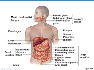
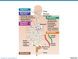
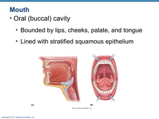
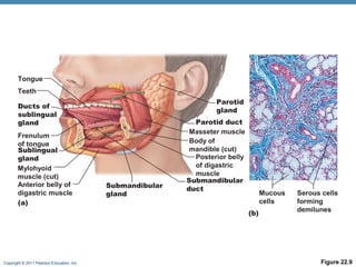
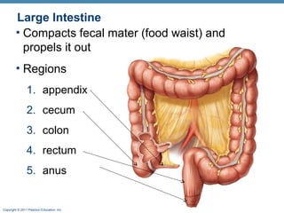
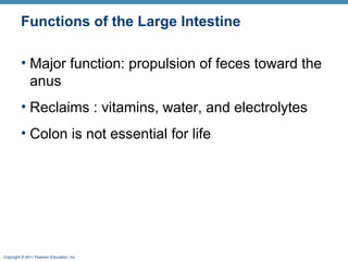
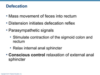
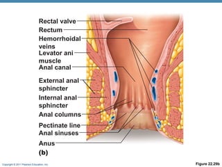
Digestive System
•

Two groups of organs
1. Alimentary canal (gastrointestinal or GI tract)
•

Digests and absorbs food

•

Mouth, pharynx, esophagus, stomach,
small intestine, and large intestine

Copyright © 2011 Pearson Education, Inc.
Digestive System
2. Accessory digestive organs
•

Teeth, tongue, gallbladder

•

Digestive glands
•

Salivary glands

•

Liver

•

pancreas

Copyright © 2011 Pearson Education, Inc.
Mouth (oral cavity)
Tongue

Esophagus

Liver
Gallbladder
Duodenum
Jejunum
Small
intestine Ileum

Anus
Copyright © 2011 Pearson Education, Inc.

Parotid gland
Sublingual gland Salivary
Submandibular
glands
gland
Pharynx
Stomach
Pancreas
(Spleen)
Transverse colon
Descending colon
Ascending colon
Large
Cecum
intestine
Sigmoid colon
Rectum
Vermiform appendix
Anal canal
Figure 22.1
Digestive Processes
•

Six essential activities
1. Ingestion
2. Propulsion
3. Mechanical digestion
4. Chemical digestion
5. Absorption
6. Defecation

Copyright © 2011 Pearson Education, Inc.
Ingestion
Mechanical
digestion
• Chewing (mouth)
• Churning (stomach)
• Segmentation
(small intestine)
Chemical
digestion

Food
Pharynx
Esophagus
Propulsion
• Swallowing
(oropharynx)
• Peristalsis
Stomach (esophagus,
stomach,
small intestine,
large intestine)
Absorption
Lymph
vessel

Small
intestine
Large
intestine

Defecation
Copyright © 2011 Pearson Education, Inc.

Blood
vessel
Mainly H2O
Feces
Anus
Figure 22.2
Blood Supply: Splanchnic Circulation
• Arteries
• Hepatic, splenic, and left gastric
• Inferior and superior mesenteric

• Hepatic portal veins
• Drains nutrient-rich blood from digestive
organs
• Delivers it to the liver

Copyright © 2011 Pearson Education, Inc.
TISSUES OF THE
ALIMENTARY CANAL

Copyright © 2011 Pearson Education, Inc.
Histology of the Alimentary Canal
• Four basic layers (tunics)
1. Mucosa
2. Submucosa
3. Muscularis externa
4. Serosa

Copyright © 2011 Pearson Education, Inc.
Mucosa
Inner layer of lumen
Three sublayers:
1. Epithelium – simple columnar and goblet cells
•

Secrete mucus and enzymes

1. Lamina propria – areolar basement membrane
2. Muscularis mucosae– thin layer of smooth muscle

Copyright © 2011 Pearson Education, Inc.
Simple Columnar with Microvilli

Copyright © 2011 Pearson Education, Inc.
Submucosa, Muscularis Externa and Serosa
Submucosa
• Connective tissue
• Blood and lymphatic vessels

Muscularis externa
• Responsible for segmentation and peristalsis
• Smooth muscle

Serosa
• Fibrous covering (visceral peritoneum)
Copyright © 2011 Pearson Education, Inc.
ORAL CAVITY

Mouth, teeth, salivary glands and tongue

Copyright © 2011 Pearson Education, Inc.
Mouth
• Oral (buccal) cavity
• Bounded by lips, cheeks, palate, and tongue
• Lined with stratified squamous epithelium

Copyright © 2011 Pearson Education, Inc.
Oral Cavity
• Lips form anterior border
• Cheeks form lateral border
• Palate forms superior border
• Hard palate: maxillae and palatine bones
• Ridges help create friction

• Soft palate: closes the nasopharynx for swallowing
• Uvula projects down from free edge

Copyright © 2011 Pearson Education, Inc.
Tongue

Oral cavity muscle
• Assists in chewing
• forming bolus
• swallowing, speech, and taste

Contains taste buds
Frenulum attaches tongue to the floor of oral cavity

Copyright © 2011 Pearson Education, Inc.
Salivary Glands
Extrinsic salivary glands
•Secrete saliva, contains digestive enzymes
1.Parotid gland – buccal area
•

Duct opens near 2nd molar

1.Submandibular – medial mandible
•

Duct opens under tongue

1.Sublingual – anterior to submandibular,
under tongue
Copyright © 2011 Pearson Education, Inc.
Tongue
Teeth
Parotid
gland

Ducts of
sublingual
gland
Frenulum
of tongue
Sublingual
gland
Mylohyoid
muscle (cut)
Anterior belly of
digastric muscle
(a)

Submandibular
gland

Parotid duct
Masseter muscle
Body of
mandible (cut)
Posterior belly
of digastric
muscle
Submandibular
duct

Mucous
cells
(b)

Copyright © 2011 Pearson Education, Inc.

Serous cells
forming
demilunes

Figure 22.9
Teeth
• 32 permanent teeth
• Covered by enamel – hardest substance in body
• Periodontal ligament
• Forms fibrous joint called a gomphosis

• Incisors - chisel shaped for cutting
• Canine - fanglike
• Premolars (bicuspids) and molars
• Have broad crowns for grinding or crushing
Copyright © 2011 Pearson Education, Inc.
Tooth Structure

Copyright © 2011 Pearson Education, Inc.
Pharynx and Esophagus
Muscular tubes allow passage of water and
food
Esophagus uses peristalsis to move food
• Travels through diaphragm
• Esophageal sphincter

Copyright © 2011 Pearson Education, Inc.
STOMACH AND INTESTINES

Copyright © 2011 Pearson Education, Inc.
Stomach Regions
• Cardiac region
• Connects to esophagus

• Fundus
• Dome region below
diaphragm

• Body
• midregion

Copyright © 2011 Pearson Education, Inc.

• Pyloric region:
• antrum, pyloric canal,
and pylorus
• Connects to
duodenum via pyloric
sphincter

• Greater curvature
• Lesser curvature
Cardia
Esophagus

Fundus

Body
Lesser
curvature

Lumen

Greater
curvature
Duodenum
(a)
Copyright © 2011 Pearson Education, Inc.

Pyloric
Pyloric
canal
antrum
Pyloric sphincter
(valve) at pylorus
Figure 22.14a
Stomach: Layers
• Four tunics
• Three muscular layers and outer tunic (serosa)
• Muscularis externa
1.longitudinal
2.circular
3.oblique
• Covered by omentums
• Greater & lesser
Copyright © 2011 Pearson Education, Inc.
Stomach: Tissues
• Mucosa
• Simple columnar epithelium with mucus covering

• Contain gastric pits
• Produce gastric acid = HCl

Copyright © 2011 Pearson Education, Inc.
Digestive Processes so Far…
• Physical digestion (chewing)
• Teeth and tongue
• Creates bolus

• Enzymatic digestion
• Enzymes in saliva
• Gastric acid

• Delivers chyme to the small intestine
• Creamy food mass
Copyright © 2011 Pearson Education, Inc.
Small Intestine
•

Major organ of absorption

•

Pyloric sphincter to ileocecal valve (2-4 m long)

•

Subdivisions
1. Duodenum : beginning portion
•

connects to stomach via pyloric sphincter

1. Jejunum : intermediate portion(~8ft)
2. Ileum : end portion (~12ft)
•

Connects to large intestine via ileocecal valve

Copyright © 2011 Pearson Education, Inc.
Small Intestine

Copyright © 2011 Pearson Education, Inc.
Duodenum
• Ducts for gallbladder and pancreases empty into
the hepatopancreatic ampulla
• Bile duct
• Main pancreatic duct

• Enter at the duodenal papilla
• Are controlled by the hepatopancreatic
sphincter

Copyright © 2011 Pearson Education, Inc.
Absorption of Small Intestine
• Modification help increase surface area
(absorption):
1. Circular folds (plicae circulares)
2. Villi
3. Microvilli

Copyright © 2011 Pearson Education, Inc.
Villi and Microvilli
Villi
• fingerlike extensions of mucosa
• Simple columnar absorptive cells
• With microvilli – increase surface area

• Goblet cells – produce mucus

Copyright © 2011 Pearson Education, Inc.
Large Intestine
• Compacts fecal mater (food waist) and
propels it out
• Regions
1. appendix
2. cecum
3. colon
4. rectum
5. anus

Copyright © 2011 Pearson Education, Inc.
Colon
• Regions of colon
• Ascending colon
• Right (hepatic) flexure

• Transverse colon
• Left (splenic) flexure

• Descending colon
• Sigmoid colon (S – shaped)

• Contains pocketlike sacks haustrum

Copyright © 2011 Pearson Education, Inc.
Large Intestine

Copyright © 2011 Pearson Education, Inc.
Rectum and Anus
• Rectum
• Inferior holding chamber

• Anal canal
• Last segment of the large intestine

• Sphincters
• Internal anal sphincter —smooth muscle
• involuntary
• External anal sphincter —skeletal muscle
• voluntary
Copyright © 2011 Pearson Education, Inc.
Functions of the Large Intestine
• Major function: propulsion of feces toward the
anus
• Reclaims : vitamins, water, and electrolytes
• Colon is not essential for life

Copyright © 2011 Pearson Education, Inc.
Motility of the Large Intestine
• Haustral contractions
• Slow segmenting movements
• Haustra sequentially contract in response to
distension

• Gastrocolic reflex
• Initiated by presence of food in the stomach
• Three slow powerful peristaltic waves per day
in the colon (mass movements)
Copyright © 2011 Pearson Education, Inc.
Defecation
• Mass movement of feces into rectum
• Distension initiates defecation reflex
• Parasympathetic signals
• Stimulate contraction of the sigmoid colon and
rectum
• Relax internal anal sphincter

• Conscious control relaxation of external anal
sphincter

Copyright © 2011 Pearson Education, Inc.
Rectal valve
Rectum
Hemorrhoidal
veins
Levator ani
muscle
Anal canal
External anal
sphincter
Internal anal
sphincter
Anal columns
Pectinate line
Anal sinuses
Anus
(b)
Copyright © 2011 Pearson Education, Inc.

Figure 22.29b

Digest anat online

  • 1.
    Digestive System • Two groupsof organs 1. Alimentary canal (gastrointestinal or GI tract) • Digests and absorbs food • Mouth, pharynx, esophagus, stomach, small intestine, and large intestine Copyright © 2011 Pearson Education, Inc.
  • 2.
    Digestive System 2. Accessorydigestive organs • Teeth, tongue, gallbladder • Digestive glands • Salivary glands • Liver • pancreas Copyright © 2011 Pearson Education, Inc.
  • 3.
    Mouth (oral cavity) Tongue Esophagus Liver Gallbladder Duodenum Jejunum Small intestineIleum Anus Copyright © 2011 Pearson Education, Inc. Parotid gland Sublingual gland Salivary Submandibular glands gland Pharynx Stomach Pancreas (Spleen) Transverse colon Descending colon Ascending colon Large Cecum intestine Sigmoid colon Rectum Vermiform appendix Anal canal Figure 22.1
  • 4.
    Digestive Processes • Six essentialactivities 1. Ingestion 2. Propulsion 3. Mechanical digestion 4. Chemical digestion 5. Absorption 6. Defecation Copyright © 2011 Pearson Education, Inc.
  • 5.
    Ingestion Mechanical digestion • Chewing (mouth) •Churning (stomach) • Segmentation (small intestine) Chemical digestion Food Pharynx Esophagus Propulsion • Swallowing (oropharynx) • Peristalsis Stomach (esophagus, stomach, small intestine, large intestine) Absorption Lymph vessel Small intestine Large intestine Defecation Copyright © 2011 Pearson Education, Inc. Blood vessel Mainly H2O Feces Anus Figure 22.2
  • 6.
    Blood Supply: SplanchnicCirculation • Arteries • Hepatic, splenic, and left gastric • Inferior and superior mesenteric • Hepatic portal veins • Drains nutrient-rich blood from digestive organs • Delivers it to the liver Copyright © 2011 Pearson Education, Inc.
  • 7.
    TISSUES OF THE ALIMENTARYCANAL Copyright © 2011 Pearson Education, Inc.
  • 8.
    Histology of theAlimentary Canal • Four basic layers (tunics) 1. Mucosa 2. Submucosa 3. Muscularis externa 4. Serosa Copyright © 2011 Pearson Education, Inc.
  • 9.
    Mucosa Inner layer oflumen Three sublayers: 1. Epithelium – simple columnar and goblet cells • Secrete mucus and enzymes 1. Lamina propria – areolar basement membrane 2. Muscularis mucosae– thin layer of smooth muscle Copyright © 2011 Pearson Education, Inc.
  • 10.
    Simple Columnar withMicrovilli Copyright © 2011 Pearson Education, Inc.
  • 11.
    Submucosa, Muscularis Externaand Serosa Submucosa • Connective tissue • Blood and lymphatic vessels Muscularis externa • Responsible for segmentation and peristalsis • Smooth muscle Serosa • Fibrous covering (visceral peritoneum) Copyright © 2011 Pearson Education, Inc.
  • 12.
    ORAL CAVITY Mouth, teeth,salivary glands and tongue Copyright © 2011 Pearson Education, Inc.
  • 13.
    Mouth • Oral (buccal)cavity • Bounded by lips, cheeks, palate, and tongue • Lined with stratified squamous epithelium Copyright © 2011 Pearson Education, Inc.
  • 14.
    Oral Cavity • Lipsform anterior border • Cheeks form lateral border • Palate forms superior border • Hard palate: maxillae and palatine bones • Ridges help create friction • Soft palate: closes the nasopharynx for swallowing • Uvula projects down from free edge Copyright © 2011 Pearson Education, Inc.
  • 15.
    Tongue Oral cavity muscle •Assists in chewing • forming bolus • swallowing, speech, and taste Contains taste buds Frenulum attaches tongue to the floor of oral cavity Copyright © 2011 Pearson Education, Inc.
  • 16.
    Salivary Glands Extrinsic salivaryglands •Secrete saliva, contains digestive enzymes 1.Parotid gland – buccal area • Duct opens near 2nd molar 1.Submandibular – medial mandible • Duct opens under tongue 1.Sublingual – anterior to submandibular, under tongue Copyright © 2011 Pearson Education, Inc.
  • 17.
    Tongue Teeth Parotid gland Ducts of sublingual gland Frenulum of tongue Sublingual gland Mylohyoid muscle(cut) Anterior belly of digastric muscle (a) Submandibular gland Parotid duct Masseter muscle Body of mandible (cut) Posterior belly of digastric muscle Submandibular duct Mucous cells (b) Copyright © 2011 Pearson Education, Inc. Serous cells forming demilunes Figure 22.9
  • 18.
    Teeth • 32 permanentteeth • Covered by enamel – hardest substance in body • Periodontal ligament • Forms fibrous joint called a gomphosis • Incisors - chisel shaped for cutting • Canine - fanglike • Premolars (bicuspids) and molars • Have broad crowns for grinding or crushing Copyright © 2011 Pearson Education, Inc.
  • 19.
    Tooth Structure Copyright ©2011 Pearson Education, Inc.
  • 20.
    Pharynx and Esophagus Musculartubes allow passage of water and food Esophagus uses peristalsis to move food • Travels through diaphragm • Esophageal sphincter Copyright © 2011 Pearson Education, Inc.
  • 21.
    STOMACH AND INTESTINES Copyright© 2011 Pearson Education, Inc.
  • 22.
    Stomach Regions • Cardiacregion • Connects to esophagus • Fundus • Dome region below diaphragm • Body • midregion Copyright © 2011 Pearson Education, Inc. • Pyloric region: • antrum, pyloric canal, and pylorus • Connects to duodenum via pyloric sphincter • Greater curvature • Lesser curvature
  • 23.
    Cardia Esophagus Fundus Body Lesser curvature Lumen Greater curvature Duodenum (a) Copyright © 2011Pearson Education, Inc. Pyloric Pyloric canal antrum Pyloric sphincter (valve) at pylorus Figure 22.14a
  • 24.
    Stomach: Layers • Fourtunics • Three muscular layers and outer tunic (serosa) • Muscularis externa 1.longitudinal 2.circular 3.oblique • Covered by omentums • Greater & lesser Copyright © 2011 Pearson Education, Inc.
  • 25.
    Stomach: Tissues • Mucosa •Simple columnar epithelium with mucus covering • Contain gastric pits • Produce gastric acid = HCl Copyright © 2011 Pearson Education, Inc.
  • 26.
    Digestive Processes soFar… • Physical digestion (chewing) • Teeth and tongue • Creates bolus • Enzymatic digestion • Enzymes in saliva • Gastric acid • Delivers chyme to the small intestine • Creamy food mass Copyright © 2011 Pearson Education, Inc.
  • 27.
    Small Intestine • Major organof absorption • Pyloric sphincter to ileocecal valve (2-4 m long) • Subdivisions 1. Duodenum : beginning portion • connects to stomach via pyloric sphincter 1. Jejunum : intermediate portion(~8ft) 2. Ileum : end portion (~12ft) • Connects to large intestine via ileocecal valve Copyright © 2011 Pearson Education, Inc.
  • 28.
    Small Intestine Copyright ©2011 Pearson Education, Inc.
  • 29.
    Duodenum • Ducts forgallbladder and pancreases empty into the hepatopancreatic ampulla • Bile duct • Main pancreatic duct • Enter at the duodenal papilla • Are controlled by the hepatopancreatic sphincter Copyright © 2011 Pearson Education, Inc.
  • 30.
    Absorption of SmallIntestine • Modification help increase surface area (absorption): 1. Circular folds (plicae circulares) 2. Villi 3. Microvilli Copyright © 2011 Pearson Education, Inc.
  • 31.
    Villi and Microvilli Villi •fingerlike extensions of mucosa • Simple columnar absorptive cells • With microvilli – increase surface area • Goblet cells – produce mucus Copyright © 2011 Pearson Education, Inc.
  • 32.
    Large Intestine • Compactsfecal mater (food waist) and propels it out • Regions 1. appendix 2. cecum 3. colon 4. rectum 5. anus Copyright © 2011 Pearson Education, Inc.
  • 33.
    Colon • Regions ofcolon • Ascending colon • Right (hepatic) flexure • Transverse colon • Left (splenic) flexure • Descending colon • Sigmoid colon (S – shaped) • Contains pocketlike sacks haustrum Copyright © 2011 Pearson Education, Inc.
  • 34.
    Large Intestine Copyright ©2011 Pearson Education, Inc.
  • 35.
    Rectum and Anus •Rectum • Inferior holding chamber • Anal canal • Last segment of the large intestine • Sphincters • Internal anal sphincter —smooth muscle • involuntary • External anal sphincter —skeletal muscle • voluntary Copyright © 2011 Pearson Education, Inc.
  • 36.
    Functions of theLarge Intestine • Major function: propulsion of feces toward the anus • Reclaims : vitamins, water, and electrolytes • Colon is not essential for life Copyright © 2011 Pearson Education, Inc.
  • 37.
    Motility of theLarge Intestine • Haustral contractions • Slow segmenting movements • Haustra sequentially contract in response to distension • Gastrocolic reflex • Initiated by presence of food in the stomach • Three slow powerful peristaltic waves per day in the colon (mass movements) Copyright © 2011 Pearson Education, Inc.
  • 38.
    Defecation • Mass movementof feces into rectum • Distension initiates defecation reflex • Parasympathetic signals • Stimulate contraction of the sigmoid colon and rectum • Relax internal anal sphincter • Conscious control relaxation of external anal sphincter Copyright © 2011 Pearson Education, Inc.
  • 39.
    Rectal valve Rectum Hemorrhoidal veins Levator ani muscle Analcanal External anal sphincter Internal anal sphincter Anal columns Pectinate line Anal sinuses Anus (b) Copyright © 2011 Pearson Education, Inc. Figure 22.29b