Digestive System: A Concise Guide
•
5 likes•238 views

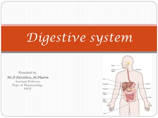
Digestive system Mouth Buccal cavity Tongue Teeth Salivary glands Pharynx Oesophagus Stomach Small intestine Large intestine Rectum Anus Liver Gall bladder Pancreas Absorption Digestion

Recommended
More Related Content
What's hot
What's hot (20)
Similar to Digestive System: A Concise Guide
Similar to Digestive System: A Concise Guide (20)
Recently uploaded
Recently uploaded (20)
Digestive System: A Concise Guide
- 1. Digestive system Presented by Ms.P.Pavithra.,M.Pharm Assistant Professor, Dept. of Pharmacology, SVCP
- 2. Introduction The digestive system consists of gastrointestinal tract (alimentary canal) and its glands. The functions of gastrointestinal tract are ingestion, digestion and absorption of food and excretion of waste products. 1.Ingestion: taking in food 2.Digestion: breaking down food into nutrients 3.Absorption: taking in nutrients by cells 4.Egestion: removing any leftover wastes
- 3. Parts of digestive system Mouth Pharynx Oesophagus Stomach Small intestine Large intestine Rectum Anus
- 4. Mouth(Buccal cavity) It is the upper expanded portion which forms the beginning of alimentary canal. It can be divided into two parts: i.) Vestibule, an outer part. It lies between lips and inner lining of cheeks externally and gums and teeth internally. ii.) Cavity of mouth an inner part. It is bounded by teeth and mastoid bone at the sides, palate above and tongue below.
- 5. Palate forms the roof of mouth cavity. It consists of hard palate which is in front and soft palate which is behind. Uvula is a conical process which hangs from the middle of soft palate.Two folds of mucous membrane called anterior and posterior pillars of fauces lie on either side of uvula. Tonsils lie in between these folds.
- 6. The important structure of mouth are Tongue Teeth Salivary glands
- 7. Tongue Tongue lies in the floor of the mouth and it is attached to hyoid bone. Tongue mixes food with saliva (contains amylase, which helps break down starch). Tongue contains: A root at which blood vessels and nerves pass. A tip which is pointed when the tongue is protruded and rounded when the tongue is in the mouth. Two margins which are in contact with lower teeth. An upper surface which contains a small elevation called dorsum A lower surface which contains a soft ligamentous structure called frenulum
- 8. The two important structures of tongue are: i.) Taste buds which are on the lateral aspects of tongue. ii.) Three types of papillae present on the upper surface. They are circumvallate papillae, fungiform papillae and filiform papillae.
- 10. Teeth Teeth mechanically break down food into small pieces (Mastication). Depending on the age at which they arise, teeth can be classified into two types i.) Permanent teeth-the teeth of adult life ii.) Temporary teeth or milk teeth –the teeth of childhood Permanent teeth: They are 32 in number and 16 are present in each jaw. Each half of the upper and lower jaw contains 8 teeth.They are: 2 incisors, 1 canine, 2 premolars and 3 molars. Temporary teeth: They are 20 in number and each jaw has 10 teeth. Each half of the jaw has 2 incisors, 1 canine and 2 molars.
- 12. Eruption of teeth: Even at birth, all the permanent and temporary teeth are buried in the alveolar process of jaws. The first tooth appears in a child at the 7th month. Later, the full set of temporary teeth is completed at the age of two years. The first permanent tooth to appear is the first molar which appears at the age of 6 years. The set of permanent teeth is completed at the age of 12 years. But the third molar (wisdom tooth) appears between 17 and 25 years.
- 14. Structure of Tooth 1.) Crown-which projects above the gum 2.) Neck-which is surrounded by gum 3.) Root-which lies beneath the gum Tooth is made of Dentine-the main mass and it is a hard material Pulp-the central cavity which contains connective tissues, blood vessels and nerves. Enamel-the covering part of which projects above the gums.
- 15. Salivary glands There are three pairs of salivary glands in the mouth. They are parotid, submandibular and sublingual glands. Parotid gland: One on each side is present below and in front of each ear. Each gland has a duct called Stenson’s duct. This duct opens on the inner side of cheek opposite to the second upper molar tooth. Submandibular glands (Submaxillar glands): They are smaller than parotid glands. One on each side lies under the angle of jaw. Each gland has a duct called Wharton’s duct which opens near the midline under the tongue.
- 16. Sublingual glands: They are the smallest salivary glands which lie under the tongue. They pour the secretions directly into the mouth through several small openings.
- 17. Saliva: It is a mixed secretion of all the three pairs of salivary glands. It is an alkaline fluid containing water to the extent of 99%. The solid contents of saliva are: Mucin which is a glycoprotein Pytalin, an enzyme which converts starch into maltose. Also, it contains salts of sodium, potassium, calcium and magnesium. Functions: It converts cooked starch into a soluble sugar called maltose. It acts as a solvent for food and helps in the swallowing. It moistens, lubricates and cleans the mouth. It excretes organic and inorganic substances and some drugs.
- 18. Pharynx Pharynx lies between the mouth and oesophagus. Pharynx consists of three parts: Nasopharynx Oropharynx Laryngopharynx Nasopharynx: It lies behind the nasal cavity. It extends from the base of skull to the level of soft palate. On either side, it has an opening for Eustachian tube. Oropharynx: It lies behind the mouth. It extends between soft palate above and upper opening of larynx below.The lateral walls of oropharynx contain tonsil. Laryngopharynx: It is the lowest part and it lies behind the larynx.
- 19. Oropharynx and laryngopharynx serve as a common channel for the passage of food and air.Through both these parts, food is conducted from mouth to oesophagus and air from nasopharynx to larynx.
- 20. Oesophagus It is a muscular tube which extends between pharynx above and cardiac orifice of stomach below. It lies between trachea in front and vertebral column at the back. From the thorax, it enters the abdomen through the oesophageal opening of diaphragm. The oesophagus contains sphincters at its upper and lower ends.These sphincters relax during swallowing.
- 21. Deglutition (the act of swallowing) In the mouth, food is masticated and mixed well with saliva.The action of tongue and cheeks convert food into a round mass called bolus.This bolus is swallowed. Swallowing occurs in the following three stages Stage:1 Closing of the lips and raising of the tongue against the palate forces the bolus into oropharynx. Now the nasopharynx is closed by soft palate and larynx is closed by epiglottis.This prevents the entry of food into respiratory passages.
- 22. Stage:2 By the contraction of the muscles of pharynx, the bolus is forced into oesophagus. Stage:3 In the oesophagus, contraction of its muscular walls carries the food down to stomach. It must be noted that the first stage is a voluntary act but it is performed automatically. But the second stage and third stage are involuntary acts.
- 24. Abdominal cavity Abdomen is the largest cavity in the body. It is oval in shape and contains a variety of organs. It can be divided into two parts: Abdomen proper-an upper larger cavity Pelvis-a lower small cavity Boundaries of abdomen: Abdomen is bounded Above by the lower surface of diaphragm Below by the brim of true pelvis In the front and sides by abdominal muscles, ribs and iliac bones. At the back by the vertebral column, psoas and quadratus lumborum muscles.
- 25. Contents of abdomen: The abdomen contains stomach, intestines, liver, spleen, pancreas, kidneys, adrenal glands, etc.
- 26. Stomach Stomach is the dilated portion of alimentary canal and it receives food from oesophagus. It lies in the upper part of abdominal cavity below the left half of diaphragm. Parts of Stomach: 1.) Two surfaces: an anterior and a posterior surface 2.) Two borders: an upper border called lesser curvature, a lower border called greater curvature. 3.) Two ends: Upper end called cardiac end, it is guarded by cardiac sphincter. Lower end called pyloric end, it is guarded by pyloric sphincter.
- 27. 4.) Fundus: A dome shaped upper part lying to the left of cardiac end. 5.) Body: The main part of stomach 6.) Pyloric antrum: The lower part
- 28. Structure of stomach: Stomach consists of the following four coats: Peritoneal coat(made of serous covering) Muscular coat (made of longitudinal, circular and oblique fibres) Submucous coat (made of areolar tissue) Mucous coat (made of mucous membrane)
- 29. Secretions of stomach: The mucous membrane of stomach contains glands which secrete gastric juice continuously. The secretion of gastric juice occurs due to: A reflex mechanism through vagus nerve Gastrin, a hormone secreted by the action of food stuffs on gastric mucous membrane. Psychological effects produced by taste or smell of food. Gastric juice contains pepsin, rennin, hydrochloric acid and intrinsic factor.
- 31. Pepsin: It is an enzyme produced by glands present in the fundus and body of stomach. In presence of hydrochloric acid, pepsin converts protein into peptone. Rennin: It is the enzyme which curdles milk. It involves the conversion of caseinogen, the soluble protein of milk into insoluble caesin.
- 32. Hydrochloric acid: It is secreted by parietal cells of gastric glands. Its concentration is about 0.4 % in gastric juice. The functions of Hcl are: Neutralisation of saliva and acidification of food. Helping the action of pepsin in converting protein into peptone. Antiseptic action by killing bacteria. Intrinsic factor: It is a content of gastric juice which is necessary for the absorption of Vitamin B12 This vitamin is necessary for the development of RBC. Chyme: It is the product of digested food in the stomach. Chyme is in a semi-liquid form and it is passed on to duodenum.
- 33. Small intestine Small intestine is the part of alimentary canal which extends from the pyloric end of stomach to caecum (the first part of large intestine) Parts: Small intestine consist of three parts Duodenum Jejunum Ileum
- 34. Duodenum: It is a C-shaped fixed part which is attached to posterior abdominal wall by peritoneum. The head of pancreas lies in the concavity of duodenum. Also the bile duct and pancreatic duct open together at the concave surface. A small eminence at this opening is called ampula of vater.
- 35. Jejunum: It is the continuation of duodenum and it is the middle portion of small intestine. Ileum: It forms the last part of small intestine.
- 36. Structure: Small intestine consists of the same four coats which are present in stomach.They are: Peritoneal coat (made of serous membrane) Muscular coat (made of only circular and longitudinal fibres) Submucous coat(made of areolar tissue) Mucous coat (which is the inner lining)
- 37. The mucous coat contains: Plicae circulares which are a number of folds of the mucous membrane. Villi which absorb carbohydrates, proteins and fat.
- 38. Digestion in small intestine: The acidic chyme from the stomach enters into the duodenum. There it mixes with 1.)The alkaline intestinal juice called succus entericus. 2.)Alkaline secretions from liver and pancreas. In the small intestine, digestion is carried out by the following enzymes of intestinal juice. 1.) Enterokinase (Enteropeptidase) which converts trypsinogen into trypsin. 2.) Erepsin which converts polypeptides into amino acids. 3.) Sucrase, maltase and lactase which convert the corresponding disaccharides into monosaccharides.
- 39. Absorption in small intestine: The absorption of digested food occurs in small intestine through villi. Villi: Villi are minute projections which are present in the inner mucous coat of the intestine. The villi give a velvetty appearance to the intestinal mucous membrane.
- 40. Each villus has: A central lymphatic vessel called lacteal. Fats are absorbed into lacteal and carried to thoracic duct. A network of capillaries surrounding the lacteal. Digested products of carbohydrates and proteins are absorbed into these capillaries.They are carried to liver by portal vein. Lymphoid tissue which holds together the lacteals and capillaries.
- 41. Large intestine Large intestine (colon) extends from the end of ileum to rectum. Large intestine consists of the following parts: Caecum, appendix, ascending colon, transverse colon, descending colon and sigmoid colon.
- 42. 1.) Caecum: It is a short rounded sac which lies in the right iliac fossa. It commences at iliacaecal valve where the ileum joins the caecum.
- 43. 2.)Vermiform appendix: It springs out from the caecum at about an inch from the ileocaecal junction. It is present in the right iliac fossa. The lumen of appendix communicates with that of caecum. The appendix is composed of the same four coats as intestine but the submucous coat contains lymphoid tissue.
- 44. 3.) Ascending colon: It ascends upwards from caecum and in front of right kidney. It turns to the left below the liver and forms the transverse colon. 4.)Transverse colon: It is the loop of large intestine which extends between the lower surfaces of liver and spleen. At the lower surface of spleen, it turns downwards to form descending colon.
- 45. 5.) Descending colon: It extends from the lower surface of spleen to brim of pelvis. It lies in the left lumbar region. 6.) Sigmoid colon: It is the continuation of descending colon and it continues below with rectum.
- 46. Structure of large intestine: Large intestine has the same four coats (Peritoneal, muscular, submucous and mucous) as small intestine. But the only difference is longitudinal muscles are arranged in three bands.The mucous membrane does not have villi. Functions of large intestine: Digestion: This is carried out by microorganisms of colon. They act on the undigested and unabsorbed residue from small intestine. Absorption: All carbohydrates, proteins and fat are already absorbed in small intestine. Only water and glucose are absorbed in the colon. Secretion: Mucin is the only secretion. It lubricates the colon and facilitates the passage of fecal matter. Excretion: Iron and some purgatives are excreted in large intestine.
- 47. Rectum It occupies the lower posterior part of pelvis. It extends between sigmoid colon and anus. The lower part of rectum is dilated and it is called rectal ampulla. Anus It is a small canal measuring about one inch in length. The opening of anus is guarded by a sphincter called anal sphincter. This sphincter is under voluntary control.
- 48. Defecation It is defined as evacuation of the fecal matter of the rectum. Defecation is a reflex mechanism. But this reflex is under voluntary control. The reflex for defecation occurs when a sufficient quantity of feces accumulates in the rectum. This produces stretching of rectal walls and also increases pressure in the rectum. When the pressure exceeds 40 mm Hg the nerve endings of rectum are stimulated. The impulses reach the spinal cord. From the spinal cord, impulses for defecation are carried to the rectum through motor nerves.
- 49. The act of defecation involves the following events: Relaxation of the anal sphincter which is the first voluntary act. This is followed by contraction of: i. Muscles of rectum ii.Muscles of pelvic floor iii.Muscles of abdominal wall iv.Diaphragm These acts increase the pressure in abdomen and pelvic cavity and help in defecation.
- 50. Digestion of food in the alimentary canal Food contains: 1)Carbohydrates 2)Proteins 3)Fat All these constituents of food are digested in the alimentary canal as follows: 1.) Carbohydrates: Ptyalin (salivary amylase) present in saliva converts cooked starches in food into a sugar called maltose. This conversion occurs in the mouth. All sugars are converted to simple monosaccharides like glucose by the action of enzymes(sucrase, maltase, lactase) in the small intestine. Glucose is absorbed through the capillaries of villi in the small intestine. It is then carried to liver by portal vein where it is stored as glycogen.
- 51. 2.) Proteins: The digestive enzymes (pepsin of stomach, trypsin and erepsin of small intestine) convert proteins into peptones, polypeptides and finally into amino acids. The amino acids are absorbed through villi of small intestine and carried to liver. 3.) Fats: Lipase, an enzyme of pancreas which is poured into small intestine converts fats into fatty acids and glycerol. These two products are absorbed through lacteals of villi. They are carried to thoracic duct through cysterna chyli. From the thoracic duct they enter into blood through left brachiocephalic vein.
- 52. Absorption of food in the alimentary canal 1.) Mouth Absorption of food does not occur in the mouth. Only drugs like nitroglycerine and isoprenaline are absorbed through buccal mucosa. 2.) Stomach No absorption of food occurs in stomach except glucose. But water and alcohol can be absorbed in the stomach.
- 53. 3.) Small intestine: Products of carbohydrates digestion like glucose and other simple sugars are absorbed through capillaries of villi. Products of protein digestion like amino acids are also absorbed through capillaries of villi. Products of fat metabolism like fatty acids and glycerol are also absorbed through lacteals of villi. 4.) Large intestine: Water, glucose and certain salts are absorbed through the mucous membrane of large intestine.
- 54. Peritoneum Peritoneum is a serous membrane which lines the abdomen and covers the abdominal organs. It consists of the following two layers. 1.) Parietal peritoneum which lines the walls of abdominal cavity.
- 55. 2.) Visceral peritoneum which covers the abdominal organs. The space between these two layers is called as peritoneal space. Organs completely covered by peritoneum are stomach, liver and intestines. Organs partly covered by peritoneum are kidneys.
- 56. Omenta: The folds of peritoneum connected to the stomach are called omenta.They are divided into: 1.) Greater omentum which hangs from the lower border of stomach to the front surface of small intestine.
- 57. 2.) Lesser omentum which extends from the lower border of liver to the lesser curvature of stomach.
- 58. Mesentry: It is the fold of peritoneum which attaches the different parts of small intestine to the posterior abdominal wall. Blood vessels, nerves and lymphatics enter the intestines only through mesentry.
- 59. Peritoneal ligaments: They are folds of peritoneum which connect organs (like liver and uterus) to the posterior abdominal wall. Pelvic peritoneum: It is the part of peritoneum present in the pelvic region. The pelvic peritoneum is actually the continuation of peritoneum in the abdominal cavity. The arrangement of pelvic peritoneum is different in males and females due to the presence of uterus and fallopian tubes in females.
- 60. Arrangement in males: In males, the peritoneum covers the upper part of rectum. Then it passes over the posterior and upper surface of bladder. Later, it becomes continuous with the peritoneum of anterior abdominal wall.
- 61. Arrangement in females: In females, the peritoneum covers the rectum as in males. But, it covers the anterior and posterior surfaces of uterus before reaching the bladder. The sac of peritoneum between the rectum and uterus is called the pouch of douglas. In the females, the peritoneum covers the fallopian tubes also. The fallopian tubes open directly into the peritoneal cavity. The mucous membrane of fallopian tubes is continuous with peritoneum.
- 62. Functions of Peritoneum: It forms a complete or partial covering for abdominal organs. It forms a smooth lining which enables the abdominal organs to move over each other without friction. The ligaments and mesentries of peritoneum hold the abdominal organs in position. Omentum and mesentry serve as store house for fat. The fat of peritoneum prevents infections being carried to abdominal organs. The peritoneum contains some fluid which absorbs shock and prevents it from getting transmitted to abdominal organs. The peritoneum itself can absorb large quantities of fluids.
- 63. ACCESSORY ORGANS OF DIGESTION Liver Gall bladder Pancreas
- 64. Liver is the largest abdominal organ. It lies in the upper part of abdominal cavity below the diaphragm and under the cover of lower ribs. The liver is a reddish-brown, wedge-shaped organ with two lobes of unequal size and shape. A human liver normally weighs approximately 1.5 kg and has a width of about 15 cm. External features: Externally, the liver contains two lobes and four surfaces. Lobes:They are right lobe and left lobe LIVER
- 65. Surfaces:They are i.) Superior surface which is in contact with the under surface of diaphragm. ii.) Inferior surface which is facing the abdominal viscera. The hylum or portal fissure is present in the inferior surface. The blood vessels of liver and bile duct pass through the hylum. iii.) Anterior surface which is separated from ribs and costal cartilages by the diaphragm. iv.) Posterior surface which lies in front of vertebral column, aorta, inferior vena cava and lower end of oesophagus.
- 66. Internal (minute or microscopic structure) The liver consists of a large number of liver cells called lobules. Each lobule has a central vein or intralobular vein. The connective tissue lying in between the lobules contains the branches of: Portal vein Hepatic artery Bile duct
- 67. Blood supply: Blood is brought to liver by hepatic artery and portal vein. But blood is carried from liver to inferior vena cava through hepatic veins. i.) Hepatic artery: It supplies oxygenated blood to liver. It is a branch of coeliac plexus which in turn arises from abdominal aorta. ii.) Portal vein: Portal vein also brings blood to liver. It carries blood from stomach, spleen and intestines (which blood contains nourishment) to the liver. It divides into interlobular veins which lie in between the lobules of liver. They subdivide and ultimately form central veins. One central vein lies in the centre of each lobule.
- 68. iii.) Hepatic veins: They carry the impure blood of liver and drain into inferior vena cava. The central veins of each lobule unite to form sublobular veins. The sublobular veins unite to form several hepatic veins. The hepatic veins join with inferior vena cava.
- 69. Bile ducts The secretion of liver (bile) is carried through bile ducts which are formed by the union of biliary canaliculi. The biliary canaliculi are small biliary channels present in between the lobules of liver. The bile ducts from the right and left lobes of liver unite to form common hepatic duct. The hepatic duct unites with cystic duct of gall bladder to form common bile duct. Later, the common bile duct unites with pancreatic duct in the duodenum at a papilla called ampulla of vater.
- 70. Functions of liver: Secretion of bile. Synthesis and storage of glycogen Formation of urea by the de-amination of amino acids Synthesis of plasma proteins like albumin and globulin. Conversion of unsaturated fats into saturated fats. Storage of iron and vitamin B12 (which are necessary for the formation of RBC) Synthesis of prothrombin and fibrinogen which are necessary for blood coagulation. Synthesis of heparin, the natural anticoagulant Production of heat as a result of metabolic reaction Inactivation of toxic substances and drugs Storage of vitamins A,D, E and K
- 71. GALL BLADDER It is a pear shaped storage sac for bile. It is situated in the under surface of the right lobe of liver. It consists of a fundus, body and neck.
- 72. Coats of gall bladder: Gall bladder consists of three coats: i.) Outer serous or peritoneal coat (which is continuous with the peritoneum covering the liver) ii.) Middle muscular coat (made of unstriped muscles) iii.) Inner mucous coat (which is continuous with the lining of bile ducts)
- 73. Duct of gall bladder: The duct through which gall bladder opens is called cystic duct. It arises at the neck of gall bladder. The cystic duct unites with common hepatic duct and forms common bile duct. The common bile duct joins with pancreatic duct and opens into the duodenum. A sphincter present in the bile duct at this termination in the duodenum is called sphincter of oddi (hepatopancreatic sphincter or Glisson's sphincter)
- 74. Functions of gall bladder: The gall bladder stores the bile that is secreted in liver. Also it concentrates the bile stored in it.
- 75. Bile: It is a dark-green-to-yellowish-brown fluid. It is an alkaline fluid secreted by the liver and stored in gall bladder. About 500 to 1000 ml of bile is secreted by liver per day. But the capacity of gall bladder is only 30 ml. so bile is concentrated in gall bladder. Bile contains 86% of water, bile salts, bile pigments, mucin, cholesterol and other substances.
- 76. Bile salts: Bile salts are sodium taurocholate and sodium glycocholate. The bile salts increase the digestive activity of lipase, a pancreatic enzyme. Also they help in the absorption of fats (like glycerol, fatty acids and cholesterol) and fat soluble vitamins(A, D, E and K) Bile pigments: They are bilirubin and biliverdin. They are formed from hemoglobin which is released in the destruction of worn out RBC in the spleen. Bilirubin which is orange or yellow and its oxidized form Biliverdin, which is green.
- 77. Gallstones are hardened deposits of digestive fluid. Gallstones can vary in size and number and may or may not cause symptoms. People who experience symptoms usually require gallbladder removal surgery. Gallstones that don't cause symptoms usually don't need treatment.
- 78. PANCREAS It is a long, slender gland which lies transversely across the posterior abdominal wall. It lies behind the stomach at the level of 1st and 2nd lumbar vertebrae. Parts: Pancreas consists of a head, body and tail. Head lies in the C-shaped curve of duodenum Body lies in front of the bodies of lumbar vertebrae. Tail lies in contact with the hylum of spleen.
- 79. Structure: Pancreas contains a number of lobules of secretory cells called acini. In between the acini there are groups of endocrine cells called islets of Langerhans. Small ducts emerge from these lobules. These ducts unite and reunite to form the pancreatic duct (duct of wirsung). This duct begins at the tail and emerges from the head of pancreas. It entres the duodenum along with common bile duct.
- 80. Secretions: The secretions of pancreas can be classified into---Exocrine secretion and Endocrine secretion Exocrine secretion: It is pancreatic juice which is digestive in function. It is conveyed to duodenum through pancreatic duct. Pancreatic juice contains the following digestive enzymes: i.) Lipase, which converts fats into fatty acids and glycerol ii.)Amylase which converts starch into maltose iii.)Trypsin which converts peptones into amino acids.
- 81. Endocrine secretion: It is secreted by the islets of Langerhans and directly poured into circulation. This secretion contains three different hormones which are secreted by the three different cells of islets of Langerhans. These hormones are Glucagon-secreted by alpha cells Insulin-secreted by beta cells Somatostatin-secreted by delta cells