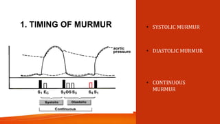
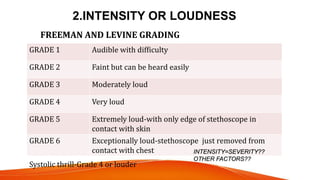
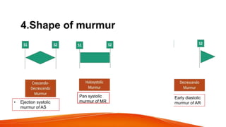
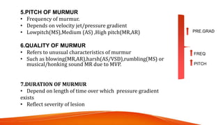
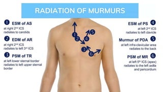
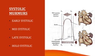
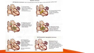
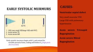
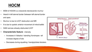
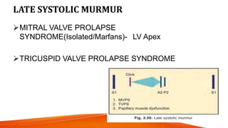
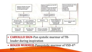
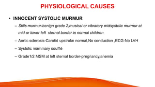
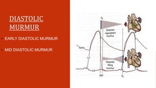
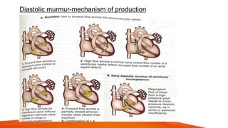
APPROACHTOA
PATIENTWITH
HEARTMURMUR
DR ANUJA JACOB
DNB RESIDENT
GENERAL MEDICINE
BMH CALICUT
DEFINITION
A cardiac murmur is defined as a relatively prolonged
series of auditory vibrations .
Characterised by timing in cardiac
cycle,intensity,pitch,configuration,duration and radiation.
MECHANISM OF
MURMUR
PRODUCTION
TURBULENT BLOOD
FLOW
 Forward flow through a constricted or irregular orifice or into a dilated
vessel or chamber
 Backward or regurgitant flow through an incompetent valve/septal defect
 Vibration of loose structure or vegetation
 Increased velocity or high rate of blood flow (hyperkinetic murmur)
HOW TO DESCRIBE A MURMUR
1. TIMING
2. LOCATION
3. DURATION
4. INTENSITY
5. FREQUENCY(PITCH)
6. CONFIGURATION
7. RADIATION
8. DYNAMIC AUSCULTATION
1. TIMING OF MURMUR • SYSTOLIC MURMUR
• DIASTOLIC MURMUR
• CONTINUOUS
MURMUR
2.INTENSITY OR LOUDNESS
GRADE 1 Audible with difficulty
GRADE 2 Faint but can be heard easily
GRADE 3 Moderately loud
GRADE 4 Very loud
GRADE 5 Extremely loud-with only edge of stethoscope in
contact with skin
GRADE 6 Exceptionally loud-stethoscope just removed from
contact with chest
FREEMAN AND LEVINE GRADING
Systolic thrill-Grade 4 or louder
INTENSITY=SEVERITY??
OTHER FACTORS??
3.LOCATION OF
MURMURS
4.Shape of murmur
• Ejection systolic
murmur of AS
Pan systolic
murmur of MR
Mid systolic
murmur of
MVP
Early diastolic
murmur of AR
5.PITCH OF MURMUR
• Frequency of murmur.
• Depends on velocity jet/pressure gradient
• Lowpitch(MS),Medium (AS) ,High pitch(MR,AR)
6.QUALITY OF MURMUR
• Refers to unusual characteristics of murmur
• Such as blowing(MR,AR),harsh(AS/VSD),rumbling(MS) or
musical/honking sound MR due to MVP.
7.DURATION OF MURMUR
• Depend on length of time over which pressure gradient
exists
• Reflect severity of lesion
FREQ
PITCH
PRE.GRAD
• DURATION
• RADIATION
SYSTOLIC
MURMURS
• EARLY SYSTOLIC
• MID SYSTOLIC
• LATE SYSTOLIC
• HOLO SYSTOLIC
EARLY SYSTOLIC MURMURS
CAUSES-
Ventricular septal defect
Very small muscular VSD
Large VSD with pulmonary
hypertension
Acute severe Tricuspid
Regurgitation
Acute severe Mitral
Regurgitation
Early systolic murmurs begin with S1 and extend for
variable period of time, ending well before S2 (high pitch
,decrescendo)
ACUTE SEVERE MR CAUSES
• Pappillary muscle rupture-MI
• Chordae tendinae-MV disease
• IE
• Blunt trauma
ACUTE SEVERE TR
•IE
•Blunt trauma
•Lower LSB
•Increases with inspiration
MID (EJECTION) SYSTOLIC
MURMUR
Begin at a short interval following S1,
end before S2 .
AORTIC STENOSIS
PULMONARY STENOSIS
HOCM
ASD
Idiopathic Dilatation of Pulmonary
Artery(pulmonary ejection sound+MSM(Upper L sternal
border)
AORTIC STENOSIS
• Ejection click f/b harsh, medium pitch,
crescendo-decrescendo systolic
murmur
• Loudest in aortic area
• Radiates along the carotid arteries
• Breath in expiration in a leaning
patient
• Reversed splitting of S2/soft or absent
S2-SEVERE
• Gallavardian phenomenon-
Musical apical murmur due to periodic
high frequency vibrations of the
fibrocalcific aortic cusps
PULMONARY STENOSIS
• Congenital
• ESM heard best in 2 &3rd Left ICS
• With increasing degree of stenosis-intensity decreases
And duration increases.
• Severe PS-RVH Signs,ECG,CXR
HOCM
• MSM of HOCM is a crescendo decrescendo murmur
• Heard in left sternal border /between left sternal border
and apex.
• Murmur is due to LVOT obstruction and MR
• It is due to systolic anterior movement of mitral leaflet
• SAM narrows already obstructed LVOT
• Characteristic feature : intensity
– Increases in Valsalva / standing /Ionotropes –all
increase degree of obs
– Decreases during squatting / handgrip/beta blockers
ASD
• Due to increased pulmonary blood flow
• Grade 2-3
• Upper to middle left sternal border
• Mostly a/w ostium secundum ASD
• Ostium primum-coexistent MR due to cleft in anterior MV
leaflet
• Sinus venosus ASD- Shunt is not large enough to
produce murmur
LATE SYSTOLIC MURMUR
MITRAL VALVE PROLAPSE
SYNDROME(Isolated/Marfans)- LV Apex
TRICUSPID VALVE PROLAPSE SYNDROME
MVP
• Posterior leaflet-Jet of MR directed anteriorly and murmur
radiates to base of heart
• Anterior leaflet –Jetof MR directed posteriorly and
murmur radiates to axilla/left infrascapular region.
MVP
Pansystolic
(Holosystolic)
Murmurs
1. Mitral valve
regurgitation
2. Tricuspid
valve
regurgitation
3. Ventricular
septal defect
Murmur begins immediately with S1 and continues up to S2
CHRONIC MITRAL REGURGITATION
CAUSES
• Rheumatic scarring of leaflet
• Mitral annular calcification
• Post infarction LV remodelling
• MR begets MR
CHRONIC TR
• Primary
Myxomatous d/s,IE,RHD,Radiation,Carcinoid,Ebstein
Anomaly,Leaflet trauma due to intracardiac device
leads,chordal detachment due to biopsy
• Secondary
From annular enlargement due to RV dilatation secondary
To RV remodelling due to pressure/volume overload.
• CARVALLO SIGN-Pan systolic murmur of TR-
louder during inspiration
• ROGER MURMUR-Pansystolic murmur of VSD-4th
ICS
PHYSIOLOGICAL CAUSES
• INNOCENT SYSTOLIC MURMUR
– Stills murmur-benign grade 2,musical or vibratory midsystolic murmur at
mid or lower left sternal border in normal children
– Aortic sclerosis-Carotid upstroke normal,No conduction ,ECG-No LVH
– Systolic mammary soufflé
– Grade1/2 MSM at left sternal border-pregnancy,anemia
DIASTOLIC
MURMUR
• EARLY DIASTOLIC MURMUR
• MID DIASTOLIC MURMUR
Diastolic murmur-mechanism of production
EARLY
DIASTOLIC
MURMUR
• AORTIC REGURGITATION
• PULMONARY
REGURGITATION
AORTIC REGURGITATION
• 1.Early diastolic high pitch,blowing,decrescendo murmur after A2 –Left 3rd
ICS(valvular)/Right 3rd ICS(Aortic root diseases)-expiration in a patient leaning
forward
• 2.Mid systolic ejection murmur –due to increased flow across aortic valve during
systole
• 3.Austin Flint murmur-soft ,low pitch rumbling mid to late diastolic murmur-at apex-
due to displacement of anterior leaflet of mitral valve by AR stream(c/c)
• 4.Cole Cecil murmur-rare –early diastolic murmur –heard over axilla
AR
• Vasodilators-murmur soft & short
• In absence of heart failure-C/C Severe AR – Peripheral
signs of AR +
PULMONARY REGURGITATION
• GRAHAM STEEL MURMUR- High pitched EDM –left sternal
edge -2nd ICS,Increases with inspiration.
• MCC:Dilatation of valve annulus from c/c elevation in
pulmonary artery pressure.(PAH signs+)
• IE/congenitally deformed
• After repair of TOF
• In absence of PAH,Murmur is soft,lower pitch than classical
Graham steel murmur
•
AR PR
Right 2nd
intercostal space
and Erbs area
Left 2nd
intercostal
space
On expiration On inspiration
DOCK MURMUR-EDM heard in left
anterior descending artery stenosis
MID DIASTOLIC MURMUR
COMMON CAUSES-
• Mitral stenosis
• Tricuspid stenosis
UNCOMMON CAUSES-
• LA Myxoma
• Carey Coombs murmur in acute rheumatic valvulitis-apex
• Austin Flint murmur of chronic AR
• Acute severe AR-( due to diastolic flutter of anterior mitral leaflet by regurgitant
blood stream)
• Ritan ’s murmur in complete heart block
• Organic pulmonary regurgitation
Flow MDM may be heard
across tricuspid valve on
right side of the heart in-
• ASD(largewith shunt)
• Isolated severe Tricuspid
regurgitation
Flow MDM may be heard across
mitral valve on left side of the
heart in-
• VSD(large)
• Severe isolated Mitral regurgitation
Mitral stenosis
• Loud S1
• S2 followed by high pitch
OS
• Low pitch rumbling mid
diastolic murmur at apex
in left decubitus position
• Pre systolic accentuation
• Length-severity
TS
• MDM
• Best heard at lower left sternal border
• Increases with inspiration
• Prolonged y descend in JVP
CONTINUOUS
MURMUR
• Begin in systolic and continue without
interruption through the timing of S2
into all or part of diastole without
change in character
HIGH TO LOW PRESSURE SHUNTS
Systemic to pulmonary communication
• Patent ductus arteriosus- GIBBSON MURMUR(UPPER LSB)
(if PAH develops-obliteration of diastolic
component,shunt reversal and differential cyanosis)
• Aortopulmonary window
• Tricuspid atresia
• Pulmonary atresia
• Anomalous origin of left coronary artery from pulmonary
artery
Systemic to right heart connection
• Coronary AV Fistula(LSB)
• Rupture sinus of Valsalva(right heart chamber)(Abrupt
onset,UPPER RSB)
Left atrium to right atrium connection
• Lutembacher syndrome(ASD+Acquired MS)
Arteriovenous fistula
• Systemic(HD)
• Pulmonary
Venovenous shunts
• Portosystemic shunts
• NORMAL FLOW THROUGH
CONSTRICTED ARTERIES
• Coarctation of aorta
• Peripheral pulmonary
artery stenosis
• Carotid stenosis(High
grade cervical bruit with
sys &dias components)
• Coeliac artery stenosis
• Mesentric artery stenosis
• Renal artery stenosis
• INCREASED FLOW THROUGH
NORMAL VESSELS
VENOUS
• Umblical vein(Cruveilheir
–Baumgarten murmur)
• Right supraclavicular area-
children,young
adults,pregnancy-hum
obliterated by pressure on
R- IJV
ARTERIAL
• Mammary souffle
• Uterine souffle
• Hepatoma
• Nephroma
To and fro murmur
DYNAMIC AUSCULTATION.
INSPIRATION Blood flow rt side increase TS,TR,PS,PR
EXPIRATION Blood flow left side increase MS,MR,AS,AR
MVP,HOCM
VALSALVA EFFECT
(Persistent expiration)
Blood flow decrease in rt f/b left ALL R + L WILL DECREASE
MVP,HOCM
STANDING POSITION Blood flow decrease on rt+left ALL MURMUR WILL
DECREASE
MVP,HOCM
SUPINE POSITION
(Passive leg raising)
Blood flow increase on rt+left ALL MURMUR WILL
INCREASE
MVP,HOCM
SQUATTING Blood flow increase rt f/b left ALL MURMUR WILL
INCREASE
MVP,HOCM
AS Pressure gradient=LV PRESSURE-AORTIC PRESSURE
AR Pressure gradient=AORTIC PRESSURE-LV PRESSURE
• HAND GRIP /INCREASE BP
AND PHENYLEPHRINE
• Increase total peripheral
resistance (afterload)
• Increase AR,MR,VSD
• Decrease AS,MVP,HOCM
• INHALED AMYL NITRATE
• Vasodilation-decrease TPR
and afterload
• Increases –AS,MVP,HOCM
• Decreases-AR,MR,VSD
All MURMURS TO BE EVALUATED ?
APPROACH
• HISTORY
• EXAMINATION OF PULSE,BP,JVP,APEX,RVH
SIGNS,PALPABLE EVENTS
• INVESTIGATIONS
THANK YOU

MURMUR Cardio vascular system ready.pptx

  • 1.
    APPROACHTOA PATIENTWITH HEARTMURMUR DR ANUJA JACOB DNBRESIDENT GENERAL MEDICINE BMH CALICUT
  • 2.
    DEFINITION A cardiac murmuris defined as a relatively prolonged series of auditory vibrations . Characterised by timing in cardiac cycle,intensity,pitch,configuration,duration and radiation.
  • 3.
  • 4.
     Forward flowthrough a constricted or irregular orifice or into a dilated vessel or chamber  Backward or regurgitant flow through an incompetent valve/septal defect  Vibration of loose structure or vegetation  Increased velocity or high rate of blood flow (hyperkinetic murmur)
  • 5.
    HOW TO DESCRIBEA MURMUR 1. TIMING 2. LOCATION 3. DURATION 4. INTENSITY 5. FREQUENCY(PITCH) 6. CONFIGURATION 7. RADIATION 8. DYNAMIC AUSCULTATION
  • 6.
    1. TIMING OFMURMUR • SYSTOLIC MURMUR • DIASTOLIC MURMUR • CONTINUOUS MURMUR
  • 7.
    2.INTENSITY OR LOUDNESS GRADE1 Audible with difficulty GRADE 2 Faint but can be heard easily GRADE 3 Moderately loud GRADE 4 Very loud GRADE 5 Extremely loud-with only edge of stethoscope in contact with skin GRADE 6 Exceptionally loud-stethoscope just removed from contact with chest FREEMAN AND LEVINE GRADING Systolic thrill-Grade 4 or louder INTENSITY=SEVERITY?? OTHER FACTORS??
  • 8.
  • 9.
    4.Shape of murmur •Ejection systolic murmur of AS Pan systolic murmur of MR Mid systolic murmur of MVP Early diastolic murmur of AR
  • 10.
    5.PITCH OF MURMUR •Frequency of murmur. • Depends on velocity jet/pressure gradient • Lowpitch(MS),Medium (AS) ,High pitch(MR,AR) 6.QUALITY OF MURMUR • Refers to unusual characteristics of murmur • Such as blowing(MR,AR),harsh(AS/VSD),rumbling(MS) or musical/honking sound MR due to MVP. 7.DURATION OF MURMUR • Depend on length of time over which pressure gradient exists • Reflect severity of lesion FREQ PITCH PRE.GRAD
  • 11.
  • 12.
    SYSTOLIC MURMURS • EARLY SYSTOLIC •MID SYSTOLIC • LATE SYSTOLIC • HOLO SYSTOLIC
  • 14.
    EARLY SYSTOLIC MURMURS CAUSES- Ventricularseptal defect Very small muscular VSD Large VSD with pulmonary hypertension Acute severe Tricuspid Regurgitation Acute severe Mitral Regurgitation Early systolic murmurs begin with S1 and extend for variable period of time, ending well before S2 (high pitch ,decrescendo)
  • 15.
    ACUTE SEVERE MRCAUSES • Pappillary muscle rupture-MI • Chordae tendinae-MV disease • IE • Blunt trauma ACUTE SEVERE TR •IE •Blunt trauma •Lower LSB •Increases with inspiration
  • 16.
    MID (EJECTION) SYSTOLIC MURMUR Beginat a short interval following S1, end before S2 . AORTIC STENOSIS PULMONARY STENOSIS HOCM ASD Idiopathic Dilatation of Pulmonary Artery(pulmonary ejection sound+MSM(Upper L sternal border)
  • 17.
    AORTIC STENOSIS • Ejectionclick f/b harsh, medium pitch, crescendo-decrescendo systolic murmur • Loudest in aortic area • Radiates along the carotid arteries • Breath in expiration in a leaning patient • Reversed splitting of S2/soft or absent S2-SEVERE • Gallavardian phenomenon- Musical apical murmur due to periodic high frequency vibrations of the fibrocalcific aortic cusps
  • 18.
    PULMONARY STENOSIS • Congenital •ESM heard best in 2 &3rd Left ICS • With increasing degree of stenosis-intensity decreases And duration increases. • Severe PS-RVH Signs,ECG,CXR
  • 19.
    HOCM • MSM ofHOCM is a crescendo decrescendo murmur • Heard in left sternal border /between left sternal border and apex. • Murmur is due to LVOT obstruction and MR • It is due to systolic anterior movement of mitral leaflet • SAM narrows already obstructed LVOT • Characteristic feature : intensity – Increases in Valsalva / standing /Ionotropes –all increase degree of obs – Decreases during squatting / handgrip/beta blockers
  • 20.
    ASD • Due toincreased pulmonary blood flow • Grade 2-3 • Upper to middle left sternal border • Mostly a/w ostium secundum ASD • Ostium primum-coexistent MR due to cleft in anterior MV leaflet • Sinus venosus ASD- Shunt is not large enough to produce murmur
  • 21.
    LATE SYSTOLIC MURMUR MITRALVALVE PROLAPSE SYNDROME(Isolated/Marfans)- LV Apex TRICUSPID VALVE PROLAPSE SYNDROME
  • 22.
    MVP • Posterior leaflet-Jetof MR directed anteriorly and murmur radiates to base of heart • Anterior leaflet –Jetof MR directed posteriorly and murmur radiates to axilla/left infrascapular region.
  • 23.
  • 24.
    Pansystolic (Holosystolic) Murmurs 1. Mitral valve regurgitation 2.Tricuspid valve regurgitation 3. Ventricular septal defect Murmur begins immediately with S1 and continues up to S2
  • 25.
    CHRONIC MITRAL REGURGITATION CAUSES •Rheumatic scarring of leaflet • Mitral annular calcification • Post infarction LV remodelling • MR begets MR
  • 26.
    CHRONIC TR • Primary Myxomatousd/s,IE,RHD,Radiation,Carcinoid,Ebstein Anomaly,Leaflet trauma due to intracardiac device leads,chordal detachment due to biopsy • Secondary From annular enlargement due to RV dilatation secondary To RV remodelling due to pressure/volume overload.
  • 27.
    • CARVALLO SIGN-Pansystolic murmur of TR- louder during inspiration • ROGER MURMUR-Pansystolic murmur of VSD-4th ICS
  • 28.
    PHYSIOLOGICAL CAUSES • INNOCENTSYSTOLIC MURMUR – Stills murmur-benign grade 2,musical or vibratory midsystolic murmur at mid or lower left sternal border in normal children – Aortic sclerosis-Carotid upstroke normal,No conduction ,ECG-No LVH – Systolic mammary soufflé – Grade1/2 MSM at left sternal border-pregnancy,anemia
  • 29.
    DIASTOLIC MURMUR • EARLY DIASTOLICMURMUR • MID DIASTOLIC MURMUR
  • 30.
  • 31.
  • 32.
    AORTIC REGURGITATION • 1.Earlydiastolic high pitch,blowing,decrescendo murmur after A2 –Left 3rd ICS(valvular)/Right 3rd ICS(Aortic root diseases)-expiration in a patient leaning forward • 2.Mid systolic ejection murmur –due to increased flow across aortic valve during systole • 3.Austin Flint murmur-soft ,low pitch rumbling mid to late diastolic murmur-at apex- due to displacement of anterior leaflet of mitral valve by AR stream(c/c) • 4.Cole Cecil murmur-rare –early diastolic murmur –heard over axilla
  • 33.
    AR • Vasodilators-murmur soft& short • In absence of heart failure-C/C Severe AR – Peripheral signs of AR +
  • 34.
    PULMONARY REGURGITATION • GRAHAMSTEEL MURMUR- High pitched EDM –left sternal edge -2nd ICS,Increases with inspiration. • MCC:Dilatation of valve annulus from c/c elevation in pulmonary artery pressure.(PAH signs+) • IE/congenitally deformed • After repair of TOF • In absence of PAH,Murmur is soft,lower pitch than classical Graham steel murmur
  • 35.
    • AR PR Right 2nd intercostalspace and Erbs area Left 2nd intercostal space On expiration On inspiration DOCK MURMUR-EDM heard in left anterior descending artery stenosis
  • 36.
    MID DIASTOLIC MURMUR COMMONCAUSES- • Mitral stenosis • Tricuspid stenosis UNCOMMON CAUSES- • LA Myxoma • Carey Coombs murmur in acute rheumatic valvulitis-apex • Austin Flint murmur of chronic AR • Acute severe AR-( due to diastolic flutter of anterior mitral leaflet by regurgitant blood stream) • Ritan ’s murmur in complete heart block • Organic pulmonary regurgitation
  • 37.
    Flow MDM maybe heard across tricuspid valve on right side of the heart in- • ASD(largewith shunt) • Isolated severe Tricuspid regurgitation Flow MDM may be heard across mitral valve on left side of the heart in- • VSD(large) • Severe isolated Mitral regurgitation
  • 38.
    Mitral stenosis • LoudS1 • S2 followed by high pitch OS • Low pitch rumbling mid diastolic murmur at apex in left decubitus position • Pre systolic accentuation • Length-severity
  • 40.
    TS • MDM • Bestheard at lower left sternal border • Increases with inspiration • Prolonged y descend in JVP
  • 41.
    CONTINUOUS MURMUR • Begin insystolic and continue without interruption through the timing of S2 into all or part of diastole without change in character HIGH TO LOW PRESSURE SHUNTS Systemic to pulmonary communication • Patent ductus arteriosus- GIBBSON MURMUR(UPPER LSB) (if PAH develops-obliteration of diastolic component,shunt reversal and differential cyanosis) • Aortopulmonary window • Tricuspid atresia • Pulmonary atresia • Anomalous origin of left coronary artery from pulmonary artery Systemic to right heart connection • Coronary AV Fistula(LSB) • Rupture sinus of Valsalva(right heart chamber)(Abrupt onset,UPPER RSB) Left atrium to right atrium connection • Lutembacher syndrome(ASD+Acquired MS) Arteriovenous fistula • Systemic(HD) • Pulmonary Venovenous shunts • Portosystemic shunts
  • 42.
    • NORMAL FLOWTHROUGH CONSTRICTED ARTERIES • Coarctation of aorta • Peripheral pulmonary artery stenosis • Carotid stenosis(High grade cervical bruit with sys &dias components) • Coeliac artery stenosis • Mesentric artery stenosis • Renal artery stenosis • INCREASED FLOW THROUGH NORMAL VESSELS VENOUS • Umblical vein(Cruveilheir –Baumgarten murmur) • Right supraclavicular area- children,young adults,pregnancy-hum obliterated by pressure on R- IJV ARTERIAL • Mammary souffle • Uterine souffle • Hepatoma • Nephroma
  • 43.
    To and fromurmur
  • 44.
    DYNAMIC AUSCULTATION. INSPIRATION Bloodflow rt side increase TS,TR,PS,PR EXPIRATION Blood flow left side increase MS,MR,AS,AR MVP,HOCM VALSALVA EFFECT (Persistent expiration) Blood flow decrease in rt f/b left ALL R + L WILL DECREASE MVP,HOCM STANDING POSITION Blood flow decrease on rt+left ALL MURMUR WILL DECREASE MVP,HOCM SUPINE POSITION (Passive leg raising) Blood flow increase on rt+left ALL MURMUR WILL INCREASE MVP,HOCM SQUATTING Blood flow increase rt f/b left ALL MURMUR WILL INCREASE MVP,HOCM
  • 45.
    AS Pressure gradient=LVPRESSURE-AORTIC PRESSURE AR Pressure gradient=AORTIC PRESSURE-LV PRESSURE • HAND GRIP /INCREASE BP AND PHENYLEPHRINE • Increase total peripheral resistance (afterload) • Increase AR,MR,VSD • Decrease AS,MVP,HOCM • INHALED AMYL NITRATE • Vasodilation-decrease TPR and afterload • Increases –AS,MVP,HOCM • Decreases-AR,MR,VSD
  • 46.
    All MURMURS TOBE EVALUATED ?
  • 47.
    APPROACH • HISTORY • EXAMINATIONOF PULSE,BP,JVP,APEX,RVH SIGNS,PALPABLE EVENTS • INVESTIGATIONS
  • 48.