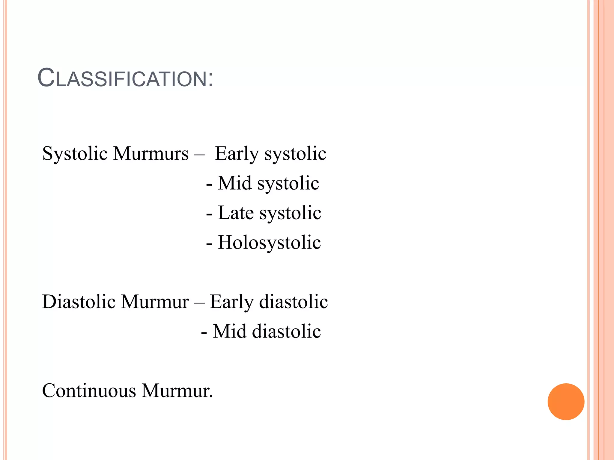
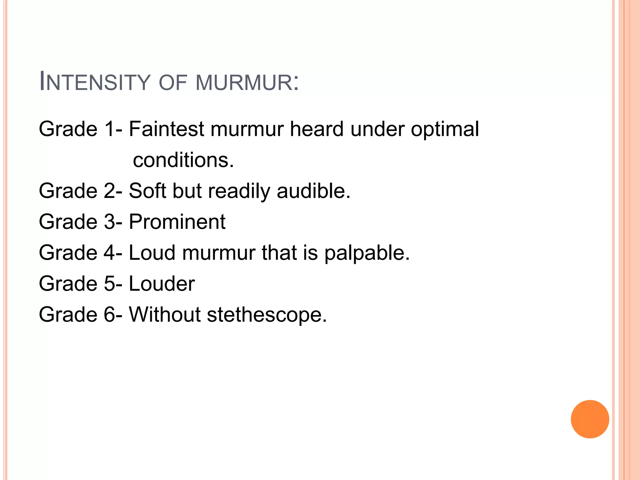
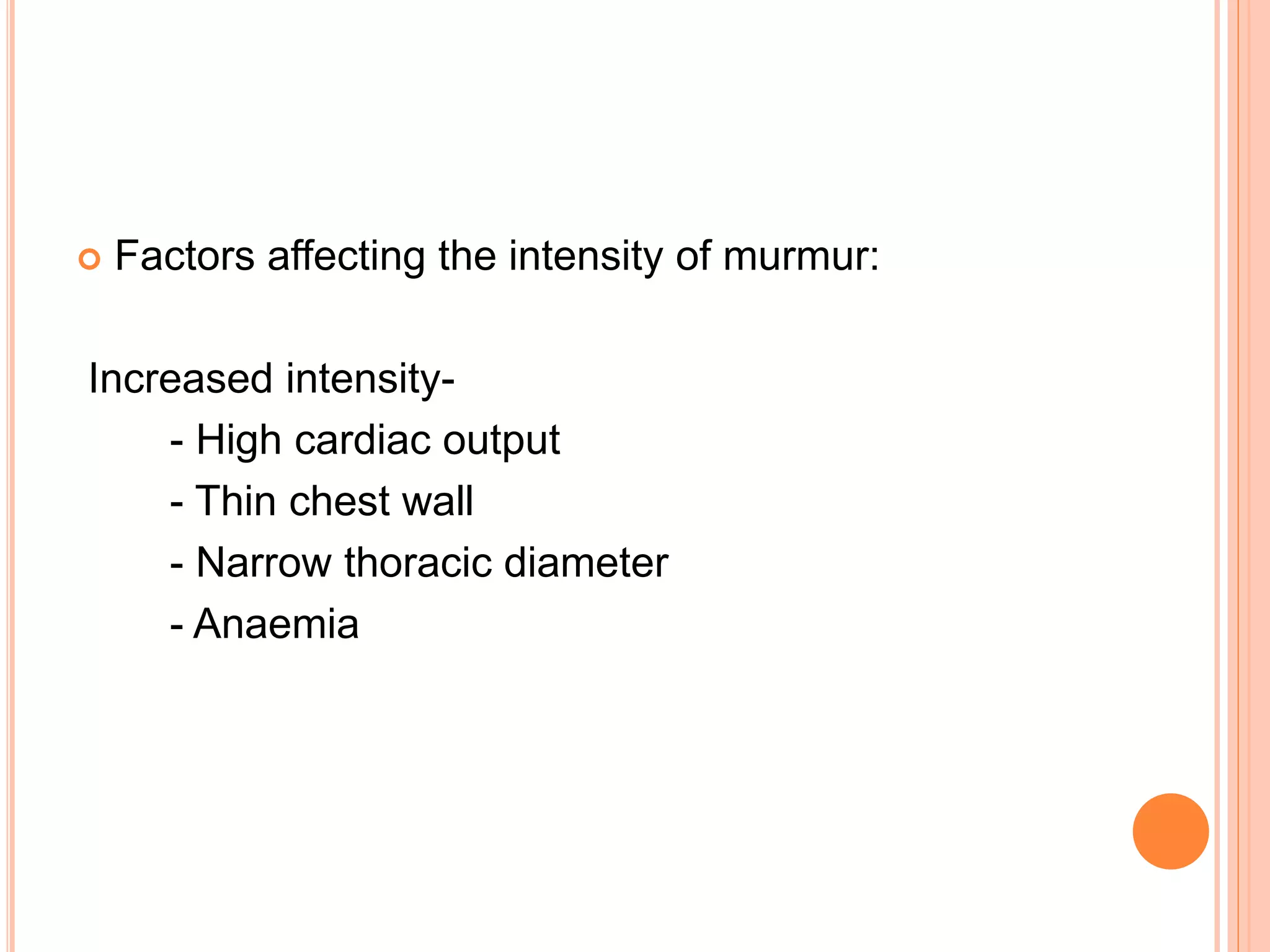
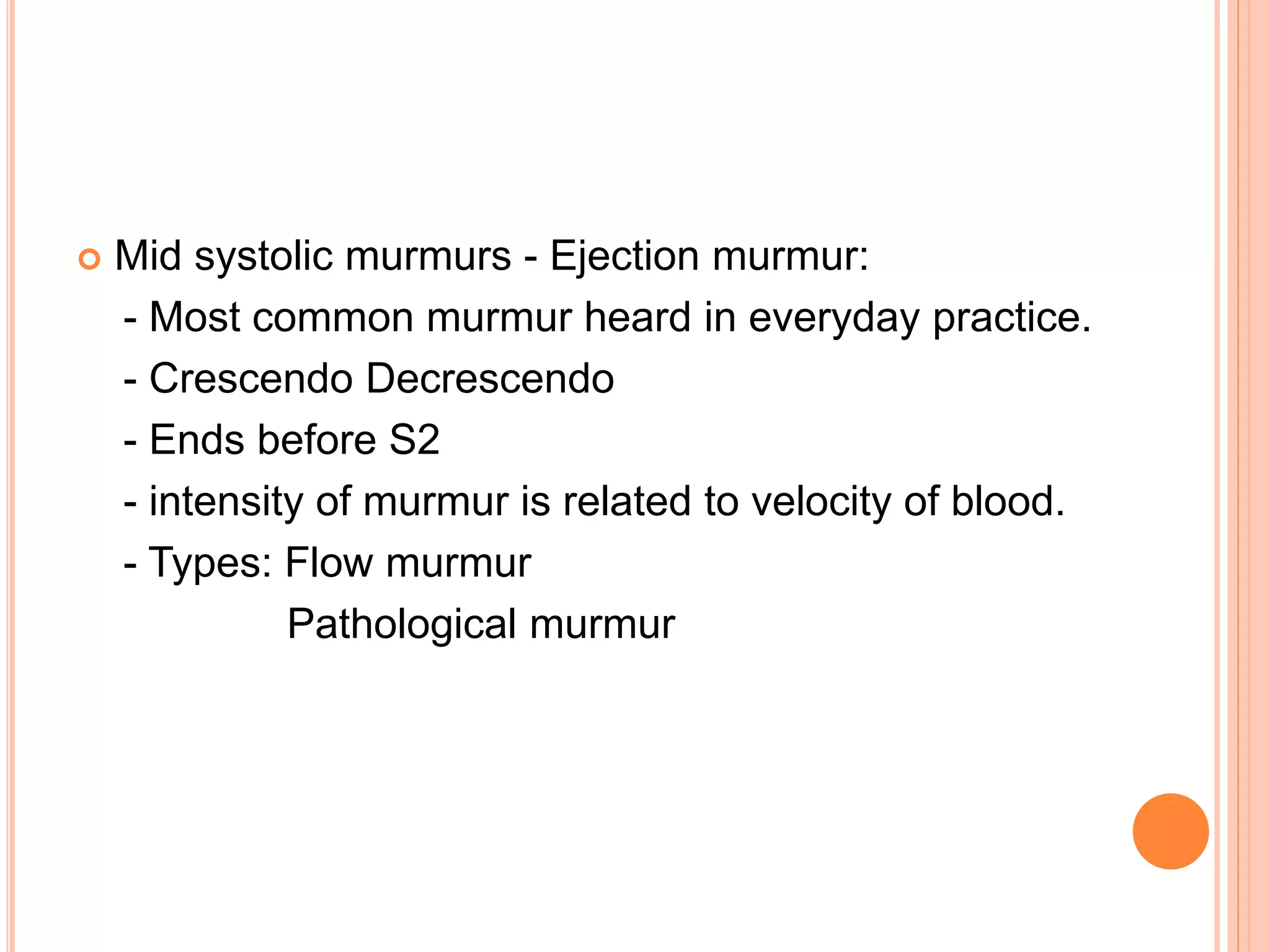
This document describes various types of cardiac murmurs and added heart sounds. It defines different types of murmurs such as systolic, diastolic, and continuous murmurs. It also describes how to characterize murmurs based on timing, location, intensity, and other factors. Specific heart conditions are discussed in relation to the murmurs and sounds they may cause, such as mid-systolic murmurs indicating aortic stenosis or holosystolic murmurs indicating mitral regurgitation. Grading scales for murmur intensity and examples of maneuvers that may modify murmurs are also provided.
In cases of right atrial enlargement the duration of the P wave hardly changes, but the P-R interval increases, so that the P--R segment ratio falls below the normal range.Left atrial enlargement, on the other hand,does not affect the P-R interval, but the P wave lengthens at the expense of the P-R segment.The result is a- ratio above P-R segment the normal maximal limit of 1.6.In combined atrial enlargement, both P-R interval and P wave are prolonged. It follows that in such cases the ratio may P-R segment
be normal.
Patent Ductus Arteroisus, PDA, Cardiology, Paediatrics, Pedicatrics, Critical Care, Emergency medicine, Medicine, Internal Medicine, MBBD, MD, India, CMC Vellore, Christian Medical College
Local Advanced Lung Cancer: Artificial Intelligence, Synergetics, Complex Sys...Oleg Kshivets
Overall life span (LS) was 1671.7±1721.6 days and cumulative 5YS reached 62.4%, 10 years – 50.4%, 20 years – 44.6%. 94 LCP lived more than 5 years without cancer (LS=2958.6±1723.6 days), 22 – more than 10 years (LS=5571±1841.8 days). 67 LCP died because of LC (LS=471.9±344 days). AT significantly improved 5YS (68% vs. 53.7%) (P=0.028 by log-rank test). Cox modeling displayed that 5YS of LCP significantly depended on: N0-N12, T3-4, blood cell circuit, cell ratio factors (ratio between cancer cells-CC and blood cells subpopulations), LC cell dynamics, recalcification time, heparin tolerance, prothrombin index, protein, AT, procedure type (P=0.000-0.031). Neural networks, genetic algorithm selection and bootstrap simulation revealed relationships between 5YS and N0-12 (rank=1), thrombocytes/CC (rank=2), segmented neutrophils/CC (3), eosinophils/CC (4), erythrocytes/CC (5), healthy cells/CC (6), lymphocytes/CC (7), stick neutrophils/CC (8), leucocytes/CC (9), monocytes/CC (10). Correct prediction of 5YS was 100% by neural networks computing (error=0.000; area under ROC curve=1.0).
Adv. biopharm. APPLICATION OF PHARMACOKINETICS : TARGETED DRUG DELIVERY SYSTEMSAkankshaAshtankar
MIP 201T & MPH 202T
ADVANCED BIOPHARMACEUTICS & PHARMACOKINETICS : UNIT 5
APPLICATION OF PHARMACOKINETICS : TARGETED DRUG DELIVERY SYSTEMS By - AKANKSHA ASHTANKAR
NVBDCP.pptx Nation vector borne disease control programSapna Thakur
NVBDCP was launched in 2003-2004 . Vector-Borne Disease: Disease that results from an infection transmitted to humans and other animals by blood-feeding arthropods, such as mosquitoes, ticks, and fleas. Examples of vector-borne diseases include Dengue fever, West Nile Virus, Lyme disease, and malaria.
Recomendações da OMS sobre cuidados maternos e neonatais para uma experiência pós-natal positiva.
Em consonância com os ODS – Objetivos do Desenvolvimento Sustentável e a Estratégia Global para a Saúde das Mulheres, Crianças e Adolescentes, e aplicando uma abordagem baseada nos direitos humanos, os esforços de cuidados pós-natais devem expandir-se para além da cobertura e da simples sobrevivência, de modo a incluir cuidados de qualidade.
Estas diretrizes visam melhorar a qualidade dos cuidados pós-natais essenciais e de rotina prestados às mulheres e aos recém-nascidos, com o objetivo final de melhorar a saúde e o bem-estar materno e neonatal.
Uma “experiência pós-natal positiva” é um resultado importante para todas as mulheres que dão à luz e para os seus recém-nascidos, estabelecendo as bases para a melhoria da saúde e do bem-estar a curto e longo prazo. Uma experiência pós-natal positiva é definida como aquela em que as mulheres, pessoas que gestam, os recém-nascidos, os casais, os pais, os cuidadores e as famílias recebem informação consistente, garantia e apoio de profissionais de saúde motivados; e onde um sistema de saúde flexível e com recursos reconheça as necessidades das mulheres e dos bebês e respeite o seu contexto cultural.
Estas diretrizes consolidadas apresentam algumas recomendações novas e já bem fundamentadas sobre cuidados pós-natais de rotina para mulheres e neonatos que recebem cuidados no pós-parto em unidades de saúde ou na comunidade, independentemente dos recursos disponíveis.
É fornecido um conjunto abrangente de recomendações para cuidados durante o período puerperal, com ênfase nos cuidados essenciais que todas as mulheres e recém-nascidos devem receber, e com a devida atenção à qualidade dos cuidados; isto é, a entrega e a experiência do cuidado recebido. Estas diretrizes atualizam e ampliam as recomendações da OMS de 2014 sobre cuidados pós-natais da mãe e do recém-nascido e complementam as atuais diretrizes da OMS sobre a gestão de complicações pós-natais.
O estabelecimento da amamentação e o manejo das principais intercorrências é contemplada.
Recomendamos muito.
Vamos discutir essas recomendações no nosso curso de pós-graduação em Aleitamento no Instituto Ciclos.
Esta publicação só está disponível em inglês até o momento.
Prof. Marcus Renato de Carvalho
www.agostodourado.com
Title: Sense of Taste
Presenter: Dr. Faiza, Assistant Professor of Physiology
Qualifications:
MBBS (Best Graduate, AIMC Lahore)
FCPS Physiology
ICMT, CHPE, DHPE (STMU)
MPH (GC University, Faisalabad)
MBA (Virtual University of Pakistan)
Learning Objectives:
Describe the structure and function of taste buds.
Describe the relationship between the taste threshold and taste index of common substances.
Explain the chemical basis and signal transduction of taste perception for each type of primary taste sensation.
Recognize different abnormalities of taste perception and their causes.
Key Topics:
Significance of Taste Sensation:
Differentiation between pleasant and harmful food
Influence on behavior
Selection of food based on metabolic needs
Receptors of Taste:
Taste buds on the tongue
Influence of sense of smell, texture of food, and pain stimulation (e.g., by pepper)
Primary and Secondary Taste Sensations:
Primary taste sensations: Sweet, Sour, Salty, Bitter, Umami
Chemical basis and signal transduction mechanisms for each taste
Taste Threshold and Index:
Taste threshold values for Sweet (sucrose), Salty (NaCl), Sour (HCl), and Bitter (Quinine)
Taste index relationship: Inversely proportional to taste threshold
Taste Blindness:
Inability to taste certain substances, particularly thiourea compounds
Example: Phenylthiocarbamide
Structure and Function of Taste Buds:
Composition: Epithelial cells, Sustentacular/Supporting cells, Taste cells, Basal cells
Features: Taste pores, Taste hairs/microvilli, and Taste nerve fibers
Location of Taste Buds:
Found in papillae of the tongue (Fungiform, Circumvallate, Foliate)
Also present on the palate, tonsillar pillars, epiglottis, and proximal esophagus
Mechanism of Taste Stimulation:
Interaction of taste substances with receptors on microvilli
Signal transduction pathways for Umami, Sweet, Bitter, Sour, and Salty tastes
Taste Sensitivity and Adaptation:
Decrease in sensitivity with age
Rapid adaptation of taste sensation
Role of Saliva in Taste:
Dissolution of tastants to reach receptors
Washing away the stimulus
Taste Preferences and Aversions:
Mechanisms behind taste preference and aversion
Influence of receptors and neural pathways
Impact of Sensory Nerve Damage:
Degeneration of taste buds if the sensory nerve fiber is cut
Abnormalities of Taste Detection:
Conditions: Ageusia, Hypogeusia, Dysgeusia (parageusia)
Causes: Nerve damage, neurological disorders, infections, poor oral hygiene, adverse drug effects, deficiencies, aging, tobacco use, altered neurotransmitter levels
Neurotransmitters and Taste Threshold:
Effects of serotonin (5-HT) and norepinephrine (NE) on taste sensitivity
Supertasters:
25% of the population with heightened sensitivity to taste, especially bitterness
Increased number of fungiform papillae
Rasamanikya is a excellent preparation in the field of Rasashastra, it is used in various Kushtha Roga, Shwasa, Vicharchika, Bhagandara, Vatarakta, and Phiranga Roga. In this article Preparation& Comparative analytical profile for both Formulationon i.e Rasamanikya prepared by Kushmanda swarasa & Churnodhaka Shodita Haratala. The study aims to provide insights into the comparative efficacy and analytical aspects of these formulations for enhanced therapeutic outcomes.
8. SYSTOLIC MURMURS:
Early Systolic Murmur:
- Acute severe MR
- Small VSD
- TR with normal PA pressure
9. Mid systolic murmurs - Ejection murmur:
- Most common murmur heard in everyday practice.
- Crescendo Decrescendo
- Ends before S2
- intensity of murmur is related to velocity of blood.
- Types: Flow murmur
Pathological murmur
10. Flow / Functional / Physiological / Innocent murmur:
- Occurs due to abnormally increased blood
flow across structurally normal heart valves.
- common causes are exercise, excitement,
tachycardia, anemia, pregnancy
- better heard at 2-4 ICS along left sternal
border.
- low-moderate intensity
- never extend into late 1/3rd of systole.
11. Pathological ejection murmur:
- Valvular, subvalvular, supravalvular narrowing
of right or left ventricular outflow tract.
- dilatation of aorta and pulmanory artery.
12.
13. Holosystolic murmurs:
- begins with S1 and end with S2
- constant amplitude and shape throughout the
systole.
eg; MR
- TR
- VSD.
14. Late systoilc murmur:
- Begins in later half of systole and extend into S2
- typically reflects milder degree of MR
15.
16. DIASTOLIC MURMURS
Early diastolic Murmur:
- Signify regurgitant blood flow through
incompetant semilunar valves. Eg: AR
- Begins with S2
- Shape and length of the murmur reflects the
pressure gradient betwwen greater vessels and
respective ventricles.
- high frequency sounds.
17. Mid diastolic Murmur:
- AV valve stenosis. Eg: MS
- low pitched murmurs
Increased AV valve flow without valvular stenosis-
- filling murmurs
- eg; Austin flint murmur of AR
ASD, VSD, PDA
MR, TR
18. Pre systolic murmur:
- late diastolic
- heard in mild-moderate MS, TS
- occurs due to augmented AV flow following
atrial contraction.
- extend into S1
- always associated with mid diastolic murmur.
19.
20. CONTINUOUS MURMURS
Results from persistent gradient between high
pressure site and a lower pressure site.
- begins in systole and spill over into early diastole
- peak in mid to late systole
eg; PDA
21.
22. MANEUVERS
Respiration –
Right sided heart murmurs – louder on inspiration.
Exception : Severe PAH with right sided heart failure
Left sided heart murmurs – louder on expiration.
23. Postural changes –
Recumbant – standing : all murmurs of right and
left side decrease in intensity except HC and MVP.
Squatting - all murmurs of right and
left side increase in intensity except HC and MVP.
24. Valsalva maneuver-
all murmurs of right and left side decrease in
intensity during strain phase of valsalva.
Upon release of valsalva murmurs of right side
return to baseline intensity in 2-3 heart beats and that
of left side return to baseline intensity in 5-10 heart
beats.
25. Isometric handgrip-
Causes increased arterial pressure, cardiac
contractility and cardiac output.
Increased – AR, MR, MS, VSD
Unchanged – right sided murmurs, AS, HC
26. Amyl nitrite
causes systemic vasodilatation and reduces systemic
BP resulting in reflux increase in HR and CO.
Increaseas – AS, MS, PS, TS, HC, TR
Decreases – MR, AR, VSD, PDA
27. EJECTION SOUNDS
High frequency transient sounds that occur in early
systole immidietely followng S1.
Mechanism-
The snapping open or doming of a stenotic thickened
malformed semilunar valves.
Sound produced by sudden tensing or reverberation
of proximal aorta or PA
28. - high frequency, sharp, discrete sounds
- equal in intensity to S1
- aortic ejection sounds are better heard at aortic area
- pulmonic ejection sounds are better heard at left
2-3 ICS at sternal border
29. Aortic Conditions associated with ejection sound;
- congenital valvular AS
- Bicuspid aortiv valve
- aortic aneurysm
- aortic root dilatation
- syst HTN
- severe TOF
30. Aortic stenosis – an ejection click is almost always
present in congenital abnormalities of aortic valves.
Eg; bicuspid valve
In aquired AS ejection clicks are less common and
A2 is poorly heard.
31. Pulmonic Conditions associated with ejection
sound;
- PV stenosis
- idiopathic dilatation of PA
- ASD
- chronic pulmanory HTN
- TOF
32. OPENING SNAP
It results from maximal opening excursion of the
mitral valve cusps into left ventricular cavity in the
early diastole after LV pressure falls below that in
the left atrium.
High frequency, distinct, sharp sound
- initiates diastolic rumble
- better heard medial to the apex
33. OS in MS ;
- marker of pliability of valves
- does not represent the severity of MS
- A2 – OS duration is an indicator of severity of lesion
- severity of MS lesion is inversely related to A2-OS
34. Decreaseed intensity of OS seen in ;
- extensive calcifacation
- mild MS
- severe PAH
- CHF
- associated with MR
- AS / AR
- dilated RV
36. AORTIC STENOSIS
S1 – Usually unremarkable
S2 – A2 is soft or absent
P2 is normal
as the severity of lesion increases A2 moves
to P2 causing single S2
Murmur – Systolic ejection murmur
- Crescendo decrescendo
- harsh, rough, grunting
- better heard at right second ICS
37. - radiates along the carotids
- Gallavardian phenomenon; high pitched
systolic murmur heard at the apex in aquired
aortic stenosis
- length and time to the peak intensity of
murmur indicates severity of lesion.
38.
39. HYPERTROPHIC CARDIOMYOPATHY
S1 – normal to accentuated
S2 – as the severity of lesion increases A2
approaches P2 and may cross it causing
reverse splitting.
S3 – not common in HOCM
S4- be wary of making diagnosis of HOCM in any
person who does not have S4
40. Murmur – harsh systolic ejection murmur
- crescendo decrescendo
- well heard lower left sternal border
at 3-4 ICS
- vary in intensity
41. AORTIC REGURGITATION
S1 - Normal
S2 – A2 is softer
- splitting of S2; It is often single bcz of inaudible
A2 or P2 is lost in diastolic murmur.
S3 – common in severe AR.
42. Cardiac murmur - Early diastolic
- Decrescendo
- High pitched
- Blowing quality
- Better heard in left 2nd ICS at sternal edge
- Radiation Left sternal border – Valvular pathology
Right sternal border – Root pathology
- length of the murmur depends on severity of lesion.
43. Associated murmur with AR,
Mid systolic murmur – flow murmur.
Named murmurs in AR,
- Austin flint M
- Cole celil M
- Sea gull M / cooing dove M
44.
45. MITRAL STENOSIS
S1 – lous and snappy
S2 – normal
Opening snap
Murmur –
- Mid diastolic component: Due to rapid ventricular filling
- Pre Systolic component : Due to atrial contraction
- Low pitched
46. - Rough rumbling
- better heard at apex with no radiation
- begins after opening snap
- decrescendo crescendo
- anything that increases the heart rate will
enhance the audibility of diastolic rumble.
47.
48. MITRAL REGURGITATION
S1 – soft
S2 – widely split
S3 – common
S4 – seen in acute MR
Murmur – holosystolic
- constant amplitude
- medium to high frequency
- better heard at LV apex
49. - other configuration of MR;
tapering holosystolic – trivial MR
mid systolic accentuation – severe MR
late systolic crescendo– papillary rupture
- greater the degree of reflux, louder the murmur
- radiation ; ant leaflet – to axilla
post leaflet – base
50. TRICUSPID REGURGITATION
S1 – may be diminished or normal
S2 – P2 loud if ass with pul HTN
- widely split
S3 – audible
S4 – in acute TR
51. Murmur – holosystolic
- constant amplitude
- left 4-5 ICS along sternal border
- very loud
- medium frequency
- may radiate to lower right sternal border,
upper left sternal edge, xiphoid area.
Severe TR will have associated diastolic flow
murmur.
53. VENTRICULAR SEPTAL DEFECT
S1 – normal
S2 – single S2 as A2 is lost in pan systolic murmur,
only P2 heard
Murmur – pansystolic
- lower left sternal border
- mixed frequency
- intensity vary with amount of shunting
associated murmur – mid diastolic flow murmur
54. REFERENCES
Essentials of cardiac physical diagnosis – Jonathan
abrahama
Clinical examination in cardiology-
B.N. Vijay Raghav Rao