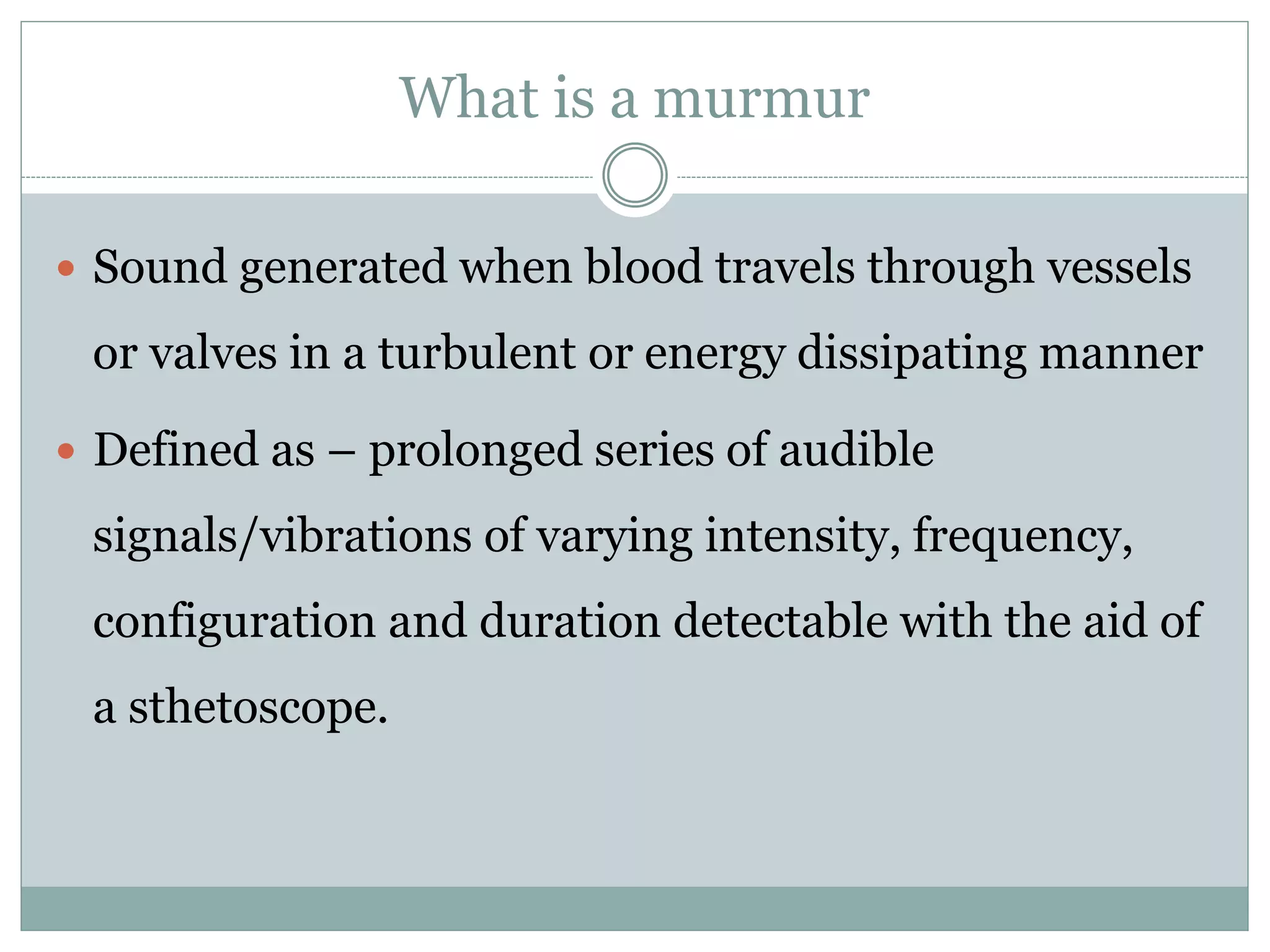
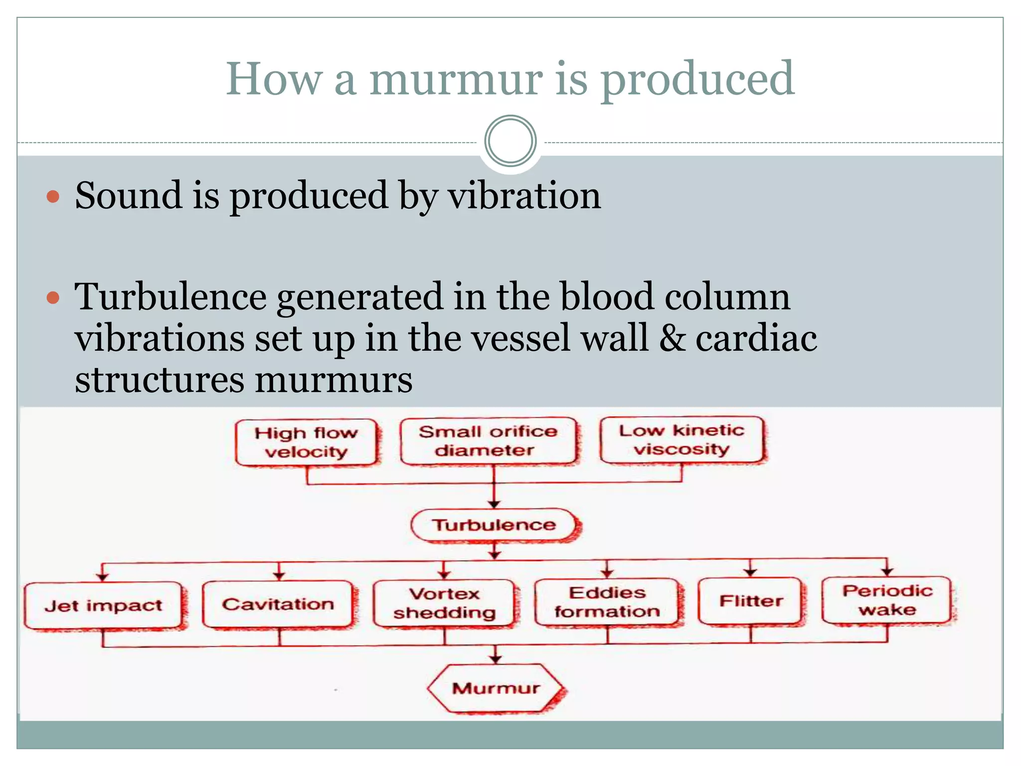
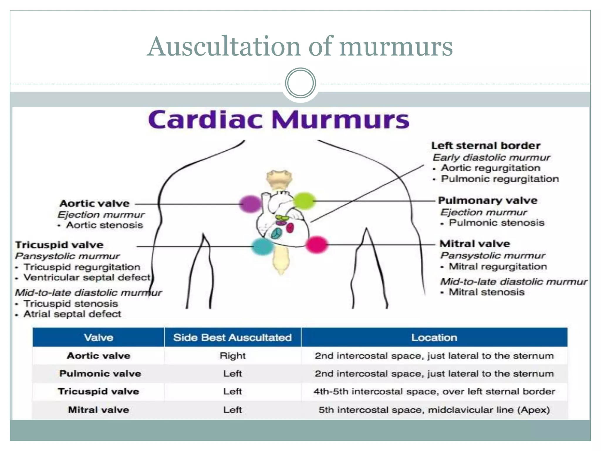
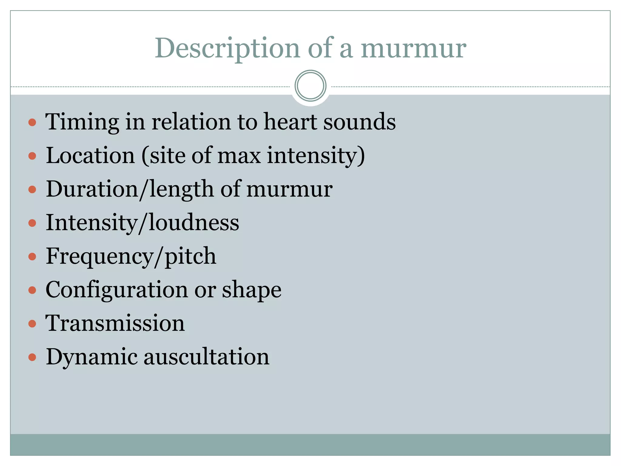
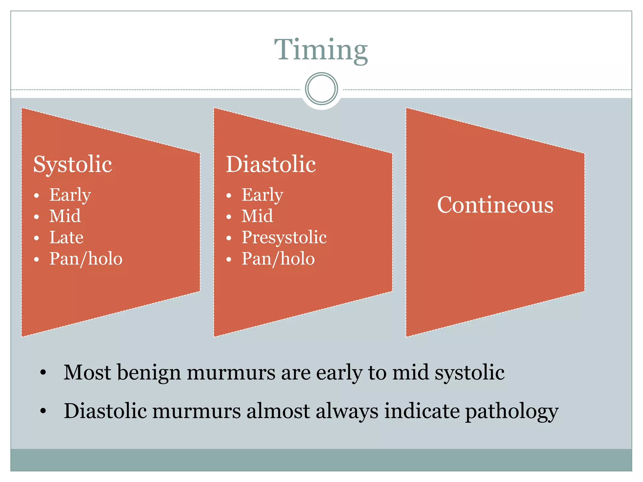
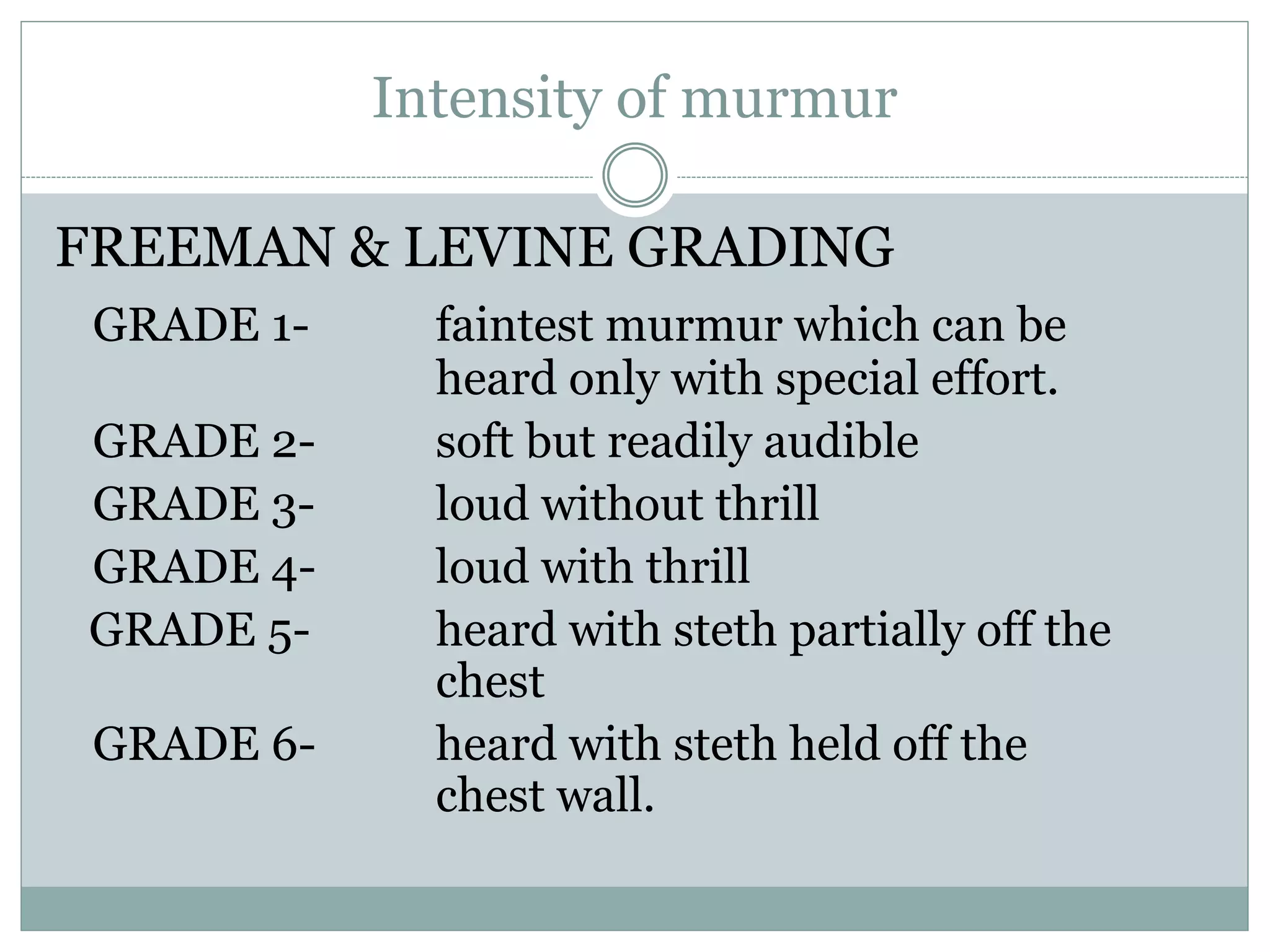
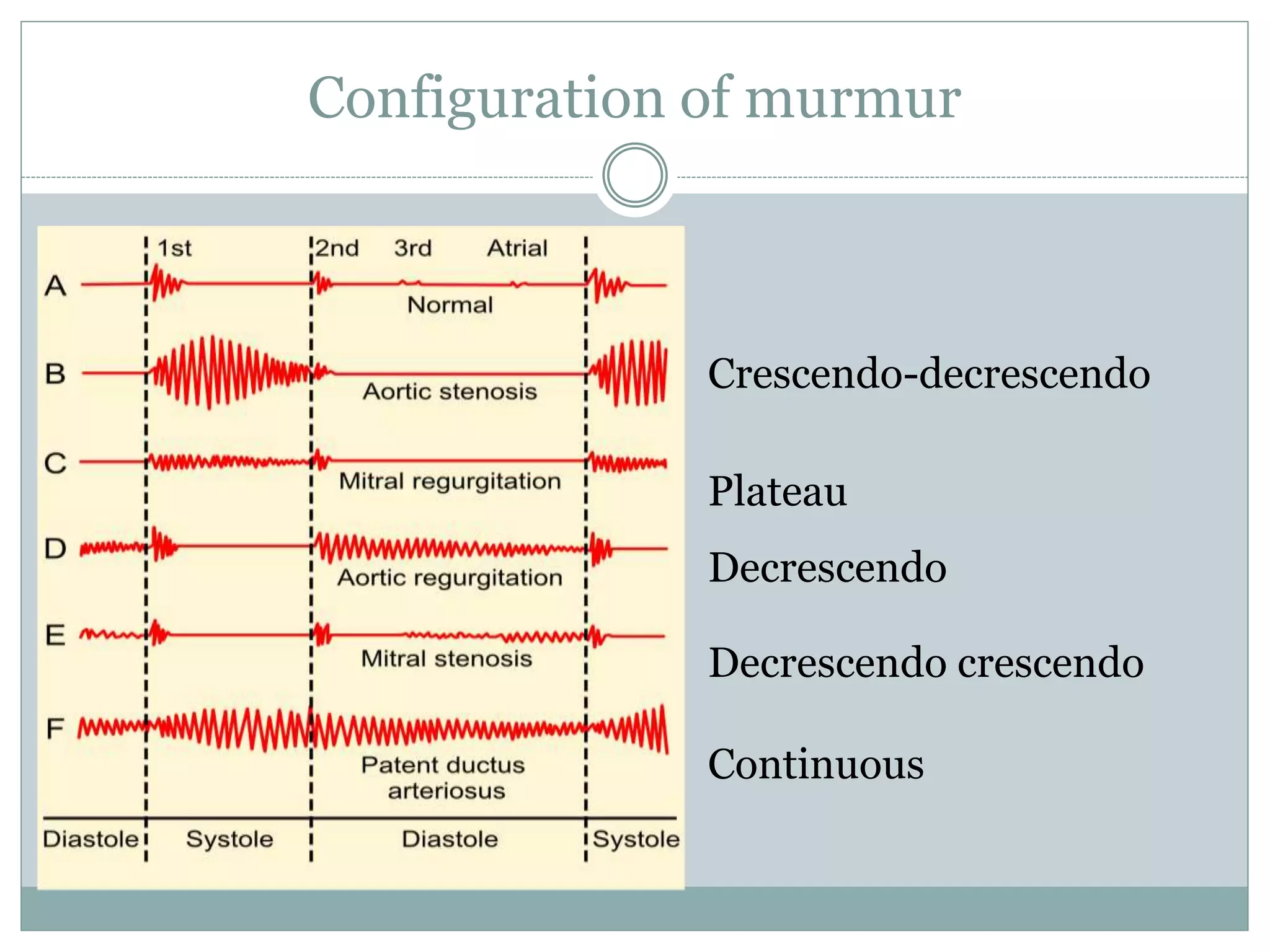
3. What is a murmur
Sound generated when blood travels through vessels
or valves in a turbulent or energy dissipating manner
Defined as – prolonged series of audible
signals/vibrations of varying intensity, frequency,
configuration and duration detectable with the aid of
a sthetoscope.
4. How a murmur is produced
Sound is produced by vibration
Turbulence generated in the blood column
vibrations set up in the vessel wall & cardiac
structures murmurs
6. Description of a murmur
Timing in relation to heart sounds
Location (site of max intensity)
Duration/length of murmur
Intensity/loudness
Frequency/pitch
Configuration or shape
Transmission
Dynamic auscultation
7. Timing
Systolic
• Early
• Mid
• Late
• Pan/holo
Diastolic
• Early
• Mid
• Presystolic
• Pan/holo
Contineous
• Most benign murmurs are early to mid systolic
• Diastolic murmurs almost always indicate pathology
8. Location
Location Murmur best heard Mummur usually not
heard
Apex MS, MR, ESM of calcified
AS
TS, TR
ESM of PS, EDM of PR
Lower sternal area
(tricuspid area)
Mild TR, MDM of TS MDM of MS
Left sternal border 3rd ICS VSD, AR, ESM of
infundibular PS
MDM of MS
Pulmonary area ESM of PS, PDA, EDM of
PR
MDM of MS and TS
Aortic area ESM of AS, EDM of AR MDM of MS
9. Intensity of murmur
FREEMAN & LEVINE GRADING
GRADE 1- faintest murmur which can be
heard only with special effort.
GRADE 2- soft but readily audible
GRADE 3- loud without thrill
GRADE 4- loud with thrill
GRADE 5- heard with steth partially off the
chest
GRADE 6- heard with steth held off the
chest wall.
10. Quality and pitch
Depends on two factors
1. Pressure difference or gradient- Gr pitch
2. Amount of Flow- Flow pitch
PITCH Hz Flow Pr Gr QUALIT
Y
E.g.:
LOW 25-
125
rumbling MDM-
MS
MEDIUM 125-
300
harsh AS
HIGH >300 blowing MR,AR
11. Transmission of murmur
Determined by origin, intensity, direction of blood
flow and physical characteristics of chest
Loud murmur transmit widely and vice versa
High frequency murmur transmited upstream and
low ones downstream
Low frequency transmits better through thoracic
wall therefore felt as thrill
MS localized to apex
MR conducted to axilla and back; LLSB in MVP-MR
AS conducted to Carotids
15. Systolic murmurs
EARLY SYSTOLIC MURMUR
High pitched, decrescendo, begin with S1 and ending
well before S2 at or before mid systole.
Regurgitant murmurs due to flow from high to low
pressure chamber
Acute severe MR
Acute TR with normal RV systolic pressure
Small VSD with PH
16. Acute MR
Best heard at or just medial to apical impulse
Causes
Papillary muscle rupture
Chordae tendineae rupture
Infective endocarditis
Blunt chest wall trauma
17. Acute TR
Soft murmur grade 1 or 2
Best heard at lower left sternal border
Increase in intensity with inspiration- Carvallo’s sign
Regurgutant c-v waves in jvp
No signs of right heart failure
18. Small VSD
Localized to left heart border, grade 4 or 5
Small muscular VSD without PH or Large and
uncorrected VSD with PH
Signs of PH- rt. Ventricle lift, loud and single or
closely split S2
20. Mid systolic murmur
High to medium pitched, crescendo decrescendo
Intensity of murmur closely parallels changes in
cardiac output
21. Aortic stenosis
Most common cause of MDM in adults
Acquired
Congenital
Valvular
Supravalvular
Subvalvular
22. AS
• Harsh, medium pitched, best heard in
2nd rt. ICS and apex
• late peaking and prolonged duration vs
early peaking and reduced duration
• High velocity jet within aortic root
therefore conducts into neck along
carotids
• Pulses parvus et tardus, soft or absent
S2, single S2 or reverse split S2
23. AS
Associated systolic ejection sound, hallmark of congenital
bicuspid AS but no relation with severity
Valvular AS Supra valvular Sub valvular
BP difference nil RUL > LUL Nil
Thrill Max 2 RICS;
Supra sternal &
carotids
Max Right carotid Mid LSB
Ejection sound Present Absent Absent
Murmur
Maximum
2 RICS 1 RICS Mid LSB
Assoc AR +/- rare +/-
24. AS
MR AS [ Gallaverdin]
Apical mid sys/
Holosystolic
Apical mid sys
A2 buried in late sys
vibrations
Clear S2 heard
P/PVC unchanged P/PVC mur =
Amyl nitrate = Amyl nitrate =
Pulse normal Pulsus parvus et
tardus
Gallaverdin phenomenon/ hourglass
phenomenon
Lower n (aortic) vs. Higher n (mitral)
periodic vibrations of stiffened non calcific
aortic valve
Differentiating from MR
26. HOCM
Mid systolic, loudest along left sternal border or
between LSB and apex
Intensity may vary beat to beat and after provocative
maneuvers but usually grade 3
Sustained left ventricular apical impulse and S4
Pulses bisference with double or triple apex
HOCM vs MVP
27. Pulmonary stenosis
Valvular
Congenital or acquired usually with an intact ventricular
septum
Best heard at left 2nd – 3rd ICS, inspitation, conduction
to Lt. supraclavicular area and neck
Begins with PES, expiration. With severity PES fuse
with S1 and S4 appear
Late peaking with duration = severity
a waves, Lt. parasternal heave RV S4
28. Pulmonary stenosis
Murmur brought on by a phasic ejection
click; radiates up & left
As severity increases length increases and
P2 becomes soft (abruptness of closure
reduced), S2 split widens, S4
Loses symmetry becomes kite shaped
May engulf A2 and P2 may be inaudible;
may be confused with VSD
29. Pulmonary stenosis
Infundibular stenosis
Asso with VSD
Best heard-left 3rd ICS
Shorter with inc severity may accompany ejection
sound due to dilated aorta
Shorten and intensity with amyl nitrate
P2 often absent
30.
31. Supra-valvular
May be associated with supra-valvular AS
Less harsh, high pitched with varying intensity
Best heard-upper left sternal border, infraclavicular
region
32. Other causes of MSM
Dilation of Aorta & Pulmonary trunk
Short soft midsystolic murmur
Left sided murmurs in marfan’s syndrome, syphilis
Right sided murmurs in idiopathic dilation of
pulmonary artery, pulmonary hypertension
MSM of Hyperdynamic circulation
Normal aorta or pulmonary trunk but increased flow
Anaemia, pregnancy, fever, thyrotoxicosis
33. OS-ASD
Rapid flow across pulmonary valve to dilated
pulmonary trunk
Pure AR
Due to Accelerated LV ejection
35. Physiological murmurs
• Still’s murmur
• Short buzzing murmur
‘twanging of a rubber band’
• Pure medium frequency by
periodic vibrations of pulmonic
leaflets at their attachment
36. Pulmonary mid systolic murmur &
Peripheral pulmonary systolic
murmur
• Angulation and disparity between
pulmonary trunk and its branches
turbulent flow
• Normally disappears with maturity
of pulmonary bed
37. Supraclavicular or
brachiocephalic systolic
murmur
• Aortic origins of major
normal brachiocephalic
arteries
• Crescendo-decrescendo,
abrupt onset, short,
sometimes radiating below
clavicle
• vs. supra valvular AS – these
murmur are softer below
clavicle and decreases with
shoulder abduction
38. Mammary Soufflé
• Late Pregnancy or puerperium
• Sometimes continuous louder in
systole, distinct gap from S1 [ time
for ejected blood to reach
mammary arteries]
• 2 or 3 RICS/ LICS
• Light Pressure augments murmur
becomes continuous; firm Pr
abolishes murmur
39. Late systolic murmurs
MVP
• Leaflets remains competent during early
ventricular contraction but overshoot in late
systole [critical V. dimensions]
• One or more mid systolic clicks precede murmur
[sudden deceleration of the column of blood
against the prolapsed leaflet or scallops]
• Longer and softer
• Prompt standing after squatting
• Valsalva II
• Short & louder
• squatting
• Sustained hand grip
• Amyl nitrate
Barlow’s syndrome refers to the
spectrum of symptoms caused by
MVP [click or murmur alone to
palpitations, chest pain, or syncope]
40. Pan/holo-systolic murmurs
Begins with S1 occupy whole systole and ends with
S2
High pitched, blowing and plateau like configuration
Regurgitant murmurs, retrograde flow from high
pressure to low pressure chamber
Causes
Chronic MR
Chronic TR
Restrictive VSD
41. Chronic MR
• S1 to S2 provided MV remains
incompetent and gradient remains
• Grade 3/6 intensity, no thrill
no/little variation
• Radiates to axilla and back becauses
jet directed posterolaterally in LA
• LLSB when jet directed against atrial
septum near base of aorta
42. Tricuspid regurgitation
• Holosystolic secondary to PH or PS
• Best heard at LLSB with no selective radiation
• May be heard at RLSB and apex when RV forms
apex
• Prominent v waves with rapid y descent in JVP
augments in expiration
• Left parasternal heave and RV S3
• Diff from organic TR
• PSM vs. ESM
• High n vs. Medium n
• Features of PAH present
• Rivero Carvallo’s sign-
Increased intensity with
inspiration
• Carvallo’s sign disappears in
RV failure and organic TR
43. VSD
• Depends on site, size and gradient
• Best heard LSB 3rd-5th ICS during expiration
• Very restrictive VSD- ESM decrescendo
pattern
• Mod and NR VSD- PSM
• Sub arterial VSD- 1 or 2 LICS similar to PS
murmur
• Septal aneurysms- click with LSM or PSM
with late Sys Accentuation
• Large shunt – MDM
• NR VSD with PAH- ESM
• PSM absent in Eissenmenger Syndrome
44. Other PSM
Aorto Pulmonary Window with PAH
Otherwise continuous murmur
Diastolic component reduced with increasing PAH
PDA with PAH
Similar mechanism
45. Differentials of holosystolic murmrur
Features MR TR VSD
Best heard Apex LLSB LSB 3rd-4th ICS
Selective
transmission
Axilla and back No selective
transmission
No selective
transmission
Thrill Rare(cordal
rupture)
Does not occur Common
Character High v, soft and
blowing
High v, soft and
blowing
Med v, rough and
harsh
Respirartion No change inspiration expiration
Accompanying
features
Eccentric LVH.
S1, LV S3
RVH, prominent v
waves, signs of PH
Biventricular
enlargement, may
have signs of PH
46.
47. Summary systolic murmurs
ESM Organic
MSM
Functional MSM Pan
systolic
Late
systolic
Acute severe
MR
AS Severe AR MR MVP
Acute TR
normal RVSP
HOCM PH TR
Small VSD PS ASD VSD
Non-restrictive
VSD with PH
MR (papillary
muscle
dysfunction)
VSD
Still’s innocent murmur
AP
window
and PDA
with PH
Idiopathic PA dilatation
Hyperkinetic circulation
50. Early diastolic
AR
A/C AR C/C AR
Short mur. -
early
equalization of
diastolic
pressures
Long mur.
Medium n –
velocity less
rapid and
pressure
gradient lower
High n
Associated S4
51. Decrescendo, soft high pitched and blowing-best
heard in sitting and leaning forward position durin
held deep expiration along LSB in 3rd-4th ICS
Valvular AR, A2 diminished or absent d/t inadequate
coaptation of cusps
Severe valvular AR with SEM in aortic area and
Austin Flint at apex
52. High pressure PR
• High pitched soft blowing decrescendo
murmur usually lasts throughout
diastole heard in the left upper sternal
border
• Associated with loud P2 and other
features of PAH
• PR vs. AR
• Loud P2, murmur begins after P2
• Normal pulse pressure
• Clinical setting
• Squatting and sustained hand grip
increases AR
53. High pressure vs normal pressure PR
High
Pressure
Normal
pressure
Decrescendo Crescendo
decrescendo
High frequency Medium to low
pitched
Onset
immediately
with p2
Delayed in onset
Usu extends
throughout
diastole
Short duration
Features of
PAH present
Usually absent
55. Causes of MDM
LV inflow
obstruction
RV inflow
obstruction
Mitral
diastolic flow
Tricuspid
diastolic
flow
MV opening
interference
PR with
no PH
MS TS Severe MR Severe TR Severe AR
(Austin Flint)
Organic
PR
Left atrial
myxoma
Right atrial
myxoma
VSD ASD Ac.rheumatic
carditis
Cor triatrium Carcinoid
synd
PDA RSOV into
Rt. Atrium
AV groove
constriction
Ebstein’s
anomaly
AP window PAPVC
RSOV into rt.
Ventricle
Coronary A
to RA
communicat
ion
Hyperkinetic
circulation
56. Mitral stenosis
• Low n rough rumbling [sound of
distant thunder] MDM
• Localized to apex, better heard in
left lateral position with bell
• Length a severity
• Long murmurs up to S1 even in
long cycles of AF- severe MS
• Late diastolic or Pre systolic
accentuation usually seen in
pliable valves and in NSR [
sometimes in AF]
57. TS
Similar to MS
Murmur usually seen associated with AF
Diff. from MS
Increases during inspiration [Augmentation of RV volume, RV
Diastolic Pr., Flow rate and gradient across valve]
LLSB
58. PR with normal PA pressures
PR assoc with Cong PS, PV IE, repair of RVOT
Negligible gradient at the start of diastole, gradient
increases especially during the IVR phase of RV when
murmur reaches maximum intensity.
•2 & 3 LICS
•Medium to low pitched
•Delayed in onset
•Short duration
•Ending before S1
59. Austin Flint Murmur
Severe AR regurgitant jet directed toward the AML
prevent the latter from opening well during diastole
generating turbulent flow
Low n MDM or late diastolic, best heard at the apex.
To differentiate from MS
No OS
Amyl nitrate inhalation AR, Austin flint murmur
60. Other Mid Diastolic Murmur
Carey Coomb’s murmurs
Acute rheumatic fever, mitral valve structures acutely inflamed with
some thickening and edema turbulence of flow during the rapid
filling phase.
+ moderate MR [increased mitral inflow in diastole]
Low pitched short MDM.
good evidence of active carditis
61. Flow Murmurs
Increased AV flow
TR, ASD, MR, VSD, PDA, hyperdynamic circulation
To differentiate from MS & TS
Short MDM
Medium Pitch- increased flow
Preceded by S3
Absence of Opening Snap
Thrill less common
62. Late Diastolic/ Pre-systolic Murmurs
MS
Higher frequency than MDM
Sometimes only PSA heard- mild MS
Generally absent in calcified valves and most of AF [
may be present during short cycle lengths in AF]
Cause-
Increased flow during atrial contraction in late systole
Increased dp/dt of LV contraction increases turbulence [ esp.
in AF short cycles]
63. Other diastolic murmurs
Cabot– Locke Murmur- [Diastolic Flow murmur]
The Cabot–Locke murmur is a diastolic murmur that sounds similar to
aortic insufficiency but does not have a decrescendo; it is heard best at the
left sternal border. [High flow thru coronary vessels, LMCA, LAD]
The murmur resolves with treatment of anaemia.
Dock’s murmur
diastolic crescendo-decrescendo, with late accentuation, [consistent with
blood flow through the coronary] in a sharply localized area, 4 cm left of the
sternum in the 3LICS, detectable only when the patient was sitting upright.
Due to stenosis of LAD
64. Key–Hodgkin murmur
EDM of AR; it has a raspy quality, [sound of a saw cutting through wood]. Hodgkin
correlated the murmur with retroversion of the aortic valve leaflets in syphilitic disease.
Rytand’s murmur in complete heart block
MDM or Late diastolic murmur
Atrial contraction coincides with the phase of rapid diastolic filling increased flow short
MDM [intermittent].
Another theory- Delayed V. contraction following A. contraction may lead to diastolic MR &
TR, because AV valve closure does not occur [unless V. systole supervenes]. When higher V
than A pressure during atrial relaxation, an incompletely closed AV valve may lead to a
reverse gradient with a considerable regurgitation volume.
65. Continuous murmur
Begin in systolic and continues without interruption
through the timing of S2 into all or part of diastole
Flow from zone of higher resistance into lower
resistance without phasic interruption b/w systole &
diastole
1. Connection b/w high pressure chamber/vessel & low
pressure chamber/vessel
2. Disturbance in flow patterns in arteries
3. Disturbances in flow patterns in veins
66. Connection b/w high pressure chamber/vessel
& low pressure chamber/vessel
1. From the aorta
a) Persistent ductus arteriosus
b) Aorto-pulmonary window
c) RSOV
2. From the coronary artery:
a) Coronary arteriovenous fistulae draining into RA, RV, PA
b) ALCAPA
3. Other arteriovenous communications
a) Broncho-pulmonary collaterals
b) Chest wall arteries–pulmonary vessels
c) Peripheral A-V Fistula
4. Others
a) Lutembacher syndrome with restricted ASD
67. PDA
• Gibson’s murmur
• At 1 or 2 LICS
• NR- high frequency soft murmur peaks
around S2
• Mod R- loud coarse machinery murmur
with eddy sounds
PDA with no continuous murmur
• Neonates- due to high PVR
• Very small ductus
• Very large ductus & large VSD- due to
equalization of pulm and sys Pr
• PAH- first dia component goes, then
sys
• AS, CoA- due to low aortic pressure
severity
S
E
V
E
R
I
T
Y
68. Continuous murmurs
APW
2 or 3 LICS
Usually associated with early devp of eissenmenger
RSOV
No peaking at S2 seen [peaks in sys or dia.]
To RA- RLSB
RV- LLSB
RVOT- 3 LICS
Lutembacher syndrome with restricted ASD
LLSB [body of RA]
69. Continuous murmurs
C-AVF
RA- RLSB or RUSB
CS- back b/w spine & Lt scapula
RV inflow- LLSB
RVOT- Upper to Mid LSB [beat to beat change in murmur may be present,
RV systolic compression, valsalva softens murmur]
PA- ULSB [no eddy sounds]
ALCAPA
Murmur louder in diastole [LV contr. I/C flow]
Do not peak at S2
Usu LUSB or RUSB
o LA- ULSB rad to Lt ant ax line
o Lt SVC- upper to mid LSB
70. Dynamic Auscultation
It refers to the technique of altering circulatory
dynamics by a variety of physiological and
pharmacological maneuvers and determining the
effects of these maneuvers on heart sounds and
murmurs .
72. Position
A. Lt Lateral Decubitus
LV impulse [apical sounds, murmurs better heard]
Act of turning increases HR[ MDM & PSA of MS ],
induces PVC [AS murmur vs. MR murmur (n/c)]
B. Sitting leaning forward full held expiration
AR & PR EDM
C. Sitting with legs dangling
Further reduces venous return
If S2 fails to fuse on sitting
D. Elbow Knee Position
Pericardial friction rub
73. Position
E. Standing to squatting and vice versa
Standing[ venous return, BP ]; [opp. in squatting]
1. All murmurs [ except HOCM , MVP earlier]
HOCM [ LV contractility, after load, preload]
MVP [ preload, afterload ]
2. A2- P2 , A2-OS , A2-S3 (n/c)
F. Hyperextension of shoulders
supraclavicular Systolic murmurs
G. Stretching of Neck
Venous hum
H. Passive elevation of both legs
Transiently increases venous return, increase S3
74. Physical Maneuvers
Inspiration
Right sided events become
more prominent
S2 split appreciable
RVs3 RVs4 prominent
Tricuspid sys & dia Mur
increased
Pulm ejection sound
reduced
Expiration
• Left sided events become
more prominent
• Diff AR & PR
• Pericardial friction rub
[exhalation & supine]
• Innocent pulm mid sys
murmur becomes more
prominent becos of
reduced AP diameter
75. Valsalva Maneuver
Inspiration followed by forced exhalation against a
closed glottis for 10 to 20 seconds
Physician has to keep flat of the hand on the abdomen
to provide the patient a force to breathe against
Not attempted in patients with IHD
Normal response has four phases
76. Valsalva Maneuver
•initial pulm VR = SV
• I/T Pr directly
transmitted to aorta.
I/T Pr = VR =
BP
sympathetic tone
HR
Sudden I/T Pr = BP
sudden return of peripherally pooled
blood to the vaso-constricted arterial
system (20 to the increased
sympathetic tone)
77. Phase II – Decrease in systemic venous return ,
systolic pressure and pulse pressure
S3 & S4 attenuated
A2-P2 interval narrows
All murmurs except MVP / HOCM decrease
Phase III- increased Left murmurs & Phase IV-
increased Right murmurs
78. Valsalva Maneuver
I/T Pr = VR = BP
sympathetic tone HR
sudden return of peripherally pooled blood
to the vaso-constricted arterial
system (20 to the increased sympathetic
tone)
PHASE
II
PHASE IV
MAXIMAL SYMPATHETIC
ACTIVATION
FLAT PART OF
STARLING’S CURVE
HEART
FAILURE
ASD
MS
79. Isometric Exercise
Calibrated Handgrip device or a handball.
Better to carryout bilaterally, sustained for 20-30 secs
Not to be done in arrhythmia / Ischemia
Transient but significant increase in SVR, BP, HR , CO , LV
filling pressure , Heart size
1. LVS3 & LVS4 increases
2. Systolic Murmur of AS reduced – reduced gradient across
aortic valve
3. AR , MR , VSD – increased
4. MDM of MS – increased
5. Syst Murmur of HOCM reduced
6. MVP murmur + click delayed
80. Isotonic Exercise
Few minutes of brisk walking sufficient
Must be auscultated quickly before effect wears off
Increases Ms murmur in low output states
Wide Split of S2 in RVF further widens after exercise
81. Pharmacological Maneuvers
Inhalation of Amyl Nitrate [Crush ampoule in towel, 3-4 deep
breaths over 10–15 s]
Lasts 2 minutes
No reduction in stroke volume as seen in NTG
First 30 secs 30 to 60 secs > 60 secs
Decreased Sys Art
Pressure
Reflex Tachycardia Increased CO, HR
82. Amyl Nitrate inhalation
AS vs. MR
TR vs. MR
PS vs. TOF
MS vs. Austin F
PR vs. AR
HOCM vs. MVP [n/c]
83. Long cycle length
Long cycle length after PVC or in long cycles of AF
Increases murmur of AS, HOCM, PS
Murmurs of MR, TR has no change