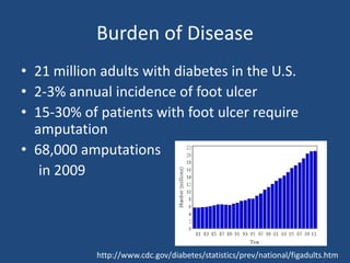
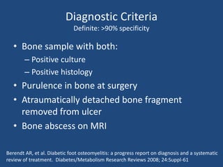
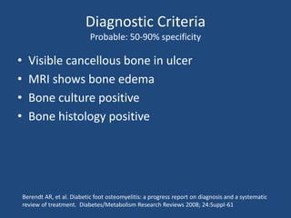
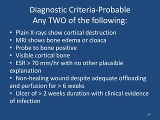
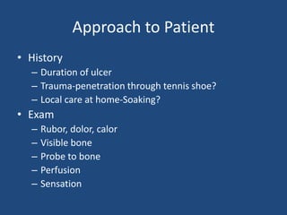
Diabetic Foot Osteomyelitis
Objectives 
• Recognize patients at risk for diabetic foot 
infections 
• Design a diagnostic work-up for diabetic foot 
osteomyelitis 
• State the principles of management of 
diabetic foot infections
Challenging Infections of Skin, Soft 
Tissue and Bone 
• Cellulitis-management in the community 
MRSA era 
• Furuncles, carbuncles, skin abscesses 
• Recurrent skin abscesses 
• Diabetic foot osteomyelitis
Disclosure 
• Very few antibiotics have FDA approval for 
osteomyelitis 
• Newer antibiotics often gain approval for skin and 
soft tissue infections 
– Amoxicillin-clavulanate 
– Daptomycin 
– Linezolid 
• Older antibiotics often lack an FDA indication for 
skin and soft tissue infection 
– Nafcillin 
– TMP-SMX
A previously healthy 40 y/o woman 
presents with uncomplicated lower 
extremity cellulitis. Which of the following 
options is the best treatment? 
A. Co-trimoxazole 
B. Amoxicillin-clavulanate 
C. Linezolid 
D. Doxycycline 
E. A and B
Skin and Soft Tissue Infections 
Purulent 
• Furuncle 
• Carbuncle 
• Abscess 
Nonpurulent 
• Cellulitis 
• Erysipelas 
• Necrotizing 
fasciitis 
DL Stevens et al; www.idsociety.org
Purulent SSTIs 
• Furuncles-purulent infection of a hair follicle 
extending through the dermis into the 
subcutaneous tissue 
• Carbuncles-coalescent infection of multiple 
hair follicles, usually larger and deeper than 
furuncles 
• Abscesses
Purulent SSTIs 
Furuncles, Carbuncles, Abscesses 
• Skin flora-may be polymicrobial 
• Staph aureus, recently USA 300 MRSA
Purulent SSTIs 
Furuncles, Carbuncles, Abscesses 
• Incision and drainage is the cornerstone of 
management, and is more effective 
ultrasound guided needle aspiration in an RCT 
• Packing may not be necessary for small, 
uncomplicated abscesses 
GF O’Malley Acad Emerg Med 16:470, 2009 
RJ Gaspari; Ann Emerg Med, 57:483, 2011
Packing versus simple I & D 
• Abscess < 5 cm on trunk or extremities 
• > 18 y/o
Open I & D versus Needle Aspiration 
• Primary endpoint = treatment failure 
– Day 0 unable to fully aspirate pus 
– Day 2 residual abscess by US, increased symptoms 
– Day 7-antibiotics and/or continued symptoms or 
additional procedure 
• Randomized by numbered packets-some 
packets disappeared! 
• TMP-SMX DS 2 tabs bid recommended 
RJ Gaspari; Ann Emerg Med, 57:483, 2011
Open I & D versus Needle Aspiration 
101 patients enrolled 
54 I & D 47 needle aspiration 
43/54 (80%) success 12/47 (26%) success 
RJ Gaspari; Ann Emerg Med, 57:483, 2011
Microbiology 
Culture Result 
Incision and 
Drainage (%) 
Ultrasound 
Guided Needle 
Aspiration (%) 
Total (%) 
MRSA 18 (35) 15 (38) 33 (36) 
MSSA 20 (39) 11 (28) 31 (34) 
Group A strep 1 (2) 1 (3) 2 (2) 
Other 3 (6) 6 (15) 9 (10) 
No culture 
4 (8) 2 (5) 6 (7) 
growth 
Mixed growth 5 (10) 5 (13) 7 (11)
Open I & D versus Needle Aspiration 
33 patients with MRSA enrolled 
54 I & D 13 needle aspiration 
11/18 (61%) success 1/13 (8%) success 
RJ Gaspari; Ann Emerg Med, 57:483, 2011
Packing versus simple I & D 
48 patients enrolled 
23 I & D + packing 25 simple I & D 
4/23 repeat procedure 
at 48 hours 
GF O’Malley Acad Emerg Med 16:470, 2009 
5/25 repeat procedure 
at 48 hours
Purulent SSTIs 
Furuncles, Carbuncles, Abscesses 
• Antibiotics-necessary only in selected cases 
– systemic signs (fever, tachycardia, tachypnea, 
leukocytosis) 
– Immunocompromised 
– Extremes of age 
– Recurrent infections 
– Failure after adequate I & D
TMP-SMX vs. placebo after I & D 
• N=220 
• Age > 16 y/o 
• Intervention TMP-SMX DS 2 tabs BID 
• Primary outcome: treatment failure at day 7 
• Secondary outcome: relapse within 30 days 
GR Schmitz; Ann Emerg Med, 56:283, 2010
Exclusions 
• Immunocompromised 
– Diabetes, HIV, malignancy 
• Fever or signs of systemic illness 
• Pregnant or breast-feeding 
• Sulfa allergy 
• Antibiotics in previous week 
• Hospitalized in previous month
Bacteriology 
Placebo (%) TMP-SMX (%) 
MRSA 47 60 
MSSA 22 15 
Coag neg staph 10 10 
Viridans strep 5 1 
No growth 8 2 
other 8 12
Antibiotics After I & D 
30% 
25% 
20% 
15% 
10% 
5% 
0% 
Failure Recurrence 
TMP-SMX 
Placebo 
P=0.12
Management of Treatment Failures 
Placebo N=27 TMP-SMX N=15 
Admit-iv antibiotics 5 2 
IV antibiotics in ED 3 1 
Repeat I & D 3 2 
Sent home w antibiotic 9 5 
I & D plus home with 
7 5 
antibiotics
RCT-Antibiotics vs. Placebo After I & D 
• N = 161 
• 3 months to 18 y/o 
• Excluded patients with fever 
• TMP-SMX 10-12 mg/kg/day TMP component 
Duong; Ann Emerg Med, 55:410, 2010
Bacteriology 
Culture Results Placebo (%) 
Trimethoprim- 
Sulfamethoxazole 
(%) 
Total (%) 
CA-MRSA 61 (81) 58 (79) 129 (80) 
MSSA 6 (8) 7 (10) 14 (9) 
Proteus mirabilis 4 (5) 2 (3) 6 (4) 
GAS 1 (1) 1 (1) 2 (1) 
Other 1 (1) 3 (4) 4 (3) 
No culture/growth 3 (4) 2 (3) 6 (3) 
Duong; Ann Emerg Med, 55:410, 2010
RCT-Antibiotics vs. Placebo 
Non-inferiority Trial, margin 7% 
35 
30 
25 
20 
15 
10 
5 
0 
Failure Relapse-10 days Relapse-90 days 
TMP-SMX 
Placebo 
95% CI ∞-6.7% 
Duong; Ann Emerg Med, 55:410, 2010
Recurrent Skin Abscesses 
• Evaluate site for local cause 
– Inadequate drainage 
– Hidradenitis 
– Pilonidal cyst 
– Foreign body 
• R/O neutrophil disorder if onset in early 
childhood 
• Consider decolonization therapy
Decolonization Therapy 
• Nasal mupirocin BID x 5 days 
• Daily bathing 
– Chlorhexidine 
– Dilute bleach 1/4 to 1/2 cup bleach in tub 
• Daily disinfection of clothing, towels, sheets, 
combs, razors,environmental surfaces 
• Consider treating family simultaneously
Purulent SSTIs 
Wrap Up 
• Incision and drainage 
• Packing not be essential for small abscesses 
• Antibiotics not necessary for most cases 
• Recurrences-look for local cause, consider 
decolonization strategies 
GF O’Malley Acad Emerg Med 16:470, 2009 
RJ Gaspari; Ann Emerg Med, 57:483, 2011
Figures 1A and 1B: Notice the integrity of the skin, the ill-described border of the 
lesion as well as the extension of erythema up medial aspect of the leg (figures 
courtesy of DermNetNZ.org).
Cellulitis 
• Low burden of organisms 
• Brisk inflammatory response 
• Culture yield is low-not recommended for 
routine practice 
• Punch biopsy cultures and serology confirm 
– Group A strep 
– Groups B, G, C, F strep
Cellulitis 
• Select agent with good activity against 
streptococci 
• If patient is improving at day 5, antimicrobial 
therapy may be discontinued 
• Coverage for S. aureus, including MRSA, is 
recommended if there is 
– Penetrating trauma 
– Purulent drainage 
– Concurrent MRSA infection elsewhere
Cellulitis 
Agents with good strep coverage 
• Penicillin G 2-4 million units Q 4-6 h 
• Clindamycin 600-900 mg Q 8 h 
• Nafcillin 1-2 grams Q 4-6 h 
• Cefazolin 1 gram Q 8 h 
• Pen VK 250-500 mg po Q 6 h 
• Cephalexin 500 mg po Q 6 h 
• Doxycycline 
• TMP-SMX
Duration of Therapy 
Uncomplicated Cellulitis 
121 subjects enrolled 
87 eligible for randomization 
at day 5 
43 received 5 more days of therapy 44 received 5 days of placebo 
42/43 resolved at day 14 and 
relapse free at day 28 
43/44 resolved at day 14 and relapse 
free at day 28 
MJ Hepburn; Arch Intern Med, 164:1669, 2004
Recurrent cellulitis 
• Annual recurrence rate 8%-20% 
• Risk factors 
– Edema 
– Obesity 
– Eczema 
– Venous insufficiency 
– Tobacco use 
– Malignancy 
– Homelessness 
– Toe web abnormalities, e.g. tinea pedis
Recurrent Cellulitis 
Antimicrobial Prophylaxis 
• 40 patients 
• > 2 episodes in previous 3 years 
• Venous insufficiency or lymphatic congestion 
• Daily pen VK 
• Decreased recurrences P< 0.06 
AC Sjoblom; Infection 21:390, 1993
Recurrent Cellulitis 
Antimicrobial Prophylaxis 
36 subjects enrolled 
18 received erythromycin 250 mg 
bid 
18 received placebo 
zero relapses over 18 months 9 relapses 
M Kremer; J. Infection, 22:37-40, 1991
Cellulitis 
Wrap-Up 
• Streptococci are the predominant pathogen 
• No need to cover MRSA unless 
– Penetrating trauma 
– Purulent drainage 
– Concurrent MRSA infection elsewhere 
• 5 days therapy is sufficient if symptoms improving 
on day 5 
• Recurrence common 
– Address risk factors 
– Consider penicillin prophylaxis if recurrences are 
frequent
Image 4. Diabetic Foot Ulcer (image courtesy antimicrobe.org)
Burden of Disease 
• 21 million adults with diabetes in the U.S. 
• 2-3% annual incidence of foot ulcer 
• 15-30% of patients with foot ulcer require 
amputation 
• 68,000 amputations 
in 2009 
http://www.cdc.gov/diabetes/statistics/prev/national/figadults.htm
Amputations per 1000 Diabetics Number of Persons with Diabetes 
http://www.cdc.gov/diabetes/statistics/lea/fig3.htm http://www.cdc.gov/diabetes/statistics/prev/national/figadults.htm
Number of Nontraumatic Lower 
Extremity Amputations among 
Diabetics 
http://www.cdc.gov/diabetes/statistics/lea/fig1.htm
Diabetic Foot Osteomyelitis 
• Risk of amputation in a patient with a diabetic 
foot ulcer rises 3 to 4-fold when osteomyelitis 
is present 
Ramsey SD; Diabetes Care, 22:382, 1999
Outcomes of Infected Diabetic Foot 
Ulcer With or Without Osteomyelitis 
70 
60 
50 
40 
30 
20 
10 
0 
Length of Stay (days) Time to Healing 
(weeks) 
14% 60% 
Amputation (%) 
Soft Tissue 
Bone 
Mutluoglu W, et. al. Scandinavian Journal of Infectious Diseases, 45: 497–503; 2013
Prevalence of Osteomyelitis in Diabetic 
Foot Ulcers 
Study N 
DFUs 
N OM Prevalence 
OM 
Comments 
Aragon-Sanchez 
2008(1) 
498 292 37% Referral center 
Ramsey 1999(2) 471 79 17% Single center HMO 
Stockl 2004 (3) 2253 855 37.9% 
1. Aragon-Sanchez FJ. Diabetologia. 51(11):1962-70, 2008. 
2. Ramsey SD. Diabetes Care. 22(3):382-7, 1999. 
3. Stockl K. Diabetes Care. 27(9):2129-34, 2004.
Image 4. Diabetic Foot Ulcer (image courtesy antimicrobe.org)
Diagnostic Criteria 
Definite: >90% specificity 
• Bone sample with both: 
– Positive culture 
– Positive histology 
• Purulence in bone at surgery 
• Atraumatically detached bone fragment 
removed from ulcer 
• Bone abscess on MRI 
Berendt AR, et al. Diabetic foot osteomyelitis: a progress report on diagnosis and a systematic 
review of treatment. Diabetes/Metabolism Research Reviews 2008; 24:Suppl-61
Diagnostic Criteria 
Probable: 50-90% specificity 
• Visible cancellous bone in ulcer 
• MRI shows bone edema 
• Bone culture positive 
• Bone histology positive 
Berendt AR, et al. Diabetic foot osteomyelitis: a progress report on diagnosis and a systematic 
review of treatment. Diabetes/Metabolism Research Reviews 2008; 24:Suppl-61
Diagnostic Criteria-Probable 
Any TWO of the following: 
• Plain X-rays show cortical destruction 
• MRI shows bone edema or cloaca 
• Probe to bone positive 
• Visible cortical bone 
• ESR > 70 mm/hr with no other plausible 
explanation 
• Non-healing wound despite adequate offloading 
and perfusion for > 6 weeks 
• Ulcer of > 2 weeks duration with clinical evidence 
of infection 
47
Approach to Patient 
• History 
– Duration of ulcer 
– Trauma-penetration through tennis shoe? 
– Local care at home-Soaking? 
• Exam 
– Rubor, dolor, calor 
– Visible bone 
– Probe to bone 
– Perfusion 
– Sensation
Approach to Patient 
• Lab 
– CBC 
– ESR 
– HbA1c 
• Imaging 
– Plain films
Approach to Patient 
• Who should undergo an MRI? 
– Ulcer present > 2 weeks PLUS signs of 
inflammation 
– Ulcer not healed after 6 weeks, despite adequate 
perfusion and unloading 
– Probe to bone positive 
– Visible cortical bone 
– ESR > 70 
– Plain film with cortical destruction
Management 
• Treat empirically with broad spectrum 
antibiotics if SIRS criteria present 
• In the absence of SIRS 
– Debride as needed 
– Send bone for culture and histology 
• Assess perfusion and re-perfuse if necessary 
• Unload the ulcer 
• Provide culture-directed antimicrobial therapy
Common Pathogens in DFO 
• Staphylococcus aureus 
• Streptococci 
– Group B strep 
– Group A, G, C strep 
• Enterobacteriaceae 
– E. coli 
– Klebsiella 
• Pseudomonas aeruginosa 
• Anaerobes 
52
Typical IV Empiric Regimen 
• Vancomycin 
– MRSA 
– Streptococci 
• Ertapenem 
– Broad coverage of enterobacteriaceae, including 
ESBL organisms, as well as anaerobes 
– Does not cover P. aeruginosa 
53
When is therapy directed at 
P. aeruginosa indicated? 
• Culture positive for P. aeruginosa 
• High “cost of failure” 
– Sepsis syndrome 
• High risk that P. aeruginosa is infecting 
pathogen 
– History of soaking the limb 
– Previous history of P. aeruginosa infection at the 
site
Intravenous agents for P. aeruginosa 
• Cefepime 
• Ceftazidime 
• Imipenem-cilastatin 
• Meropenem 
• Doripenem 
• Aztreonam
Intravenous Agents for MRSA 
Osteomyelitis 
Vancomycin 
• FDA approved for MRSA 
infection 
• Decades of clinical experience 
• Less costly 
• Requires serum level 
monitoring 
• Nephrotoxic at higher levels 
• 2.5 fold increased risk of 
failure compared to beta 
lactams 
Daptomycin 
• FDA approved for 
bacteremia and soft tissue 
infections 
• Higher drug acquisition cost 
• Once daily dosing 
• No serum level monitoring 
• Good bone penetration 
• Good outcomes in 
uncontrolled case series 
56
Cochrane Review 
Oral versus IV Therapy for DFO 
Oral IV RR 
End of treatment 
remission 
70/80 58/70 1.04 (0.92-1.18) 
12 month remission 49/64 44/54 0.94 (0.78-1.13) 
Mild adverse events 11/64 8/54 1.08 (0.49-2.42) 
Severe adverse 
3/49 4/42 0.69 (0.19-2.57) 
events 
Conterno LO, The Cochrane Library, 2013, Issue 9
Oral Empiric Regimen 
• TMP-SMX 
– Active against 97.5% of S. aureus 
• Moxifloxacin 
– 400 mg po daily 
Bacterium Bactrim Bactrim + FQ FQ 
E. coli 72% 82% 75% 
Klebsiella 
87% 90% 97% 
pneumonia 
Enterobacter 88% 93% 98% 
Staph. aureus 97.5% 54% 
58
TMP-SMX Dosing 
59 
Author N Cases Dose Outcome 
Saengnipanthkul(1) 66 Chronic OM 1 DS BID 45% cure 
Sanchez(2) 25 S. aureus 
Debrided 
Rifampin added 
7 mg/kg/day 
9 months 
100% cure 
Nguyen(5) 28 Rifampin 10 mg/kg 
bid added 
8 mg/kg/day 78% cure 
De Barros(3) 
Portuguese 
60 TMP 20 mg/kg/day 98% 
Stein(4) 39 Orthopedic implants 
8 subjects dropped 
out 
TMP 20 mg/kg/day 67% 
(1) Saengnipanthkul S. Journal of the Medical Association of Thailand 1988; 71(4):186-191. 
(2) Sanchez C. Enfermedades Infecciosas y Microbiologia Clinica 1997; 15(1):10-13. 
(3) de Barros JW. Revista Da Sociedade Brasileira de Medicina Tropical 1992; 25(4):235-239. 
(4) Stein A. Antimicrobial Agents & Chemotherapy 1998; 42(12):3086-3091.
Rifampin RCTs 
Osteomyelitis in Non-diabetics 
Author Monotherapy Rifampin P 
Norden, van der 
Auwera 
12/21 (57%) 17/20 (85%) 0.05 
Zimmerli 7/12 (58%) 12/12 (100%) <0.02 
1. Norden CW, Southern Medical Journal; 79:947-51, 1986 
2. Van der Auwera P, Antimicrobial Agents and Chemotherapy; 28:467-72, 1985 
3. Zimmerilli W, JAMA; 279:1537-41, 1998
Rifampin in Diabetic Foot Osteomyelitis 
Retrospective Study 
Monotherapy Rifampin 
Regimen 
P 
(Fisher) 
Success 15/27 (55%) 17/23 (74%) 0.24 
Senneville E, Diabetes Care 31:637–642, 2008
Antimicrobial Therapy for DFO 
Wrap-Up 
• If systemic signs absent, delay therapy until 
bone sample obtained for culture 
• IV therapy still considered standard by many 
• Oral therapy may be considered for mild to 
moderate disease 
• Rifampin looks like a promising adjunct, but 
limited data, and not FDA approved for OM
Extent of Debridement 
Study N Design Duration therapy Remission 
Bamberger 
1987 
51 > 10 weeks 53% 
Nix 
1987 
24 Cipro + mtz 115 days 29% 
Peterson 
1989 
29 Limited debridement 
cipro 
3 months 65% 
Ha Van 
1996 
35 Limited debridement Mean 246 days 57% 
Venkatesan 
1997 
22 Limited debridement Median 12 weeks 81% 
Pittet 
1999 
50 Occasional debridement IV 24 days + > 6 weeks oral 70% 
Eneroth 
1999 
112 I & D or bone resection IV 7 days + 17-18 weeks 
oral 
45% 
Senneville 
2001 
17 Rifampin + ofloxacin IV 5.5 days 
Oral 6 months 
88% 
Yadlipalli 58 “least resection possible” IV mean 40 days 79%
Debridement 
Goal of Initial Operation 
• Removal of callus, fibrinous or other acellular 
debris, and any areas of obvious/irreversible 
necrosis. 
• Draining any abscess cavities / joint space 
infections that are present 
64 
Neal Barshes, MD
Debridement 
Objectives of First or Second Stage 
• Resecting any remaining necrotic, non-viable or 
grossly-infected bone. 
• Achieving soft tissue coverage over any remaining 
bone. 
• Any further procedures needed to optimize 
subsequent foot function or surgical offloading 
(ex. Achilles tendon lengthening, completion 
transmetatarsal amputation) 
65 
Neal Barshes, MD
Standardization of Offloading 
Piaggesi A, Diabetes Care: 586, 2007 66

Diabetic Foot Osteomyelitis

  • 1.
  • 2.
    Objectives • Recognizepatients at risk for diabetic foot infections • Design a diagnostic work-up for diabetic foot osteomyelitis • State the principles of management of diabetic foot infections
  • 3.
    Challenging Infections ofSkin, Soft Tissue and Bone • Cellulitis-management in the community MRSA era • Furuncles, carbuncles, skin abscesses • Recurrent skin abscesses • Diabetic foot osteomyelitis
  • 4.
    Disclosure • Veryfew antibiotics have FDA approval for osteomyelitis • Newer antibiotics often gain approval for skin and soft tissue infections – Amoxicillin-clavulanate – Daptomycin – Linezolid • Older antibiotics often lack an FDA indication for skin and soft tissue infection – Nafcillin – TMP-SMX
  • 5.
    A previously healthy40 y/o woman presents with uncomplicated lower extremity cellulitis. Which of the following options is the best treatment? A. Co-trimoxazole B. Amoxicillin-clavulanate C. Linezolid D. Doxycycline E. A and B
  • 6.
    Skin and SoftTissue Infections Purulent • Furuncle • Carbuncle • Abscess Nonpurulent • Cellulitis • Erysipelas • Necrotizing fasciitis DL Stevens et al; www.idsociety.org
  • 7.
    Purulent SSTIs •Furuncles-purulent infection of a hair follicle extending through the dermis into the subcutaneous tissue • Carbuncles-coalescent infection of multiple hair follicles, usually larger and deeper than furuncles • Abscesses
  • 8.
    Purulent SSTIs Furuncles,Carbuncles, Abscesses • Skin flora-may be polymicrobial • Staph aureus, recently USA 300 MRSA
  • 9.
    Purulent SSTIs Furuncles,Carbuncles, Abscesses • Incision and drainage is the cornerstone of management, and is more effective ultrasound guided needle aspiration in an RCT • Packing may not be necessary for small, uncomplicated abscesses GF O’Malley Acad Emerg Med 16:470, 2009 RJ Gaspari; Ann Emerg Med, 57:483, 2011
  • 10.
    Packing versus simpleI & D • Abscess < 5 cm on trunk or extremities • > 18 y/o
  • 11.
    Open I &D versus Needle Aspiration • Primary endpoint = treatment failure – Day 0 unable to fully aspirate pus – Day 2 residual abscess by US, increased symptoms – Day 7-antibiotics and/or continued symptoms or additional procedure • Randomized by numbered packets-some packets disappeared! • TMP-SMX DS 2 tabs bid recommended RJ Gaspari; Ann Emerg Med, 57:483, 2011
  • 12.
    Open I &D versus Needle Aspiration 101 patients enrolled 54 I & D 47 needle aspiration 43/54 (80%) success 12/47 (26%) success RJ Gaspari; Ann Emerg Med, 57:483, 2011
  • 13.
    Microbiology Culture Result Incision and Drainage (%) Ultrasound Guided Needle Aspiration (%) Total (%) MRSA 18 (35) 15 (38) 33 (36) MSSA 20 (39) 11 (28) 31 (34) Group A strep 1 (2) 1 (3) 2 (2) Other 3 (6) 6 (15) 9 (10) No culture 4 (8) 2 (5) 6 (7) growth Mixed growth 5 (10) 5 (13) 7 (11)
  • 14.
    Open I &D versus Needle Aspiration 33 patients with MRSA enrolled 54 I & D 13 needle aspiration 11/18 (61%) success 1/13 (8%) success RJ Gaspari; Ann Emerg Med, 57:483, 2011
  • 15.
    Packing versus simpleI & D 48 patients enrolled 23 I & D + packing 25 simple I & D 4/23 repeat procedure at 48 hours GF O’Malley Acad Emerg Med 16:470, 2009 5/25 repeat procedure at 48 hours
  • 16.
    Purulent SSTIs Furuncles,Carbuncles, Abscesses • Antibiotics-necessary only in selected cases – systemic signs (fever, tachycardia, tachypnea, leukocytosis) – Immunocompromised – Extremes of age – Recurrent infections – Failure after adequate I & D
  • 17.
    TMP-SMX vs. placeboafter I & D • N=220 • Age > 16 y/o • Intervention TMP-SMX DS 2 tabs BID • Primary outcome: treatment failure at day 7 • Secondary outcome: relapse within 30 days GR Schmitz; Ann Emerg Med, 56:283, 2010
  • 18.
    Exclusions • Immunocompromised – Diabetes, HIV, malignancy • Fever or signs of systemic illness • Pregnant or breast-feeding • Sulfa allergy • Antibiotics in previous week • Hospitalized in previous month
  • 19.
    Bacteriology Placebo (%)TMP-SMX (%) MRSA 47 60 MSSA 22 15 Coag neg staph 10 10 Viridans strep 5 1 No growth 8 2 other 8 12
  • 20.
    Antibiotics After I& D 30% 25% 20% 15% 10% 5% 0% Failure Recurrence TMP-SMX Placebo P=0.12
  • 21.
    Management of TreatmentFailures Placebo N=27 TMP-SMX N=15 Admit-iv antibiotics 5 2 IV antibiotics in ED 3 1 Repeat I & D 3 2 Sent home w antibiotic 9 5 I & D plus home with 7 5 antibiotics
  • 22.
    RCT-Antibiotics vs. PlaceboAfter I & D • N = 161 • 3 months to 18 y/o • Excluded patients with fever • TMP-SMX 10-12 mg/kg/day TMP component Duong; Ann Emerg Med, 55:410, 2010
  • 23.
    Bacteriology Culture ResultsPlacebo (%) Trimethoprim- Sulfamethoxazole (%) Total (%) CA-MRSA 61 (81) 58 (79) 129 (80) MSSA 6 (8) 7 (10) 14 (9) Proteus mirabilis 4 (5) 2 (3) 6 (4) GAS 1 (1) 1 (1) 2 (1) Other 1 (1) 3 (4) 4 (3) No culture/growth 3 (4) 2 (3) 6 (3) Duong; Ann Emerg Med, 55:410, 2010
  • 24.
    RCT-Antibiotics vs. Placebo Non-inferiority Trial, margin 7% 35 30 25 20 15 10 5 0 Failure Relapse-10 days Relapse-90 days TMP-SMX Placebo 95% CI ∞-6.7% Duong; Ann Emerg Med, 55:410, 2010
  • 25.
    Recurrent Skin Abscesses • Evaluate site for local cause – Inadequate drainage – Hidradenitis – Pilonidal cyst – Foreign body • R/O neutrophil disorder if onset in early childhood • Consider decolonization therapy
  • 26.
    Decolonization Therapy •Nasal mupirocin BID x 5 days • Daily bathing – Chlorhexidine – Dilute bleach 1/4 to 1/2 cup bleach in tub • Daily disinfection of clothing, towels, sheets, combs, razors,environmental surfaces • Consider treating family simultaneously
  • 27.
    Purulent SSTIs WrapUp • Incision and drainage • Packing not be essential for small abscesses • Antibiotics not necessary for most cases • Recurrences-look for local cause, consider decolonization strategies GF O’Malley Acad Emerg Med 16:470, 2009 RJ Gaspari; Ann Emerg Med, 57:483, 2011
  • 28.
    Figures 1A and1B: Notice the integrity of the skin, the ill-described border of the lesion as well as the extension of erythema up medial aspect of the leg (figures courtesy of DermNetNZ.org).
  • 29.
    Cellulitis • Lowburden of organisms • Brisk inflammatory response • Culture yield is low-not recommended for routine practice • Punch biopsy cultures and serology confirm – Group A strep – Groups B, G, C, F strep
  • 30.
    Cellulitis • Selectagent with good activity against streptococci • If patient is improving at day 5, antimicrobial therapy may be discontinued • Coverage for S. aureus, including MRSA, is recommended if there is – Penetrating trauma – Purulent drainage – Concurrent MRSA infection elsewhere
  • 31.
    Cellulitis Agents withgood strep coverage • Penicillin G 2-4 million units Q 4-6 h • Clindamycin 600-900 mg Q 8 h • Nafcillin 1-2 grams Q 4-6 h • Cefazolin 1 gram Q 8 h • Pen VK 250-500 mg po Q 6 h • Cephalexin 500 mg po Q 6 h • Doxycycline • TMP-SMX
  • 32.
    Duration of Therapy Uncomplicated Cellulitis 121 subjects enrolled 87 eligible for randomization at day 5 43 received 5 more days of therapy 44 received 5 days of placebo 42/43 resolved at day 14 and relapse free at day 28 43/44 resolved at day 14 and relapse free at day 28 MJ Hepburn; Arch Intern Med, 164:1669, 2004
  • 33.
    Recurrent cellulitis •Annual recurrence rate 8%-20% • Risk factors – Edema – Obesity – Eczema – Venous insufficiency – Tobacco use – Malignancy – Homelessness – Toe web abnormalities, e.g. tinea pedis
  • 34.
    Recurrent Cellulitis AntimicrobialProphylaxis • 40 patients • > 2 episodes in previous 3 years • Venous insufficiency or lymphatic congestion • Daily pen VK • Decreased recurrences P< 0.06 AC Sjoblom; Infection 21:390, 1993
  • 35.
    Recurrent Cellulitis AntimicrobialProphylaxis 36 subjects enrolled 18 received erythromycin 250 mg bid 18 received placebo zero relapses over 18 months 9 relapses M Kremer; J. Infection, 22:37-40, 1991
  • 36.
    Cellulitis Wrap-Up •Streptococci are the predominant pathogen • No need to cover MRSA unless – Penetrating trauma – Purulent drainage – Concurrent MRSA infection elsewhere • 5 days therapy is sufficient if symptoms improving on day 5 • Recurrence common – Address risk factors – Consider penicillin prophylaxis if recurrences are frequent
  • 37.
    Image 4. DiabeticFoot Ulcer (image courtesy antimicrobe.org)
  • 38.
    Burden of Disease • 21 million adults with diabetes in the U.S. • 2-3% annual incidence of foot ulcer • 15-30% of patients with foot ulcer require amputation • 68,000 amputations in 2009 http://www.cdc.gov/diabetes/statistics/prev/national/figadults.htm
  • 39.
    Amputations per 1000Diabetics Number of Persons with Diabetes http://www.cdc.gov/diabetes/statistics/lea/fig3.htm http://www.cdc.gov/diabetes/statistics/prev/national/figadults.htm
  • 40.
    Number of NontraumaticLower Extremity Amputations among Diabetics http://www.cdc.gov/diabetes/statistics/lea/fig1.htm
  • 41.
    Diabetic Foot Osteomyelitis • Risk of amputation in a patient with a diabetic foot ulcer rises 3 to 4-fold when osteomyelitis is present Ramsey SD; Diabetes Care, 22:382, 1999
  • 42.
    Outcomes of InfectedDiabetic Foot Ulcer With or Without Osteomyelitis 70 60 50 40 30 20 10 0 Length of Stay (days) Time to Healing (weeks) 14% 60% Amputation (%) Soft Tissue Bone Mutluoglu W, et. al. Scandinavian Journal of Infectious Diseases, 45: 497–503; 2013
  • 43.
    Prevalence of Osteomyelitisin Diabetic Foot Ulcers Study N DFUs N OM Prevalence OM Comments Aragon-Sanchez 2008(1) 498 292 37% Referral center Ramsey 1999(2) 471 79 17% Single center HMO Stockl 2004 (3) 2253 855 37.9% 1. Aragon-Sanchez FJ. Diabetologia. 51(11):1962-70, 2008. 2. Ramsey SD. Diabetes Care. 22(3):382-7, 1999. 3. Stockl K. Diabetes Care. 27(9):2129-34, 2004.
  • 44.
    Image 4. DiabeticFoot Ulcer (image courtesy antimicrobe.org)
  • 45.
    Diagnostic Criteria Definite:>90% specificity • Bone sample with both: – Positive culture – Positive histology • Purulence in bone at surgery • Atraumatically detached bone fragment removed from ulcer • Bone abscess on MRI Berendt AR, et al. Diabetic foot osteomyelitis: a progress report on diagnosis and a systematic review of treatment. Diabetes/Metabolism Research Reviews 2008; 24:Suppl-61
  • 46.
    Diagnostic Criteria Probable:50-90% specificity • Visible cancellous bone in ulcer • MRI shows bone edema • Bone culture positive • Bone histology positive Berendt AR, et al. Diabetic foot osteomyelitis: a progress report on diagnosis and a systematic review of treatment. Diabetes/Metabolism Research Reviews 2008; 24:Suppl-61
  • 47.
    Diagnostic Criteria-Probable AnyTWO of the following: • Plain X-rays show cortical destruction • MRI shows bone edema or cloaca • Probe to bone positive • Visible cortical bone • ESR > 70 mm/hr with no other plausible explanation • Non-healing wound despite adequate offloading and perfusion for > 6 weeks • Ulcer of > 2 weeks duration with clinical evidence of infection 47
  • 48.
    Approach to Patient • History – Duration of ulcer – Trauma-penetration through tennis shoe? – Local care at home-Soaking? • Exam – Rubor, dolor, calor – Visible bone – Probe to bone – Perfusion – Sensation
  • 49.
    Approach to Patient • Lab – CBC – ESR – HbA1c • Imaging – Plain films
  • 50.
    Approach to Patient • Who should undergo an MRI? – Ulcer present > 2 weeks PLUS signs of inflammation – Ulcer not healed after 6 weeks, despite adequate perfusion and unloading – Probe to bone positive – Visible cortical bone – ESR > 70 – Plain film with cortical destruction
  • 51.
    Management • Treatempirically with broad spectrum antibiotics if SIRS criteria present • In the absence of SIRS – Debride as needed – Send bone for culture and histology • Assess perfusion and re-perfuse if necessary • Unload the ulcer • Provide culture-directed antimicrobial therapy
  • 52.
    Common Pathogens inDFO • Staphylococcus aureus • Streptococci – Group B strep – Group A, G, C strep • Enterobacteriaceae – E. coli – Klebsiella • Pseudomonas aeruginosa • Anaerobes 52
  • 53.
    Typical IV EmpiricRegimen • Vancomycin – MRSA – Streptococci • Ertapenem – Broad coverage of enterobacteriaceae, including ESBL organisms, as well as anaerobes – Does not cover P. aeruginosa 53
  • 54.
    When is therapydirected at P. aeruginosa indicated? • Culture positive for P. aeruginosa • High “cost of failure” – Sepsis syndrome • High risk that P. aeruginosa is infecting pathogen – History of soaking the limb – Previous history of P. aeruginosa infection at the site
  • 55.
    Intravenous agents forP. aeruginosa • Cefepime • Ceftazidime • Imipenem-cilastatin • Meropenem • Doripenem • Aztreonam
  • 56.
    Intravenous Agents forMRSA Osteomyelitis Vancomycin • FDA approved for MRSA infection • Decades of clinical experience • Less costly • Requires serum level monitoring • Nephrotoxic at higher levels • 2.5 fold increased risk of failure compared to beta lactams Daptomycin • FDA approved for bacteremia and soft tissue infections • Higher drug acquisition cost • Once daily dosing • No serum level monitoring • Good bone penetration • Good outcomes in uncontrolled case series 56
  • 57.
    Cochrane Review Oralversus IV Therapy for DFO Oral IV RR End of treatment remission 70/80 58/70 1.04 (0.92-1.18) 12 month remission 49/64 44/54 0.94 (0.78-1.13) Mild adverse events 11/64 8/54 1.08 (0.49-2.42) Severe adverse 3/49 4/42 0.69 (0.19-2.57) events Conterno LO, The Cochrane Library, 2013, Issue 9
  • 58.
    Oral Empiric Regimen • TMP-SMX – Active against 97.5% of S. aureus • Moxifloxacin – 400 mg po daily Bacterium Bactrim Bactrim + FQ FQ E. coli 72% 82% 75% Klebsiella 87% 90% 97% pneumonia Enterobacter 88% 93% 98% Staph. aureus 97.5% 54% 58
  • 59.
    TMP-SMX Dosing 59 Author N Cases Dose Outcome Saengnipanthkul(1) 66 Chronic OM 1 DS BID 45% cure Sanchez(2) 25 S. aureus Debrided Rifampin added 7 mg/kg/day 9 months 100% cure Nguyen(5) 28 Rifampin 10 mg/kg bid added 8 mg/kg/day 78% cure De Barros(3) Portuguese 60 TMP 20 mg/kg/day 98% Stein(4) 39 Orthopedic implants 8 subjects dropped out TMP 20 mg/kg/day 67% (1) Saengnipanthkul S. Journal of the Medical Association of Thailand 1988; 71(4):186-191. (2) Sanchez C. Enfermedades Infecciosas y Microbiologia Clinica 1997; 15(1):10-13. (3) de Barros JW. Revista Da Sociedade Brasileira de Medicina Tropical 1992; 25(4):235-239. (4) Stein A. Antimicrobial Agents & Chemotherapy 1998; 42(12):3086-3091.
  • 60.
    Rifampin RCTs Osteomyelitisin Non-diabetics Author Monotherapy Rifampin P Norden, van der Auwera 12/21 (57%) 17/20 (85%) 0.05 Zimmerli 7/12 (58%) 12/12 (100%) <0.02 1. Norden CW, Southern Medical Journal; 79:947-51, 1986 2. Van der Auwera P, Antimicrobial Agents and Chemotherapy; 28:467-72, 1985 3. Zimmerilli W, JAMA; 279:1537-41, 1998
  • 61.
    Rifampin in DiabeticFoot Osteomyelitis Retrospective Study Monotherapy Rifampin Regimen P (Fisher) Success 15/27 (55%) 17/23 (74%) 0.24 Senneville E, Diabetes Care 31:637–642, 2008
  • 62.
    Antimicrobial Therapy forDFO Wrap-Up • If systemic signs absent, delay therapy until bone sample obtained for culture • IV therapy still considered standard by many • Oral therapy may be considered for mild to moderate disease • Rifampin looks like a promising adjunct, but limited data, and not FDA approved for OM
  • 63.
    Extent of Debridement Study N Design Duration therapy Remission Bamberger 1987 51 > 10 weeks 53% Nix 1987 24 Cipro + mtz 115 days 29% Peterson 1989 29 Limited debridement cipro 3 months 65% Ha Van 1996 35 Limited debridement Mean 246 days 57% Venkatesan 1997 22 Limited debridement Median 12 weeks 81% Pittet 1999 50 Occasional debridement IV 24 days + > 6 weeks oral 70% Eneroth 1999 112 I & D or bone resection IV 7 days + 17-18 weeks oral 45% Senneville 2001 17 Rifampin + ofloxacin IV 5.5 days Oral 6 months 88% Yadlipalli 58 “least resection possible” IV mean 40 days 79%
  • 64.
    Debridement Goal ofInitial Operation • Removal of callus, fibrinous or other acellular debris, and any areas of obvious/irreversible necrosis. • Draining any abscess cavities / joint space infections that are present 64 Neal Barshes, MD
  • 65.
    Debridement Objectives ofFirst or Second Stage • Resecting any remaining necrotic, non-viable or grossly-infected bone. • Achieving soft tissue coverage over any remaining bone. • Any further procedures needed to optimize subsequent foot function or surgical offloading (ex. Achilles tendon lengthening, completion transmetatarsal amputation) 65 Neal Barshes, MD
  • 66.
    Standardization of Offloading Piaggesi A, Diabetes Care: 586, 2007 66

Editor's Notes

  • #29 Figures 1a & 1b: Notice the integrity of the skin, the ill-described border of the lesion as well as the extension of erythema up medial aspect of the leg (figures courtesy of DermNetNZ.org).