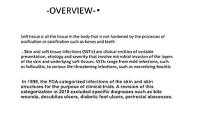
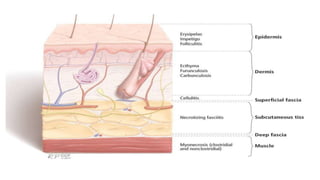
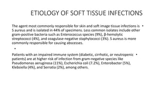
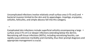
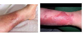
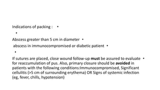
SOFT TISSUE INFECTIONS WITH
SURGICAL IMPLICATIONS
Presented by:
Tarek Yassin
Intern in the surgical department of Ziv hospital , Safed-Israel
Soft tissue is all the tissue in the body that is not hardened by the processes of
ossification or calcification such as bones and teeth
. Skin and soft tissue infections (SSTIs) are clinical entities of variable
presentation, etiology and severity that involve microbial invasion of the layers
of the skin and underlying soft tissues. SSTIs range from mild infections, such
as folliculitis, to serious life-threatening infections, such as necrotizing fasciitis
In 1998, the FDA categorized infections of the skin and skin
structures for the purpose of clinical trials. A revision of this
categorization in 2010 excluded specific diagnoses such as bite
wounds, decubitus ulcers, diabetic foot ulcers, perirectal abscesses.
•
-OVERVIEW-
ETIOLOGY OF SOFT TISSUE INFECTIONS
•
The agent most commonly responsible for skin and soft image tissue infections is
S aureus and is isolated in 44% of specimens. Less common isolates include other
gram-positive bacteria such as Enterococcus species (9%), β-hemolytic
streptococci (4%), and coagulase-negative staphylococci (3%). S aureus is more
commonly responsible for causing abscesses.
•
•
Patients with an impaired immune system (diabetic, cirrhotic, or neutropenic
patients) are at higher risk of infection from gram-negative species like
Pseudomonas aeruginosa (11%), Escherichia coli (7.2%), Enterobacter (5%),
Klebsiella (4%), and Serratia (2%), among others.
•
Uncomplicated infections involve relatively small surface area (<75 cm2) and
bacterial invasion limited to the skin and its appendages. Impetigo, erysipelas,
cellulitis, folliculitis, and simple abscess fall into this category.
•
•
Complicated skin infections include superficial cellulitis encompassing a large
surface area (>75 cm2) or deeper infections extending below the dermis.
Necrotizing soft tissue infections (NSTIs), including necrotizing fasciitis, can
rapidly cause extensive morbidity and mortality, thus their prompt diagnosis and
appropriate management is crucial.
IMPETIGO
•
Impetigo is a common skin Infection of the superficial epidermis
caused most commonly by S.Aureus
•
Most commonly in children particulary those in unhealthy living
conditions .
•
In Adults it may follow other skin or upper respiratory infection .
•
The clinical presentation of Impteigo honey-colored crusting which
can be itchy or painful
Management of Impetigo
•
Topical therapy (e.g : Mupirocin) for impetigo should be administered if there are
a limited number of lesions.
•
Extensive impetigo: requires treatment with systemic antibiotics . Unless
cultures reveal only beta-hemolytic streptococci (usually group
A Streptococcus [GAS]), the oral antibiotic prescribed for impetigo should be
effective for the treatment of both S. aureus and streptococcal infections(e.g :
Cephalexin and dicloxacillin)
•
Erysplea and cellulitis
•
-Erysipelas is a cutaneous infection localized to the upper layers of the
dermis, while cellulitis is a deeper infection, affecting the deeper dermis
and subcutaneous tissue.
•
-Erysplea and nonpurulent cellulitis are most commonly caused by strep.
Pyogenes while purulent cellulitis is most commonly caused by s.aureus
•
-Eryspleas are Sharply demarcated, erythematous skin lesion while
cellulitis is Erythematous skin lesion with indistinct margins
•
Eryspleas , unlike cellulitis . Involves the lymphatic vessels of the affected
area
•
Managhement of cellulitis and Erysplea
•
Management of nonpurulent cellulitis and Eryspleas should be
managed with oral antibiotics in the outpatient setting. For patients
with unambiguous erysipelas who do not meet criteria for parenteral
antibiotics, empiric oral antibiotics active against beta-hemolytic
streptococci should be administered(e/g Peniciilin V and Amoxicillin)
•
Patients with severe sepsis or an immunocompromising condition —
Vancomycin and Cefepime(IV)
•
Indications for MRSA coverage — Empiric coverage for MRSA is
indicated for patients with MRSA risk factors and those who have
increased morbidity if suboptimal antibiotics are administered. Conditions
that warrant MRSA coverage include the following :
•
●Systemic signs of toxicity (eg, fever >100.5°F/38°C, hypotension,
sustained tachycardia)
•
●Cellulitis with purulent drainage or exudate
•
●Immunocompromising condition (eg, neutropenia, immunosuppressive
drugs such as chemotherapy for malignancy)
•
●Presence of risk factor(s) for MRSA infection (eg, known MRSA
colonization or past infection, recent health care exposure, recent
antibiotic use, intravenous drug use)
FOLLICULITIS
•
Folliculitis refers to inflammation of the superficial or deep portion of the hair
follicle. The classic clinical findings of superficial folliculitis are folliculocentric,
inflamed papules and/or pustules on hair-bearing SkIN Nodules are a feature
of deep, follicular inflammation.
•
The most common causative agent of Folliculitis is S.aureus, risk factors :
Exposure to hot tubs or heated swimming pools – Pseudomonal folliculitis,
•
Prolonged oral antibiotic therapy – Gram-negative folliculitis
•
nasal carriage of S. aureus, hyperhidrosis, occlusion of hair follicles, and shaving-
S.aureus
•
mmunosuppression – Increases suspicion for fungal, viral, or
demodectic folliculitis, Folliculitis generally resolves with adequate
hygiene and warm soaks.
•
Furuncle – A furuncle is a well-circumscribed, painful,
suppurative inflammatory nodule involving hair follicles that
usually arises from preexisting folliculitis. A furuncle can occur
at any site that contains hair follicles, especially in regions that
are subject to friction and maceration (eg, face, neck, axillae,
groin, thighs, and buttocks). The lesion may extend into the
dermis and subcutaneous tissues and often serves as a nidus
for cellulitis and skin abscess.
•
●Carbuncle – A carbuncle is a coalescence of several inflamed
follicles into a single inflammatory mass with purulent drainage
from multiple follicle
Management
•
Indications of packing :
•
•
Abscess greater than 5 cm in diameter
•
abscess in immunocompromised or diabetic patient
•
•
If sutures are placed, close wound follow-up must be assured to evaluate
for reaccumulation of pus. Also, primary closure should be avoided in
patients with the following conditions:Immunocompromised, Significant
cellulitis (>5 cm of surrounding erythema) OR Signs of systemic infection
(eg, fever, chills, hypotension)
LOOP DRAINAGE TECHNIQUE
•
The loop drainage technique is an alternative to incision and drainage
that may be less painful for the patient and avoids packing .This
method also leaves less scarring and may prevent recurrence of the
abscess, especially in children.
•
A systematic database review (Gottlieb M, Peksa,2018) comprising
470 total patients which was conducted on CID technique failed in 25
of 265 cases (9.43%). The LDT failed in 8 of 195 cases (4.10%). There
was an odds ratio of 2.63 in favor of higher failures in the CID group.
furthermore subgroups analysis by age group demonstrated improved
efficacy of the LDT in pediatric patients
Necrotizing Fascitis
•
Necrotizing Fascitis is a necrotizing soft tissue infection , which can
be classified to 3 types:-
•
1-Type 1 -Polymicrobial : It’s caused by a polymicrobial source
including gram-positive cocci, gram-negative rods, and anaerobic
bacteria, specifically Clostridium perfringens and C septicum.
•
2-Type :It’s caused by monomicrobial source of β-
hemolytic Streptococcus or Staphylococcus species, with MRSA
contributing to the increasing number of community-acquired NSTIs.
•
Type 3 is a rare but fulminant subset resulting from a V
vulnificus infection of traumatized skin exposed to a body of salt-
water.
-
Signs and symptoms
•
-SIRS : tachycardia (heart rate >90 beats/min), tachypnea (respiratory
rate >20 breaths/min), fever or hypothermia (temperature >38 or <36
°C), and leukocytosis, leukopenia.
•
-In addition to signs of SIRS, patients can present with skin changes
like erythema(72%), bullae(38 %), necrosis, pain(out of
proportion72%), and crepitus(50%).
•
-Patients can present with a range of symptoms, from minimal skin
change to frank necrosis, and the time of progression to fulminant
disease varies in each patient.
Laboratory Risk Indicator for Necrotizing
Fasciitis
Management
•
1- source control with wide surgical debridement
•
2-broad-spectrum intravenous antibiotics
•
3- supportive care and resuscitation
•
•
Mortality rate among patients with Necrotizing infectious fasciitis who don’t
undergo surgical debridement is 100%. Debridement.
•
During debridement the following findings will be typically present :the
tissue will appear necrotic with dead muscle, thrombosed vessels, the
classic “dishwater” fluid, and a positive finger test, in which the tissue
layers can be easily separated from one another.
•
-The goal of operative management is to perform aggressive
debridement of all necrotic tissue until healthy, viable (bleeding)
tissue is reached. Inspection and debridement in the operating room
should be continued every one to two days until necrotic tissue is no
longer present.
•
-Wide spectrum antibiotic treatemt should be administered after
cultured are token (e.g :Carbapenem or Pipperacillin/tazobactam )
Plus Clindamycin.
References
•
Schwartz's Principles of Surgery, 11e Brunicardi F, Andersen DK,
Billiar TR, Dunn DL, Kao LS, Hunter JG, Matthews JB, Pollock R
•
•
UpTodate- www.uptodate.com
•
•
Long B, April MD. Is Loop Drainage Technique
More Effective for Treatment of Soft Tissue Abscess Compared With
Conventional Incision and Drainage? Ann Emerg Med. 2019
Jan;73(1):19-21. doi: 10.1016/j.annemergmed.2018.02.006. Epub
2018 Mar 9. PMID: 29530657.
•

SOFT TISSUE abscess and other....................

  • 1.
    SOFT TISSUE INFECTIONSWITH SURGICAL IMPLICATIONS Presented by: Tarek Yassin Intern in the surgical department of Ziv hospital , Safed-Israel
  • 2.
    Soft tissue isall the tissue in the body that is not hardened by the processes of ossification or calcification such as bones and teeth . Skin and soft tissue infections (SSTIs) are clinical entities of variable presentation, etiology and severity that involve microbial invasion of the layers of the skin and underlying soft tissues. SSTIs range from mild infections, such as folliculitis, to serious life-threatening infections, such as necrotizing fasciitis In 1998, the FDA categorized infections of the skin and skin structures for the purpose of clinical trials. A revision of this categorization in 2010 excluded specific diagnoses such as bite wounds, decubitus ulcers, diabetic foot ulcers, perirectal abscesses. • -OVERVIEW-
  • 4.
    ETIOLOGY OF SOFTTISSUE INFECTIONS • The agent most commonly responsible for skin and soft image tissue infections is S aureus and is isolated in 44% of specimens. Less common isolates include other gram-positive bacteria such as Enterococcus species (9%), β-hemolytic streptococci (4%), and coagulase-negative staphylococci (3%). S aureus is more commonly responsible for causing abscesses. • • Patients with an impaired immune system (diabetic, cirrhotic, or neutropenic patients) are at higher risk of infection from gram-negative species like Pseudomonas aeruginosa (11%), Escherichia coli (7.2%), Enterobacter (5%), Klebsiella (4%), and Serratia (2%), among others.
  • 5.
    • Uncomplicated infections involverelatively small surface area (<75 cm2) and bacterial invasion limited to the skin and its appendages. Impetigo, erysipelas, cellulitis, folliculitis, and simple abscess fall into this category. • • Complicated skin infections include superficial cellulitis encompassing a large surface area (>75 cm2) or deeper infections extending below the dermis. Necrotizing soft tissue infections (NSTIs), including necrotizing fasciitis, can rapidly cause extensive morbidity and mortality, thus their prompt diagnosis and appropriate management is crucial.
  • 7.
    IMPETIGO • Impetigo is acommon skin Infection of the superficial epidermis caused most commonly by S.Aureus • Most commonly in children particulary those in unhealthy living conditions . • In Adults it may follow other skin or upper respiratory infection . • The clinical presentation of Impteigo honey-colored crusting which can be itchy or painful
  • 8.
    Management of Impetigo • Topicaltherapy (e.g : Mupirocin) for impetigo should be administered if there are a limited number of lesions. • Extensive impetigo: requires treatment with systemic antibiotics . Unless cultures reveal only beta-hemolytic streptococci (usually group A Streptococcus [GAS]), the oral antibiotic prescribed for impetigo should be effective for the treatment of both S. aureus and streptococcal infections(e.g : Cephalexin and dicloxacillin) •
  • 9.
    Erysplea and cellulitis • -Erysipelasis a cutaneous infection localized to the upper layers of the dermis, while cellulitis is a deeper infection, affecting the deeper dermis and subcutaneous tissue. • -Erysplea and nonpurulent cellulitis are most commonly caused by strep. Pyogenes while purulent cellulitis is most commonly caused by s.aureus • -Eryspleas are Sharply demarcated, erythematous skin lesion while cellulitis is Erythematous skin lesion with indistinct margins • Eryspleas , unlike cellulitis . Involves the lymphatic vessels of the affected area •
  • 11.
    Managhement of cellulitisand Erysplea • Management of nonpurulent cellulitis and Eryspleas should be managed with oral antibiotics in the outpatient setting. For patients with unambiguous erysipelas who do not meet criteria for parenteral antibiotics, empiric oral antibiotics active against beta-hemolytic streptococci should be administered(e/g Peniciilin V and Amoxicillin) • Patients with severe sepsis or an immunocompromising condition — Vancomycin and Cefepime(IV)
  • 12.
    • Indications for MRSAcoverage — Empiric coverage for MRSA is indicated for patients with MRSA risk factors and those who have increased morbidity if suboptimal antibiotics are administered. Conditions that warrant MRSA coverage include the following : • ●Systemic signs of toxicity (eg, fever >100.5°F/38°C, hypotension, sustained tachycardia) • ●Cellulitis with purulent drainage or exudate • ●Immunocompromising condition (eg, neutropenia, immunosuppressive drugs such as chemotherapy for malignancy) • ●Presence of risk factor(s) for MRSA infection (eg, known MRSA colonization or past infection, recent health care exposure, recent antibiotic use, intravenous drug use)
  • 13.
    FOLLICULITIS • Folliculitis refers toinflammation of the superficial or deep portion of the hair follicle. The classic clinical findings of superficial folliculitis are folliculocentric, inflamed papules and/or pustules on hair-bearing SkIN Nodules are a feature of deep, follicular inflammation. • The most common causative agent of Folliculitis is S.aureus, risk factors : Exposure to hot tubs or heated swimming pools – Pseudomonal folliculitis, • Prolonged oral antibiotic therapy – Gram-negative folliculitis • nasal carriage of S. aureus, hyperhidrosis, occlusion of hair follicles, and shaving- S.aureus • mmunosuppression – Increases suspicion for fungal, viral, or demodectic folliculitis, Folliculitis generally resolves with adequate hygiene and warm soaks.
  • 15.
    • Furuncle – Afuruncle is a well-circumscribed, painful, suppurative inflammatory nodule involving hair follicles that usually arises from preexisting folliculitis. A furuncle can occur at any site that contains hair follicles, especially in regions that are subject to friction and maceration (eg, face, neck, axillae, groin, thighs, and buttocks). The lesion may extend into the dermis and subcutaneous tissues and often serves as a nidus for cellulitis and skin abscess. • ●Carbuncle – A carbuncle is a coalescence of several inflamed follicles into a single inflammatory mass with purulent drainage from multiple follicle
  • 17.
  • 18.
    • Indications of packing: • • Abscess greater than 5 cm in diameter • abscess in immunocompromised or diabetic patient • • If sutures are placed, close wound follow-up must be assured to evaluate for reaccumulation of pus. Also, primary closure should be avoided in patients with the following conditions:Immunocompromised, Significant cellulitis (>5 cm of surrounding erythema) OR Signs of systemic infection (eg, fever, chills, hypotension)
  • 19.
    LOOP DRAINAGE TECHNIQUE • Theloop drainage technique is an alternative to incision and drainage that may be less painful for the patient and avoids packing .This method also leaves less scarring and may prevent recurrence of the abscess, especially in children. • A systematic database review (Gottlieb M, Peksa,2018) comprising 470 total patients which was conducted on CID technique failed in 25 of 265 cases (9.43%). The LDT failed in 8 of 195 cases (4.10%). There was an odds ratio of 2.63 in favor of higher failures in the CID group. furthermore subgroups analysis by age group demonstrated improved efficacy of the LDT in pediatric patients
  • 22.
    Necrotizing Fascitis • Necrotizing Fascitisis a necrotizing soft tissue infection , which can be classified to 3 types:- • 1-Type 1 -Polymicrobial : It’s caused by a polymicrobial source including gram-positive cocci, gram-negative rods, and anaerobic bacteria, specifically Clostridium perfringens and C septicum. • 2-Type :It’s caused by monomicrobial source of β- hemolytic Streptococcus or Staphylococcus species, with MRSA contributing to the increasing number of community-acquired NSTIs. • Type 3 is a rare but fulminant subset resulting from a V vulnificus infection of traumatized skin exposed to a body of salt- water. -
  • 23.
    Signs and symptoms • -SIRS: tachycardia (heart rate >90 beats/min), tachypnea (respiratory rate >20 breaths/min), fever or hypothermia (temperature >38 or <36 °C), and leukocytosis, leukopenia. • -In addition to signs of SIRS, patients can present with skin changes like erythema(72%), bullae(38 %), necrosis, pain(out of proportion72%), and crepitus(50%). • -Patients can present with a range of symptoms, from minimal skin change to frank necrosis, and the time of progression to fulminant disease varies in each patient.
  • 24.
    Laboratory Risk Indicatorfor Necrotizing Fasciitis
  • 25.
    Management • 1- source controlwith wide surgical debridement • 2-broad-spectrum intravenous antibiotics • 3- supportive care and resuscitation • • Mortality rate among patients with Necrotizing infectious fasciitis who don’t undergo surgical debridement is 100%. Debridement. • During debridement the following findings will be typically present :the tissue will appear necrotic with dead muscle, thrombosed vessels, the classic “dishwater” fluid, and a positive finger test, in which the tissue layers can be easily separated from one another.
  • 26.
    • -The goal ofoperative management is to perform aggressive debridement of all necrotic tissue until healthy, viable (bleeding) tissue is reached. Inspection and debridement in the operating room should be continued every one to two days until necrotic tissue is no longer present. • -Wide spectrum antibiotic treatemt should be administered after cultured are token (e.g :Carbapenem or Pipperacillin/tazobactam ) Plus Clindamycin.
  • 27.
    References • Schwartz's Principles ofSurgery, 11e Brunicardi F, Andersen DK, Billiar TR, Dunn DL, Kao LS, Hunter JG, Matthews JB, Pollock R • • UpTodate- www.uptodate.com • • Long B, April MD. Is Loop Drainage Technique More Effective for Treatment of Soft Tissue Abscess Compared With Conventional Incision and Drainage? Ann Emerg Med. 2019 Jan;73(1):19-21. doi: 10.1016/j.annemergmed.2018.02.006. Epub 2018 Mar 9. PMID: 29530657.
  • 28.