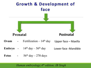
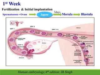
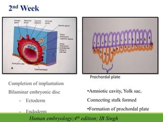
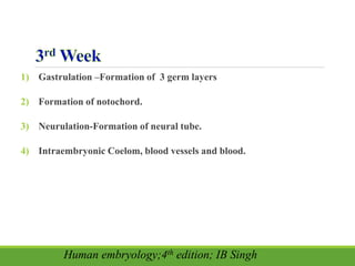
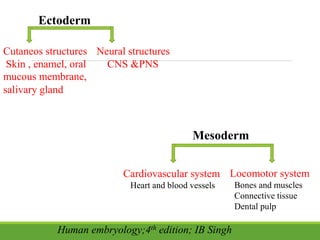
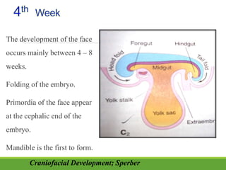
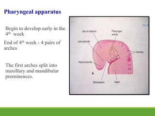
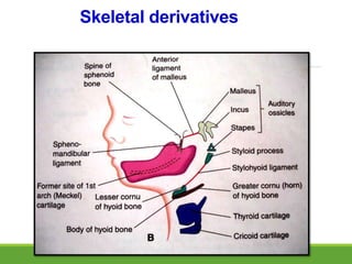
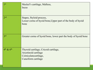
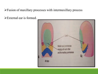
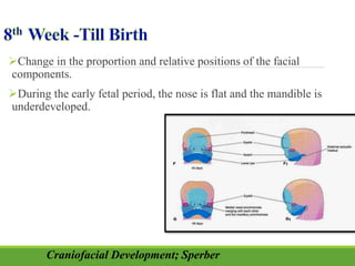
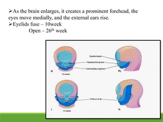
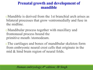
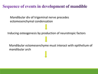
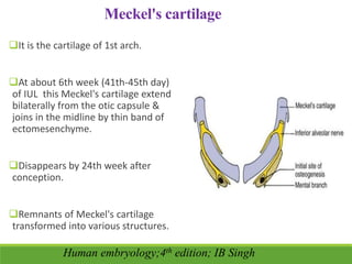
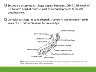
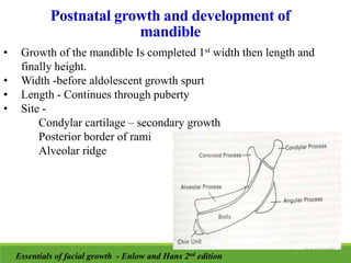
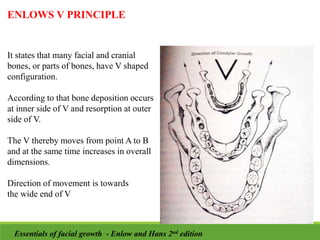
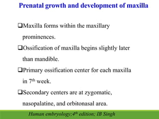
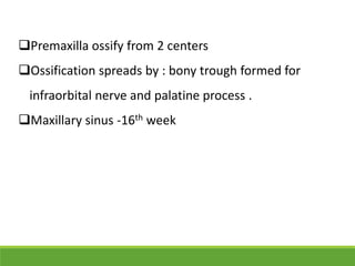
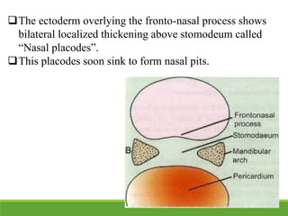
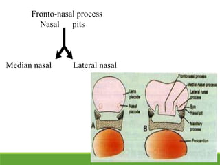
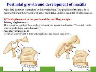
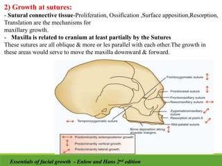
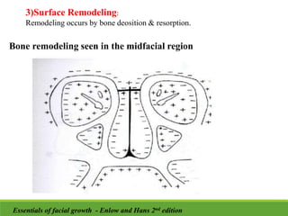
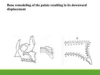
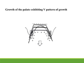
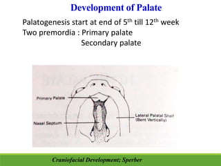
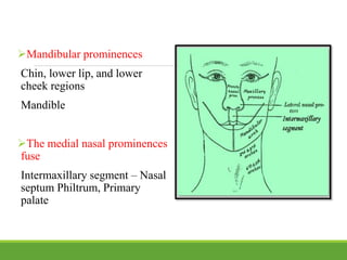
Introduction
Definitions
Growth and development of face
Embryonic devlopment of face
-Gastrulation
-Pharyngeal apparatus
Prenatal and Postnatal devlopment of mandible
Prenatal and postnatal devlopment of maxilla
Summary
References
Introduction
Human beings keep changing during their lives, they change
in size, appearance and psychological make up. The prenatal
growth and development of human embryo is consequence
of increase in number of cells, increase in individual cell
size, increase in amount of noncellular material.
Definitions
“Growth usually refers to an increase in size and number” –
Proffit W. R.
“The Self multiplication of living substance”- J.X.Huxley
“Change in any morphological parameter which is measurable”-
Melvin Moss
“Growth refers to increase in size”– Todd T. Winget
“Increase in size, change in proportion and progressive
complexity” – Krogman W.M.
Proffit W. R. Contemporary orthodontics; 4th edition
 “Development is a progress towards maturity” –
Todd T. Winget
 “Development is in complexity” - Proffit W. R.
 “Development connotes a maturational process
involving progressive differentiation at the cellular
and tissue levels” – Donald H. Enlow
 “Development refers to all naturally occurring,
progressive, unidirectional, sequential changes in life
of an individual from its elaboration as a
multifunctional unit terminating to death” – Moyer R.
E.
Proffit W. R. Contemporary orthodontics; 4th edition
Postnatal
Upper face – Maxilla
Lower face -Mandible
Prenatal
Ovum - Fertilization – 14th day
Embryo - 14th day – 56th day
Fetus - 56th day – 270 days
Human embryology;4th edition; IB Singh
1st Week
Fertilization & Initial implantation
Spermatozoon + Ovum Zygote
3days
Morula Blastula
Human embryology;4th edition; IB Singh
Completion of implantation
Bilaminar embryonic disc
- Ectoderm
- Endoderm
•Amniotic cavity, Yolk sac,
Connecting stalk formed
•Formation of prochordal plate
Prochordal plate
Human embryology;4th edition; IB Singh
1) Gastrulation –Formation of 3 germ layers
2) Formation of notochord.
3) Neurulation-Formation of neural tube.
4) Intraembryonic Coelom, blood vessels and blood.
Human embryology;4th edition; IB Singh
-Ectoderm
-Mesoderm
-Endoderm
Human embryology;4th edition; IB Singh
Ectoderm
Cutaneos structures
Skin , enamel, oral
mucous membrane,
salivary gland
Neural structures
CNS &PNS
Mesoderm
Cardiovascular system
Heart and blood vessels
Locomotor system
Bones and muscles
Connective tissue
Dental pulp
Human embryology;4th edition; IB Singh
Endoderm
Lining epithelium of respiratory system & alimentary canal
Secretory cells of liver , pancreas
Epithelium is derived from all three germ layers -
eg. endoderm - epithelial lining inside viscera, mesoderm -
mesothelium lining outside of viscera, and ectoderm - skin
epithelium
Notochord Formation
 Notochord is
flexible rod shape
body. Derive from
mesoderm.
 Form basis of axial
skeleton.
 Extend cranially to
form primitive
node to
buccopharyngeal
membrane.
Human embryology;4th edition; IB Singh
Neurulation
Neural plates bend up and later fuses to form a hollow tube called neural
tube.
It differentiate into brain and spinal cord of crntral nervous systeme
Neural tube – Brain & Spinal Cord
• Colour atlas of clinical embryology – Keith L. Moore, T.V.N. Persaud, 2nd edition.
Derivatives of Neural crest cells
 Connective tissue components including ectomesenchyme of
facial prominences and pharyngeal arches.
 Cartilage and bones of facial and visceral skeleton, ligaments in
the facial and oral regions.
 Dental papilla, portion of periodontal ligament and pituitary
gland.
 Ciliary muscles.
Craniofacial Development; Sperber
 Spinal ganglia, adrenal medulla, neurolemma sheath,
meningeal covering of brain.
 Autonomic nervous system – sympathetic and
parasympathetic ganglion.
 Blood vessels, carotid body , parafollicular cell and
adrenomedullary cells
Craniofacial Development; Sperber
The development of the face
occurs mainly between 4 – 8
weeks.
Folding of the embryo.
Primordia of the face appear
at the cephalic end of the
embryo.
Mandible is the first to form.
Craniofacial Development; Sperber
The stomodeum &
Buccopharyngeal membrane
Five processes have appeared.
-Frontonasal process (1)
-Maxillary processes (2)
-Mandibular processes (2)
Pharyngeal apparatus forms.
Two Nasal placodes
olfactory epithelium and nerve
Two Lense placodes
Ectodermal thickenings
• Colour atlas of clinical embryology – Keith L. Moore, T.V.N. Persaud, 2nd edition.
Pharyngeal apparatus
Begin to develop early in the
4th week
End of 4th week - 4 pairs of
arches
The first arches split into
maxillary and mandibular
prominences.
Pharyngeal arches
Mesodermal thickenings (1, 2, 3,
4 and 6)
Pharyngeal pouches
Endodermal extension in between
two arches
Pharyngeal grooves (clefts)
Ectoderm dips in to meet endoderm
Pharyngeal membranes. Appear
in the floor of grooves
Craniofacial Development; Sperber
Muscles, Nerves,
Arteries,
Cartilages, Bones
Pharyngeal Arch
Mesoderm, Ectoderm, Endoderm,
Neural crest cells, Cartilaginous rod,
Arterial, Muscular & Nerve
components
Craniofacial Development; Sperber
1st Muscles of mastication-
- mylohyoid
- tensor veli palitini
-tensor tympani
-anterior belly of the digastric
2nd facial muscles, stylohyoid , stapedius , posterior belly of digastric
3rd Stylopharyngeus muscle
4th
&
Muscles of soft palate muscles of pharynx cricothyroid ,
cricopharyngeus
6th Intrinsic muscles of the larynx , muscles of esophagus
Skeletal derivatives
1st Meckel’s cartilage, Malleus,
Incus
2nd Stapes, Styloid process,
Lesser cornu of hyoid bone,Upper part of the body of hyoid
bone
3rd Greater cornu of hyoid bone, lower part the body of hyoid bone
4th & 6th Thyroid cartilage, Cricoid cartilage,
Aryetinoid cartilage
Corniculatecartilage,
Cuneiform cartilage
Nerve & Arteries
Craniofacial Development; Sperber
Nerves Arteries
1st Trigeminal
(CN V)
Maxillary artery
2nd Facial
(CN VII)
Stapedial artery
3rd Glossopharyngeal
(CN IX)
Common carotid artery,internal
carotid artery
4th Vagus
(CN X) – Superior laryngeal
branch
Rt subclavian artery
Arch of aorta
6th Vagus
(CN X) –Recurrent laryngeal
branch
Pulmonary arteries
1st
Ventral - Tongue
2nd
Dorsal
Dorsal
Tubo-tympanic recess
Ventral - Tonsil
Inferior Parathyroid Gland,
Thymus
3rd
Pharyngeal pouch derivatives
Craniofacial Development; Sperber
Superior Parathyroid Gland,
Thyroid
4th
5th Ultimobranchial Pouch
Combined with 4th Pouch
1st Groove - External acoustic meatus
1st Membrane - Tympanic membrane
 Formation of nasal pit
-Lateral nasal process
-Medial nasal process
 Maxillary swellings grow
ventrally and medially.
 Nasolacrimal groove
 Eyes derived from –
Neuroectoderm, surface
ectoderm, mesoderm , and
neural crest cells.
Craniofacial Development; Sperber
Fusion of medial nasal processes.
Fusion of medial & lateral nasal processes
Maxillary prominence begin to merge with the lateral nasal
prominence
The nasal pits deepen-Nasal sac
Mandibular processes fuse.
Craniofacial Development; Sperber
• Primordia of the auricle forms
1st Pharyngeal groove forms the external acoustic meatus
• Eyelids develop
Intermaxillary process
- Nasal septum
-Philtrum of the upper lip.
-Premaxilla
Craniofacial Development; Sperber
Fusion of maxillary processes with intermaxillary process
External ear is formed.
Change in the proportion and relative positions of the facial
components.
During the early fetal period, the nose is flat and the mandible is
underdeveloped.
Craniofacial Development; Sperber
As the brain enlarges, it creates a prominent forehead, the
eyes move medially, and the external ears rise.
Eyelids fuse – 10week
Open – 26th week
Prenatal growth and development of
mandible
oMandible is derived from the 1st branchial arch arises as
bilateral processes that grow ventromedially and fuse in
the midline.
oMandibular process together with maxillary and
frontonasal process bound the
primitive mouth /stomodeum.
oThe cartilages and bones of mandibular skeleton form
from embryonic neural crest cells that originate in the
mid & hind brain region of neural folds.
Human embryology;4th edition; IB Singh
Mandibular div of trigeminal nerve precedes
ectomesenchymal condensation
Inducing osteogenesis by production of neurotropic factors
Mandibular ectomesenchyme must interact with epithelium of
mandibular arch
Intramembranous bone formation lying lateral to
Meckels cartilage
`
Ossification of osteogenic membrane (formed from
ectomesenchymal condensation at 36-38 days of
development
Meckel's cartilage
It is the cartilage of 1st arch.
At about 6th week (41th-45th day)
of IUL this Meckel's cartilage extend
bilaterally from the otic capsule &
joins in the midline by thin band of
ectomesenchyme.
Disappears by 24th week after
conception.
Remnants of Meckel's cartilage
transformed into various structures.
Human embryology;4th edition; IB Singh
 Secondary accessory cartilages appear between 10th & 14th week of
IUL to form head of condyle, part of coronoid process & mental
protuberance.
 Condylar cartilage as cone shaped structure in ramal region – 10 th
week of IUL-primordium for future condyle.
 Cartilage Cells of condylar process differentiate
& increase in number.
Ossification starts at 14th week of IUL & first
evidence of endochondral bone appears in
condyle region.
 By mid-fetal life much of
the cone shape cartilage is
replaced by bone ,but its
superior end persists into
adulthood acting as both
growth & articular
cartilage.
 In the mental region 1 or 2
small cartilages appear &
ossify in the 7th month of
post conception to form
mental ossicles on either
side of symphysis.
Postnatal growth and development of
mandible
• Growth of the mandible Is completed 1st width then length and
finally height.
• Width -before aldolescent growth spurt
• Length - Continues through puberty
• Site -
Condylar cartilage – secondary growth
Posterior border of rami
Alveolar ridge
Essentials of facial growth - Enlow and Hans 2nd edition
BODY OF MANDIBLE
• Increase in length of mandibular
body occurs by resorption at
anterior border of ramus.
• This allows growth in
dental arch to accommodate
permanent molar.
• Mandible undergoes rotational
pattern of growth.
• It is depository on outer surface
& resorptive on inferior aspect of
medial surface.
• The superior aspect of medial
surface just below teeth is
depository.
RAMUS UPRIGHTING
• Remodeling of ramus occurs in archial
pattern.
• With anterior displacement condyle
maintains contact with temporal fossa.
• Ramal angle of childhood upright
slightly in adolescence & in late
adulthood it becomes acute.
• Till uprighting, there is deposition at
posterior ramal border but after
uprighting there is selective
deposition/resorption pattern in anterior
& posterior border.
• Along with change in angulation ,there
is also an increase in height of ramus.
Essentials of facial growth - Enlow and Hans 2nd edition
Mandibular foramen
Chin
Remodelling changes in the
symphysis is very unique.
Essentials of facial growth - Enlow and Hans 2nd edition
ENLOWS V PRINCIPLE
It states that many facial and cranial
bones, or parts of bones, have V shaped
configuration.
According to that bone deposition occurs
at inner side of V and resorption at outer
side of V.
The V thereby moves from point A to B
and at the same time increases in overall
dimensions.
Direction of movement is towards
the wide end of V
Essentials of facial growth - Enlow and Hans 2nd edition
Coronoid Process
Deposits on medial surface of coronoid
leads to posterior lengthening of mandible
& also increase in height occurs.
So coronoid also
becomes posteriorly relocated.
lingual tuberosity
Condyle
Essentials of facial growth - Enlow and Hans 2nd edition
Prenatal growth and development of maxilla
Maxilla forms within the maxillary
prominences.
Ossification of maxilla begins slightly later
than mandible.
Primary ossification center for each maxilla
in 7th week.
Secondary centers are at zygomatic,
nasopalatine, and orbitonasal area.
Human embryology;4th edition; IB Singh
Premaxilla ossify from 2 centers
Ossification spreads by : bony trough formed for
infraorbital nerve and palatine process .
Maxillary sinus -16th week
Around 4th week of intra uterine life a prominent
bulge on ventral aspect of embryo appear.
Below that bulge a depression is seen called
stomodeum. Floor is formed by buccopharyngeal
membrane.
The stomodeum is overlap superiorly by frontonasal
process.
The mandibular arch of both side form lateral wall of
stomodeum. Mandibular arch gives off bud from dorsal
end called maxillary process
The ectoderm overlying the fronto-nasal process shows
bilateral localized thickening above stomodeum called
“Nasal placodes”.
This placodes soon sink to form nasal pits.
Fronto-nasal process
Nasal pits
Median nasal Lateral nasal
Postnatal growth and development of maxilla
Maxillary complex is attached to the cranial base. The position of the maxilla is
dependent upon the growth at spheno-occipital & spheno-occipital synchondroses.
1)The displacement in the position of the maxillary complex
Primary displacement –
This occurs by gowth of the maxillary tuberosity in a posterior direction .This results in the
whole maxilla being carried anteriorly.
Secondary displacement-
Occurs in a Downward & forward direction as the cranial base grow .
2) Growth at sutures:
- Sutural connective tissue-Proliferation, Ossification ,Surface apposition,Resorption,
Translation are the mechanisms for
maxillary growth.
- Maxilla is related to cranium at least partially by the Sutures
These sutures are all oblique & more or les parallel with each other.The growth in
these areas would serve to move the maxilla downward & forward.
Essentials of facial growth - Enlow and Hans 2nd edition
3)Surface Remodeling:
Remodeling occurs by bone deosition & resorption.
Bone remodeling seen in the midfacial region
Essentials of facial growth - Enlow and Hans 2nd edition
Bone remodeling of the palate resulting in its downward
displacement
Growth of the palate exhibiting V pattern of growth
Development of Palate
Palatogenesis start at end of 5th till 12th week
Two premordia : Primary palate
Secondary palate
Craniofacial Development; Sperber
Primary palate
Developes from deep part of
inter maxillary segment of
maxilla.
Develops end of 5th week
Internal merging of medial
nasal process
Secondary palate
Primordium of hard and
soft palate posterior to
incisive foramen.
Develops 6th week from
Lateral palatine processes
of maxillary prominence.
The palatine process fuse In midline and with
Nasal septum And posterior part of Primary
palate.
 Frontal prominence
Forehead ,bridge of the nose,
dorsum of the nose
Frontal and nasal bones
 Maxillary prominences
Upper cheek regions,most of
the upper lip except philtrun
Maxilla, zygomatic bone,
secondary palate
 Lateral nasal prominences
Alae of the nose
Grays anatomy;39th edition; Starding Susan
Mandibular prominences
Chin, lower lip, and lower
cheek regions
Mandible
The medial nasal prominences
fuse
Intermaxillary segment – Nasal
septum Philtrum, Primary
palate
The size of the embryo from fertilization to 8th week
Length of embryo
4th Week of IU – 5mm
8th Week – 30mm
Full term – 300mm
Human embryology;4th edition; IB Singh
Summary
•We have seen how an every individual spends first 9
months of his or her life in it’s mother’s womb, from the
day of inception as a single cell strucure to an organism
having billions of cells, numerous tissues and organs which
are functioning in a perfect harmony
References
• Colour atlas of clinical embryology – Keith L. Moore, T.V.N.
Persaud, 2nd edition.
• Human embryology;4th edition; IB Singh
• Craniofacial Development; Sperber
• Oral anatomy, histology and embryology ;3rd edition; Berkovitz
• Oral Histology,Development,Structure and Function.5th
edition;A.R.Tencate:Mosby
• Orban’s Oral Histology and Embryology.11th edition;Mosby
• Grays anatomy;39th edition; Starding Susan
• Anatomy; vol 3; 3rd edition; B.D.Chaurasia
• Proffit W. R. Contemporary orthodontics; 4th edition
• Essentials of facial growth - Enlow and Hans 2nd edition
Devlopement of face.pptx

Devlopement of face.pptx

  • 2.
    Introduction Definitions Growth and developmentof face Embryonic devlopment of face -Gastrulation -Pharyngeal apparatus Prenatal and Postnatal devlopment of mandible Prenatal and postnatal devlopment of maxilla Summary References
  • 3.
    Introduction Human beings keepchanging during their lives, they change in size, appearance and psychological make up. The prenatal growth and development of human embryo is consequence of increase in number of cells, increase in individual cell size, increase in amount of noncellular material.
  • 4.
    Definitions “Growth usually refersto an increase in size and number” – Proffit W. R. “The Self multiplication of living substance”- J.X.Huxley “Change in any morphological parameter which is measurable”- Melvin Moss “Growth refers to increase in size”– Todd T. Winget “Increase in size, change in proportion and progressive complexity” – Krogman W.M. Proffit W. R. Contemporary orthodontics; 4th edition
  • 5.
     “Development isa progress towards maturity” – Todd T. Winget  “Development is in complexity” - Proffit W. R.  “Development connotes a maturational process involving progressive differentiation at the cellular and tissue levels” – Donald H. Enlow  “Development refers to all naturally occurring, progressive, unidirectional, sequential changes in life of an individual from its elaboration as a multifunctional unit terminating to death” – Moyer R. E. Proffit W. R. Contemporary orthodontics; 4th edition
  • 6.
    Postnatal Upper face –Maxilla Lower face -Mandible Prenatal Ovum - Fertilization – 14th day Embryo - 14th day – 56th day Fetus - 56th day – 270 days Human embryology;4th edition; IB Singh
  • 7.
    1st Week Fertilization &Initial implantation Spermatozoon + Ovum Zygote 3days Morula Blastula Human embryology;4th edition; IB Singh
  • 8.
    Completion of implantation Bilaminarembryonic disc - Ectoderm - Endoderm •Amniotic cavity, Yolk sac, Connecting stalk formed •Formation of prochordal plate Prochordal plate Human embryology;4th edition; IB Singh
  • 9.
    1) Gastrulation –Formationof 3 germ layers 2) Formation of notochord. 3) Neurulation-Formation of neural tube. 4) Intraembryonic Coelom, blood vessels and blood. Human embryology;4th edition; IB Singh
  • 10.
  • 11.
    Ectoderm Cutaneos structures Skin ,enamel, oral mucous membrane, salivary gland Neural structures CNS &PNS Mesoderm Cardiovascular system Heart and blood vessels Locomotor system Bones and muscles Connective tissue Dental pulp Human embryology;4th edition; IB Singh
  • 12.
    Human embryology;4th edition;IB Singh Endoderm Lining epithelium of respiratory system & alimentary canal Secretory cells of liver , pancreas Epithelium is derived from all three germ layers - eg. endoderm - epithelial lining inside viscera, mesoderm - mesothelium lining outside of viscera, and ectoderm - skin epithelium
  • 13.
    Notochord Formation  Notochordis flexible rod shape body. Derive from mesoderm.  Form basis of axial skeleton.  Extend cranially to form primitive node to buccopharyngeal membrane. Human embryology;4th edition; IB Singh
  • 14.
    Neurulation Neural plates bendup and later fuses to form a hollow tube called neural tube. It differentiate into brain and spinal cord of crntral nervous systeme Neural tube – Brain & Spinal Cord • Colour atlas of clinical embryology – Keith L. Moore, T.V.N. Persaud, 2nd edition.
  • 15.
    Derivatives of Neuralcrest cells  Connective tissue components including ectomesenchyme of facial prominences and pharyngeal arches.  Cartilage and bones of facial and visceral skeleton, ligaments in the facial and oral regions.  Dental papilla, portion of periodontal ligament and pituitary gland.  Ciliary muscles. Craniofacial Development; Sperber
  • 16.
     Spinal ganglia,adrenal medulla, neurolemma sheath, meningeal covering of brain.  Autonomic nervous system – sympathetic and parasympathetic ganglion.  Blood vessels, carotid body , parafollicular cell and adrenomedullary cells Craniofacial Development; Sperber
  • 17.
    The development ofthe face occurs mainly between 4 – 8 weeks. Folding of the embryo. Primordia of the face appear at the cephalic end of the embryo. Mandible is the first to form. Craniofacial Development; Sperber
  • 18.
    The stomodeum & Buccopharyngealmembrane Five processes have appeared. -Frontonasal process (1) -Maxillary processes (2) -Mandibular processes (2) Pharyngeal apparatus forms.
  • 19.
    Two Nasal placodes olfactoryepithelium and nerve Two Lense placodes Ectodermal thickenings • Colour atlas of clinical embryology – Keith L. Moore, T.V.N. Persaud, 2nd edition.
  • 20.
    Pharyngeal apparatus Begin todevelop early in the 4th week End of 4th week - 4 pairs of arches The first arches split into maxillary and mandibular prominences.
  • 21.
    Pharyngeal arches Mesodermal thickenings(1, 2, 3, 4 and 6) Pharyngeal pouches Endodermal extension in between two arches Pharyngeal grooves (clefts) Ectoderm dips in to meet endoderm Pharyngeal membranes. Appear in the floor of grooves Craniofacial Development; Sperber
  • 22.
    Muscles, Nerves, Arteries, Cartilages, Bones PharyngealArch Mesoderm, Ectoderm, Endoderm, Neural crest cells, Cartilaginous rod, Arterial, Muscular & Nerve components Craniofacial Development; Sperber
  • 24.
    1st Muscles ofmastication- - mylohyoid - tensor veli palitini -tensor tympani -anterior belly of the digastric 2nd facial muscles, stylohyoid , stapedius , posterior belly of digastric 3rd Stylopharyngeus muscle 4th & Muscles of soft palate muscles of pharynx cricothyroid , cricopharyngeus 6th Intrinsic muscles of the larynx , muscles of esophagus
  • 25.
  • 26.
    1st Meckel’s cartilage,Malleus, Incus 2nd Stapes, Styloid process, Lesser cornu of hyoid bone,Upper part of the body of hyoid bone 3rd Greater cornu of hyoid bone, lower part the body of hyoid bone 4th & 6th Thyroid cartilage, Cricoid cartilage, Aryetinoid cartilage Corniculatecartilage, Cuneiform cartilage
  • 27.
    Nerve & Arteries CraniofacialDevelopment; Sperber
  • 28.
    Nerves Arteries 1st Trigeminal (CNV) Maxillary artery 2nd Facial (CN VII) Stapedial artery 3rd Glossopharyngeal (CN IX) Common carotid artery,internal carotid artery 4th Vagus (CN X) – Superior laryngeal branch Rt subclavian artery Arch of aorta 6th Vagus (CN X) –Recurrent laryngeal branch Pulmonary arteries
  • 29.
    1st Ventral - Tongue 2nd Dorsal Dorsal Tubo-tympanicrecess Ventral - Tonsil Inferior Parathyroid Gland, Thymus 3rd Pharyngeal pouch derivatives Craniofacial Development; Sperber
  • 30.
    Superior Parathyroid Gland, Thyroid 4th 5thUltimobranchial Pouch Combined with 4th Pouch 1st Groove - External acoustic meatus 1st Membrane - Tympanic membrane
  • 31.
     Formation ofnasal pit -Lateral nasal process -Medial nasal process  Maxillary swellings grow ventrally and medially.  Nasolacrimal groove  Eyes derived from – Neuroectoderm, surface ectoderm, mesoderm , and neural crest cells. Craniofacial Development; Sperber
  • 32.
    Fusion of medialnasal processes. Fusion of medial & lateral nasal processes Maxillary prominence begin to merge with the lateral nasal prominence The nasal pits deepen-Nasal sac Mandibular processes fuse. Craniofacial Development; Sperber
  • 33.
    • Primordia ofthe auricle forms 1st Pharyngeal groove forms the external acoustic meatus • Eyelids develop
  • 34.
    Intermaxillary process - Nasalseptum -Philtrum of the upper lip. -Premaxilla Craniofacial Development; Sperber
  • 35.
    Fusion of maxillaryprocesses with intermaxillary process External ear is formed.
  • 36.
    Change in theproportion and relative positions of the facial components. During the early fetal period, the nose is flat and the mandible is underdeveloped. Craniofacial Development; Sperber
  • 37.
    As the brainenlarges, it creates a prominent forehead, the eyes move medially, and the external ears rise. Eyelids fuse – 10week Open – 26th week
  • 38.
    Prenatal growth anddevelopment of mandible oMandible is derived from the 1st branchial arch arises as bilateral processes that grow ventromedially and fuse in the midline. oMandibular process together with maxillary and frontonasal process bound the primitive mouth /stomodeum. oThe cartilages and bones of mandibular skeleton form from embryonic neural crest cells that originate in the mid & hind brain region of neural folds. Human embryology;4th edition; IB Singh
  • 39.
    Mandibular div oftrigeminal nerve precedes ectomesenchymal condensation Inducing osteogenesis by production of neurotropic factors Mandibular ectomesenchyme must interact with epithelium of mandibular arch
  • 40.
    Intramembranous bone formationlying lateral to Meckels cartilage ` Ossification of osteogenic membrane (formed from ectomesenchymal condensation at 36-38 days of development
  • 41.
    Meckel's cartilage It isthe cartilage of 1st arch. At about 6th week (41th-45th day) of IUL this Meckel's cartilage extend bilaterally from the otic capsule & joins in the midline by thin band of ectomesenchyme. Disappears by 24th week after conception. Remnants of Meckel's cartilage transformed into various structures. Human embryology;4th edition; IB Singh
  • 42.
     Secondary accessorycartilages appear between 10th & 14th week of IUL to form head of condyle, part of coronoid process & mental protuberance.  Condylar cartilage as cone shaped structure in ramal region – 10 th week of IUL-primordium for future condyle.
  • 43.
     Cartilage Cellsof condylar process differentiate & increase in number. Ossification starts at 14th week of IUL & first evidence of endochondral bone appears in condyle region.
  • 44.
     By mid-fetallife much of the cone shape cartilage is replaced by bone ,but its superior end persists into adulthood acting as both growth & articular cartilage.  In the mental region 1 or 2 small cartilages appear & ossify in the 7th month of post conception to form mental ossicles on either side of symphysis.
  • 45.
    Postnatal growth anddevelopment of mandible • Growth of the mandible Is completed 1st width then length and finally height. • Width -before aldolescent growth spurt • Length - Continues through puberty • Site - Condylar cartilage – secondary growth Posterior border of rami Alveolar ridge Essentials of facial growth - Enlow and Hans 2nd edition
  • 46.
    BODY OF MANDIBLE •Increase in length of mandibular body occurs by resorption at anterior border of ramus. • This allows growth in dental arch to accommodate permanent molar. • Mandible undergoes rotational pattern of growth. • It is depository on outer surface & resorptive on inferior aspect of medial surface. • The superior aspect of medial surface just below teeth is depository.
  • 47.
    RAMUS UPRIGHTING • Remodelingof ramus occurs in archial pattern. • With anterior displacement condyle maintains contact with temporal fossa. • Ramal angle of childhood upright slightly in adolescence & in late adulthood it becomes acute. • Till uprighting, there is deposition at posterior ramal border but after uprighting there is selective deposition/resorption pattern in anterior & posterior border. • Along with change in angulation ,there is also an increase in height of ramus. Essentials of facial growth - Enlow and Hans 2nd edition
  • 48.
    Mandibular foramen Chin Remodelling changesin the symphysis is very unique. Essentials of facial growth - Enlow and Hans 2nd edition
  • 49.
    ENLOWS V PRINCIPLE Itstates that many facial and cranial bones, or parts of bones, have V shaped configuration. According to that bone deposition occurs at inner side of V and resorption at outer side of V. The V thereby moves from point A to B and at the same time increases in overall dimensions. Direction of movement is towards the wide end of V Essentials of facial growth - Enlow and Hans 2nd edition
  • 50.
    Coronoid Process Deposits onmedial surface of coronoid leads to posterior lengthening of mandible & also increase in height occurs. So coronoid also becomes posteriorly relocated.
  • 51.
    lingual tuberosity Condyle Essentials offacial growth - Enlow and Hans 2nd edition
  • 52.
    Prenatal growth anddevelopment of maxilla Maxilla forms within the maxillary prominences. Ossification of maxilla begins slightly later than mandible. Primary ossification center for each maxilla in 7th week. Secondary centers are at zygomatic, nasopalatine, and orbitonasal area. Human embryology;4th edition; IB Singh
  • 53.
    Premaxilla ossify from2 centers Ossification spreads by : bony trough formed for infraorbital nerve and palatine process . Maxillary sinus -16th week
  • 54.
    Around 4th weekof intra uterine life a prominent bulge on ventral aspect of embryo appear. Below that bulge a depression is seen called stomodeum. Floor is formed by buccopharyngeal membrane.
  • 55.
    The stomodeum isoverlap superiorly by frontonasal process. The mandibular arch of both side form lateral wall of stomodeum. Mandibular arch gives off bud from dorsal end called maxillary process
  • 56.
    The ectoderm overlyingthe fronto-nasal process shows bilateral localized thickening above stomodeum called “Nasal placodes”. This placodes soon sink to form nasal pits.
  • 57.
  • 58.
    Postnatal growth anddevelopment of maxilla Maxillary complex is attached to the cranial base. The position of the maxilla is dependent upon the growth at spheno-occipital & spheno-occipital synchondroses. 1)The displacement in the position of the maxillary complex Primary displacement – This occurs by gowth of the maxillary tuberosity in a posterior direction .This results in the whole maxilla being carried anteriorly. Secondary displacement- Occurs in a Downward & forward direction as the cranial base grow .
  • 59.
    2) Growth atsutures: - Sutural connective tissue-Proliferation, Ossification ,Surface apposition,Resorption, Translation are the mechanisms for maxillary growth. - Maxilla is related to cranium at least partially by the Sutures These sutures are all oblique & more or les parallel with each other.The growth in these areas would serve to move the maxilla downward & forward. Essentials of facial growth - Enlow and Hans 2nd edition
  • 60.
    3)Surface Remodeling: Remodeling occursby bone deosition & resorption. Bone remodeling seen in the midfacial region Essentials of facial growth - Enlow and Hans 2nd edition
  • 61.
    Bone remodeling ofthe palate resulting in its downward displacement
  • 62.
    Growth of thepalate exhibiting V pattern of growth
  • 63.
    Development of Palate Palatogenesisstart at end of 5th till 12th week Two premordia : Primary palate Secondary palate Craniofacial Development; Sperber
  • 64.
    Primary palate Developes fromdeep part of inter maxillary segment of maxilla. Develops end of 5th week Internal merging of medial nasal process Secondary palate Primordium of hard and soft palate posterior to incisive foramen. Develops 6th week from Lateral palatine processes of maxillary prominence.
  • 65.
    The palatine processfuse In midline and with Nasal septum And posterior part of Primary palate.
  • 66.
     Frontal prominence Forehead,bridge of the nose, dorsum of the nose Frontal and nasal bones  Maxillary prominences Upper cheek regions,most of the upper lip except philtrun Maxilla, zygomatic bone, secondary palate  Lateral nasal prominences Alae of the nose Grays anatomy;39th edition; Starding Susan
  • 67.
    Mandibular prominences Chin, lowerlip, and lower cheek regions Mandible The medial nasal prominences fuse Intermaxillary segment – Nasal septum Philtrum, Primary palate
  • 68.
    The size ofthe embryo from fertilization to 8th week Length of embryo 4th Week of IU – 5mm 8th Week – 30mm Full term – 300mm Human embryology;4th edition; IB Singh
  • 69.
    Summary •We have seenhow an every individual spends first 9 months of his or her life in it’s mother’s womb, from the day of inception as a single cell strucure to an organism having billions of cells, numerous tissues and organs which are functioning in a perfect harmony
  • 70.
    References • Colour atlasof clinical embryology – Keith L. Moore, T.V.N. Persaud, 2nd edition. • Human embryology;4th edition; IB Singh • Craniofacial Development; Sperber • Oral anatomy, histology and embryology ;3rd edition; Berkovitz • Oral Histology,Development,Structure and Function.5th edition;A.R.Tencate:Mosby • Orban’s Oral Histology and Embryology.11th edition;Mosby • Grays anatomy;39th edition; Starding Susan • Anatomy; vol 3; 3rd edition; B.D.Chaurasia • Proffit W. R. Contemporary orthodontics; 4th edition • Essentials of facial growth - Enlow and Hans 2nd edition