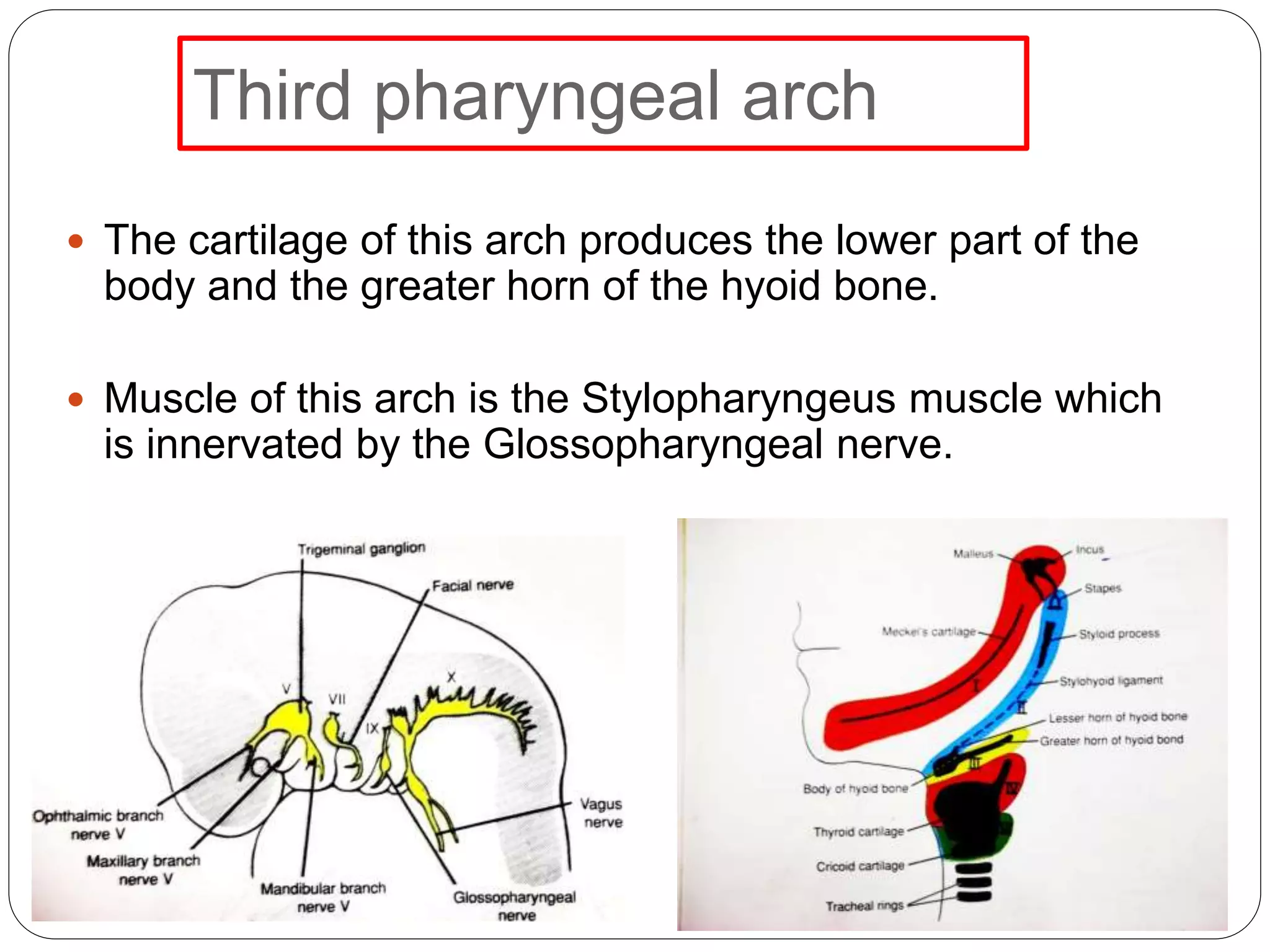
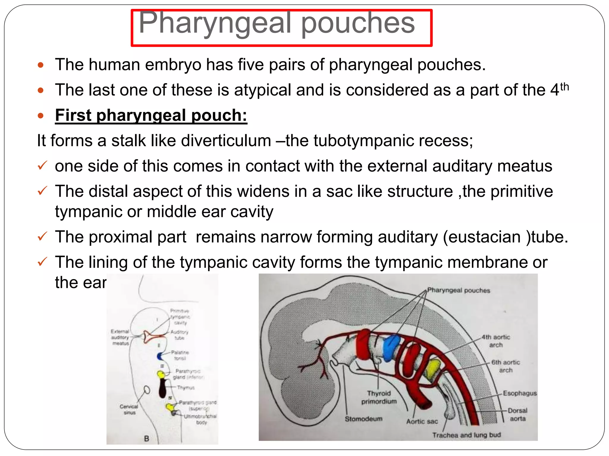
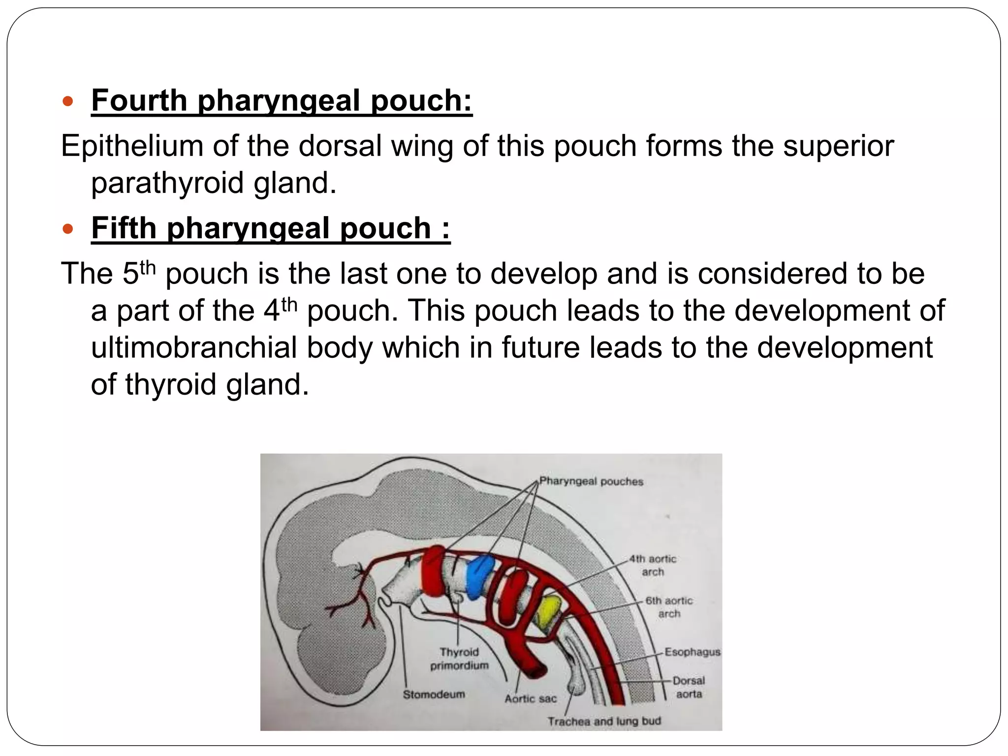
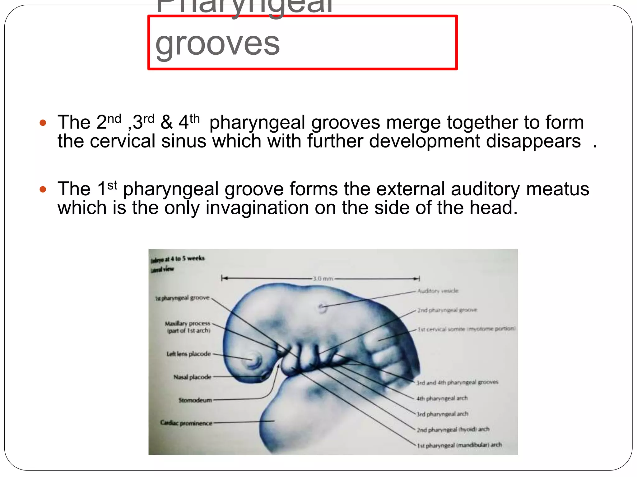
Downloaded 20 times
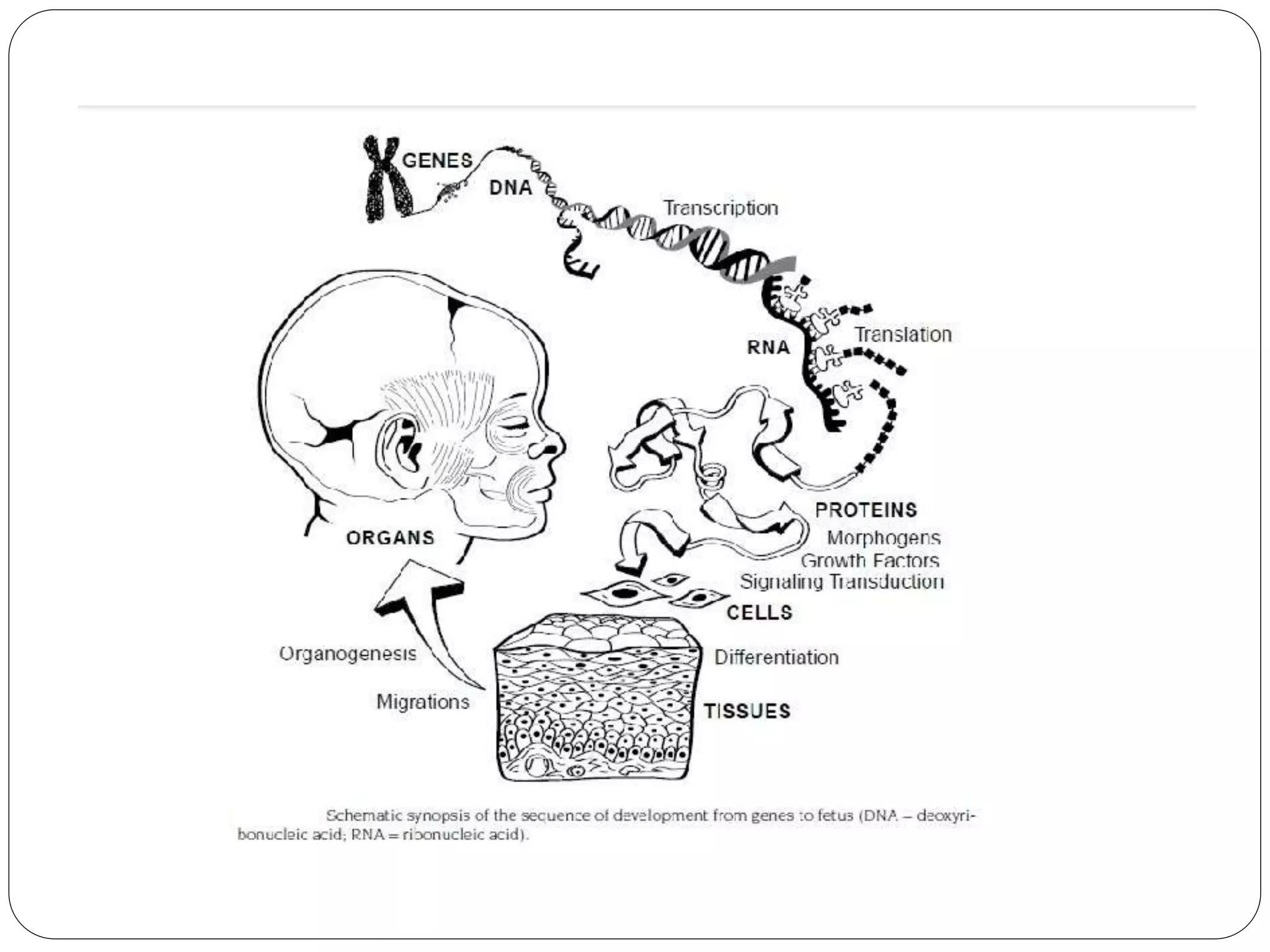
![GENETICS
Concept of gene as a unit of recombination refer as
DNA nucleiotide base pair
Embryologically gene as a unit of function is a
sequence of hundreds or thousands of nucleotides that
specify the sequence of aa [makes up the primary
structure of polypeptide]
Genome refers to the array of genes, haploid expressed
as a functional genotype in development
Physical and behavioral traits of an organism](https://image.slidesharecdn.com/sem3growthanddevelopmentoffaceparti-180520051643/75/growth-and-development-of-face-4-2048.jpg)
![ Gene controls the synthesis of protein formation
The longivity and proliferation of differentiated cells is
also genetically determined by
• continuous mitotics [short life spans]
• Intermittent mitotics
• Post mitotics [long life span]](https://image.slidesharecdn.com/sem3growthanddevelopmentoffaceparti-180520051643/75/growth-and-development-of-face-6-2048.jpg)
![SIGNAL TRANSDUCTION
Transcription regulate the identity and patterning of
embryonic structures and development of individual
organs
A signaling center or node (eg, Hensen’s node) [cell
group that regulates the behavior of surrounding cells by
producing positive and negative intercellular signaling
molecules]
Growth factors stimulate cell proliferation and
differentiation by acting through specific receptors on
responsive cells](https://image.slidesharecdn.com/sem3growthanddevelopmentoffaceparti-180520051643/75/growth-and-development-of-face-7-2048.jpg)
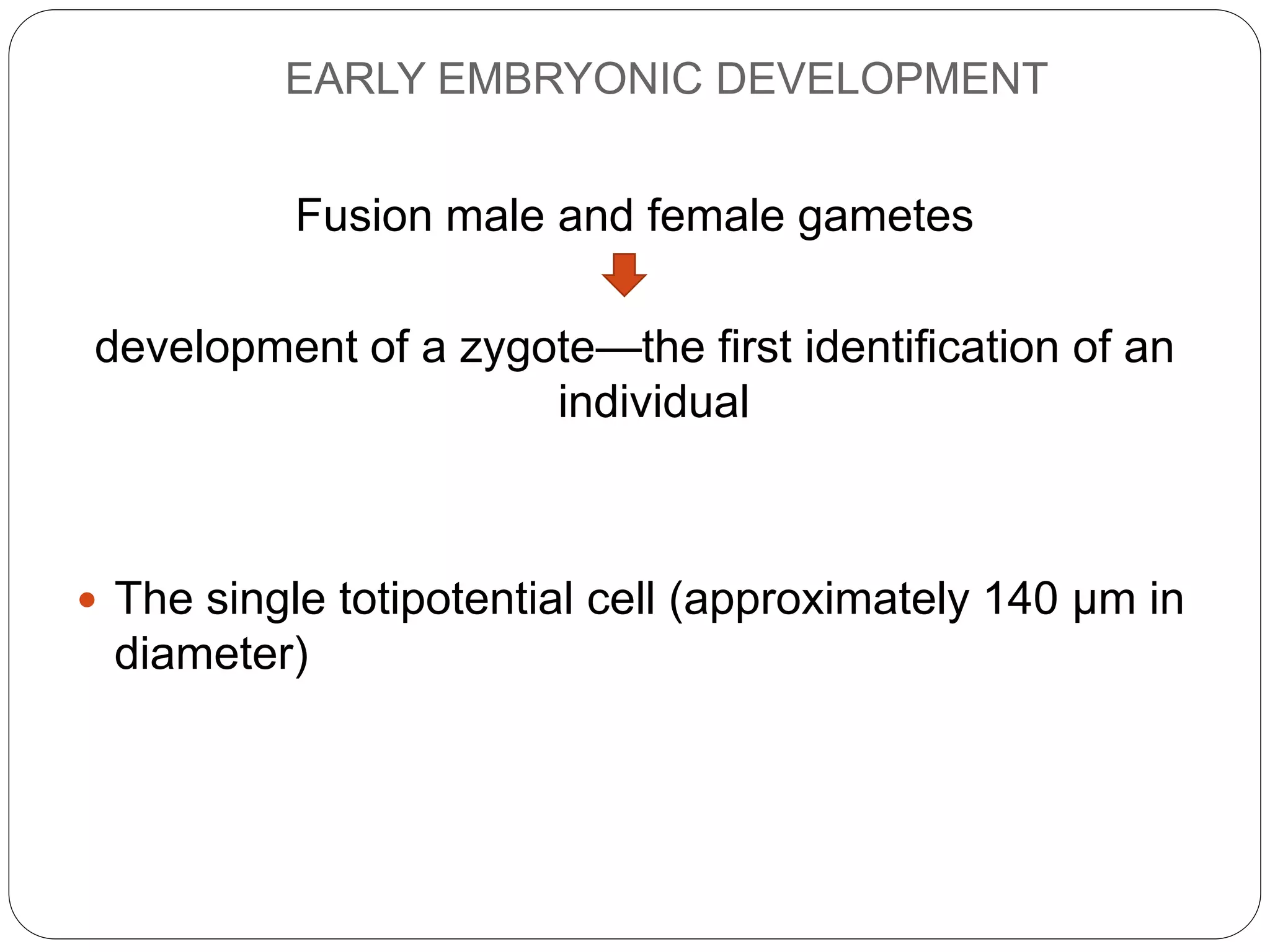
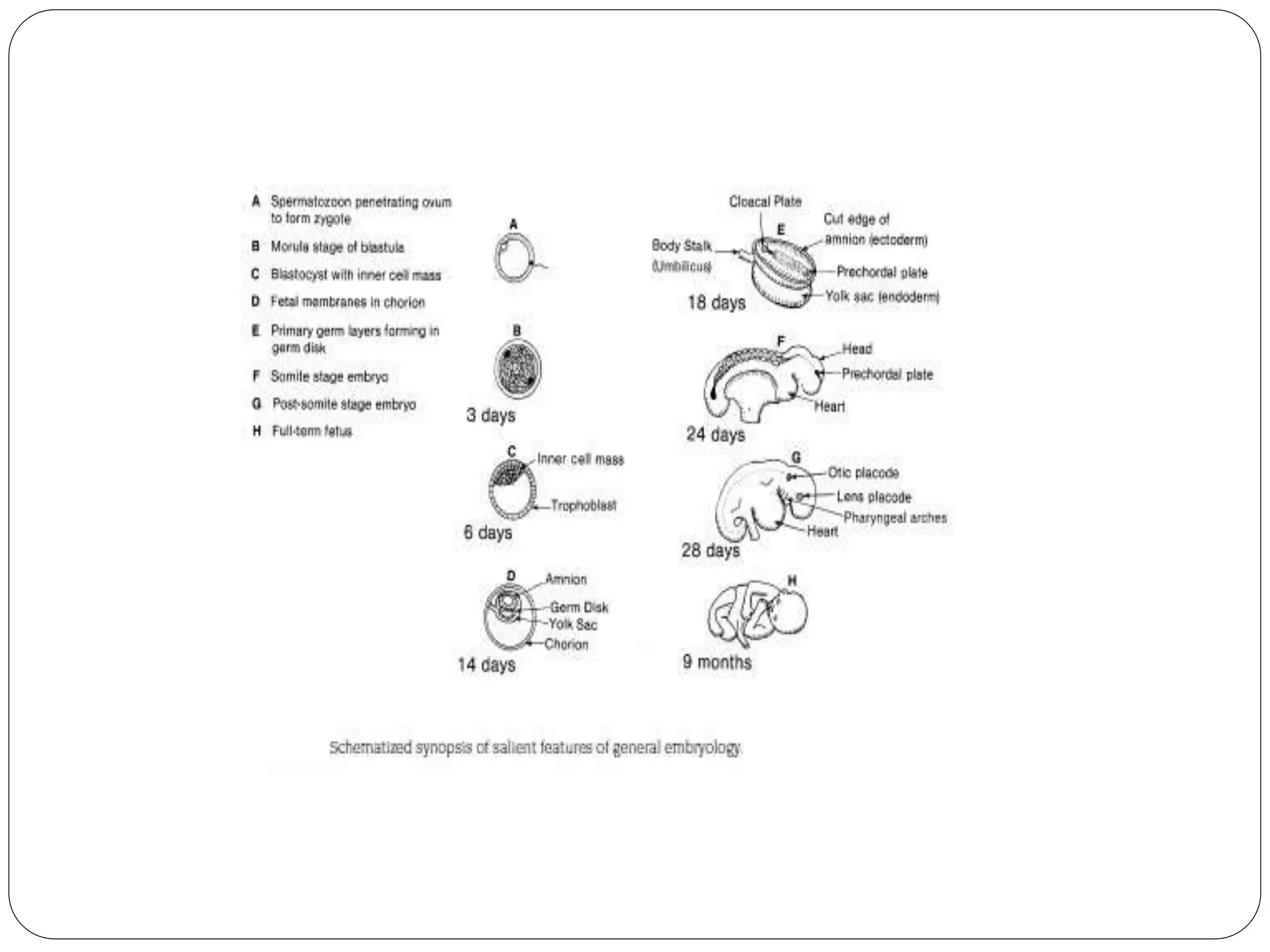
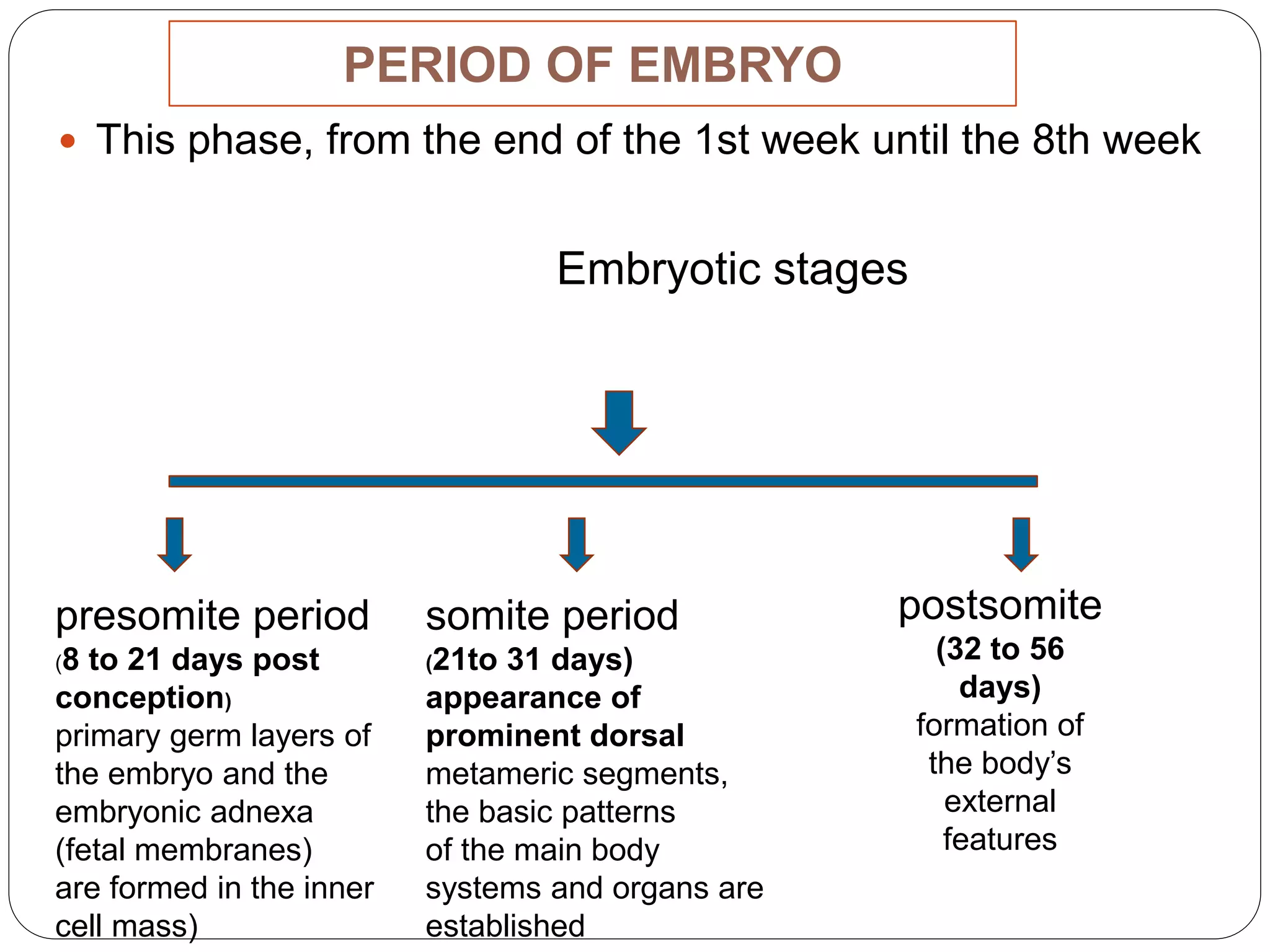
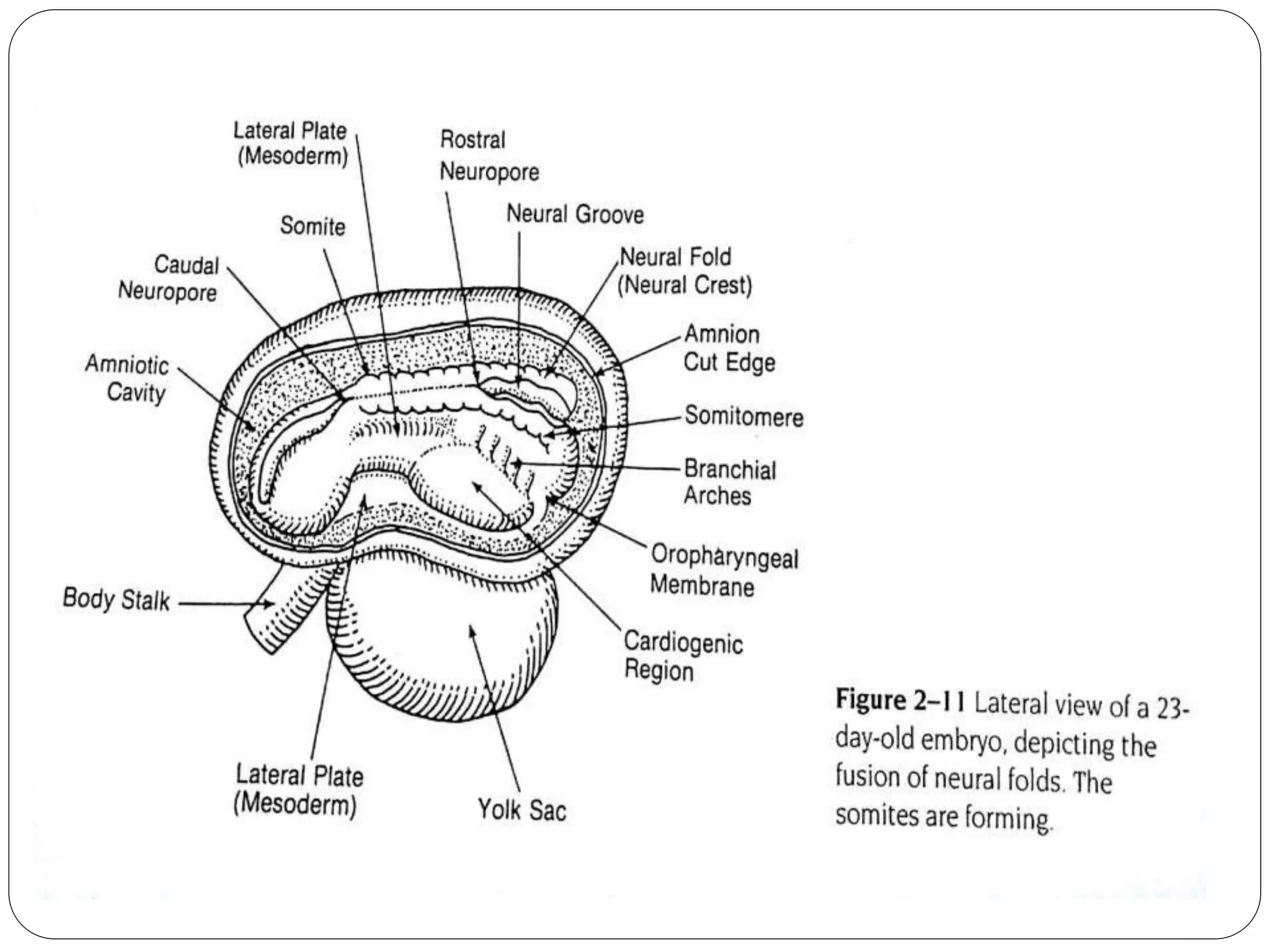
![EMBRYOGENESIS
Phases
Preimplantation
period
[first 7 days]
Fetal
period
[ the next
7
calendar
months]
Embroyoni
c period
[the next 7
weeks]](https://image.slidesharecdn.com/sem3growthanddevelopmentoffaceparti-180520051643/75/growth-and-development-of-face-12-2048.jpg)
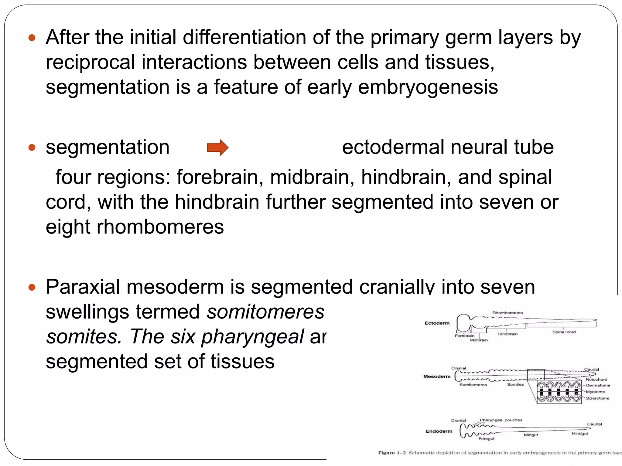
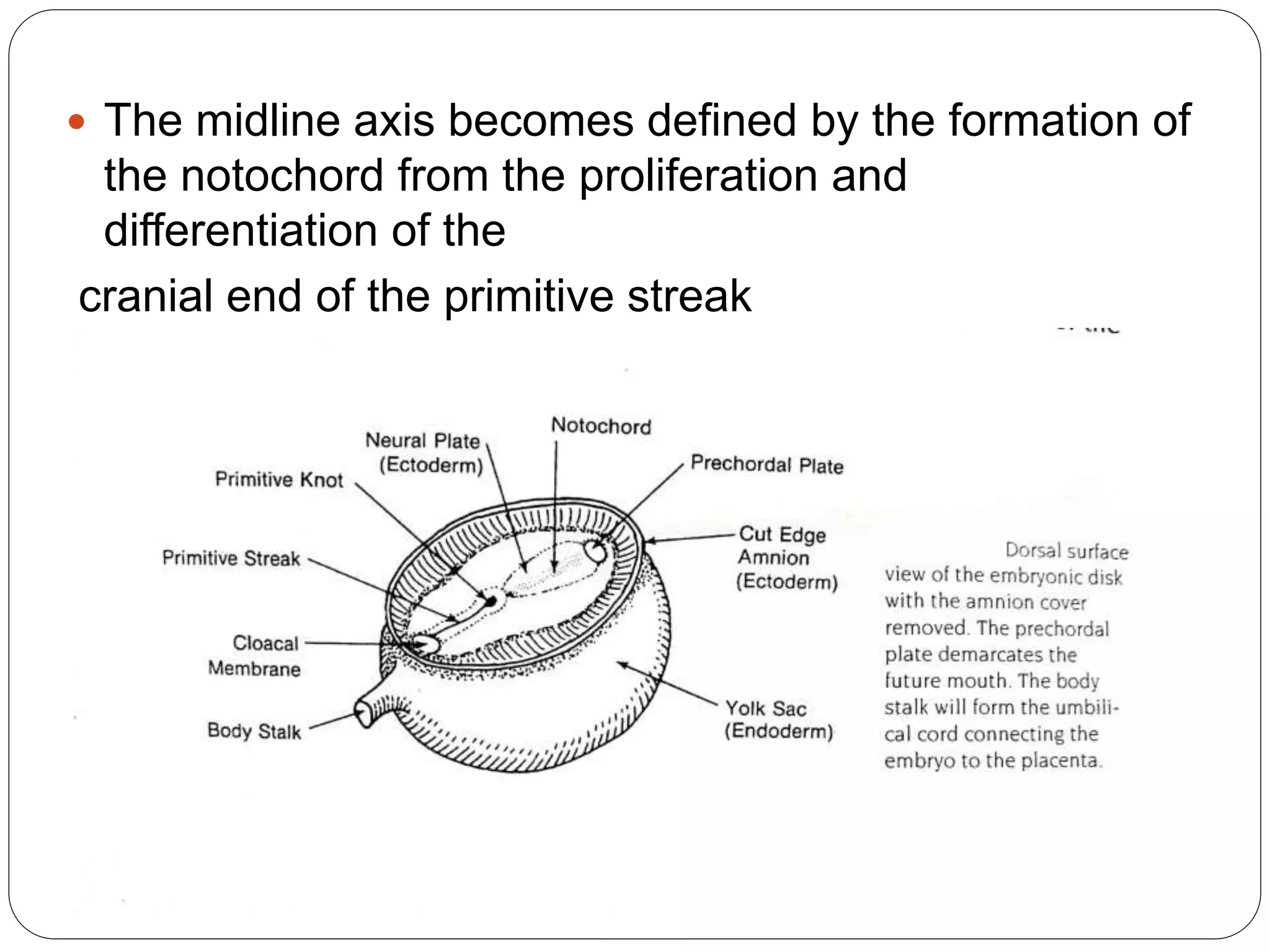
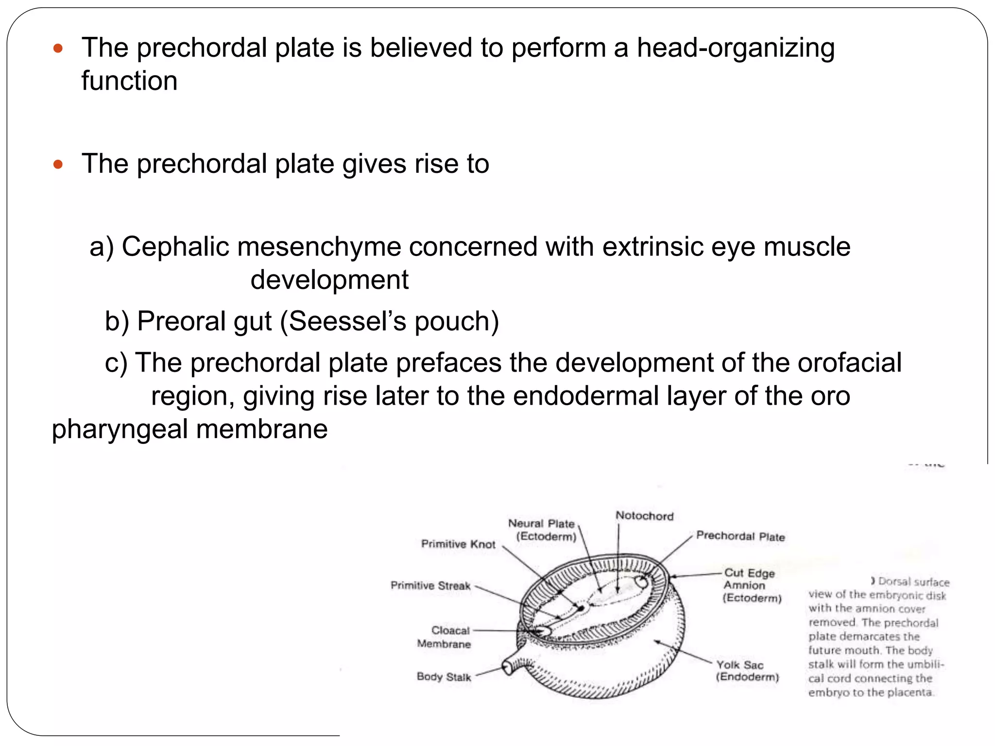
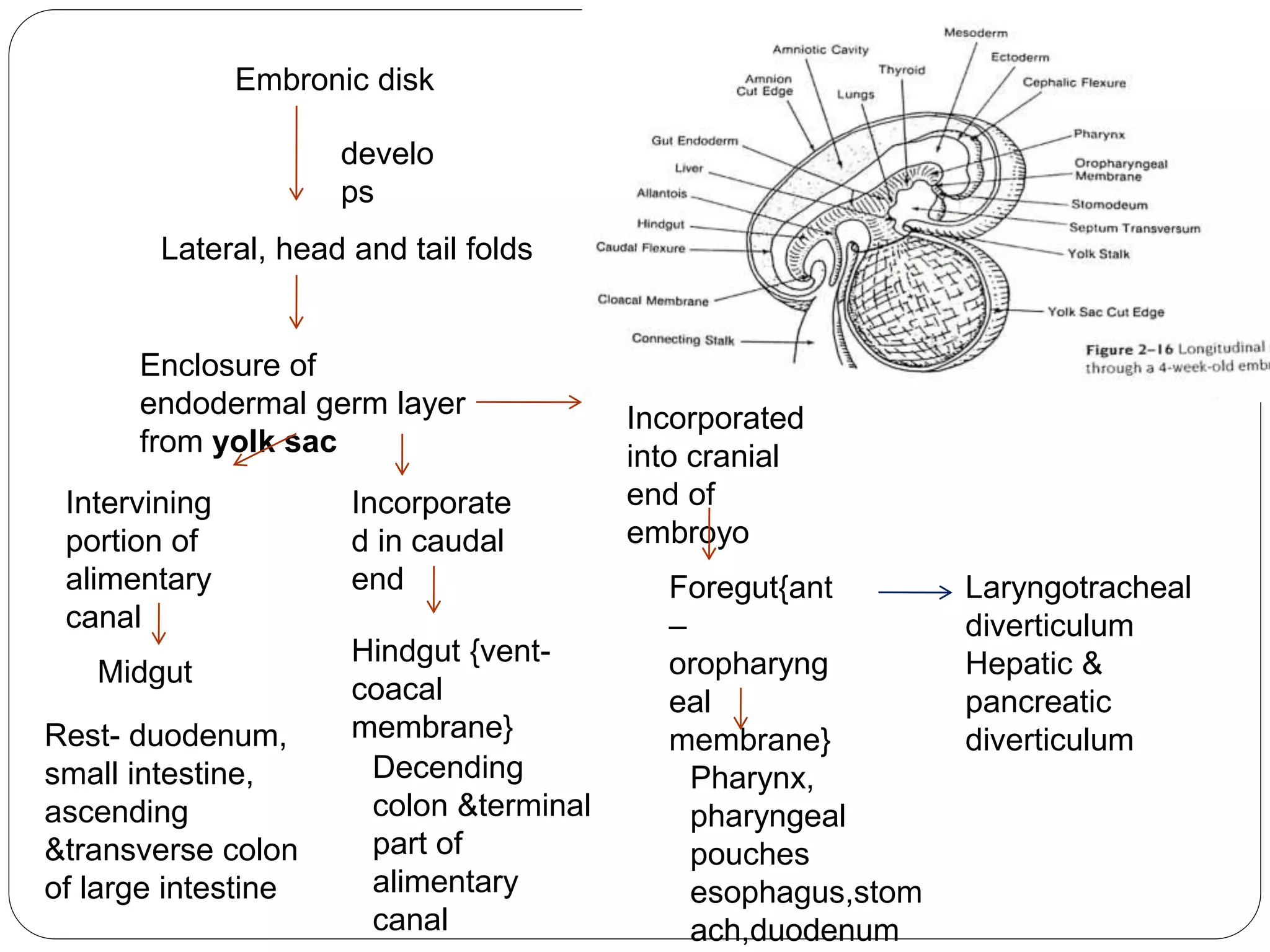
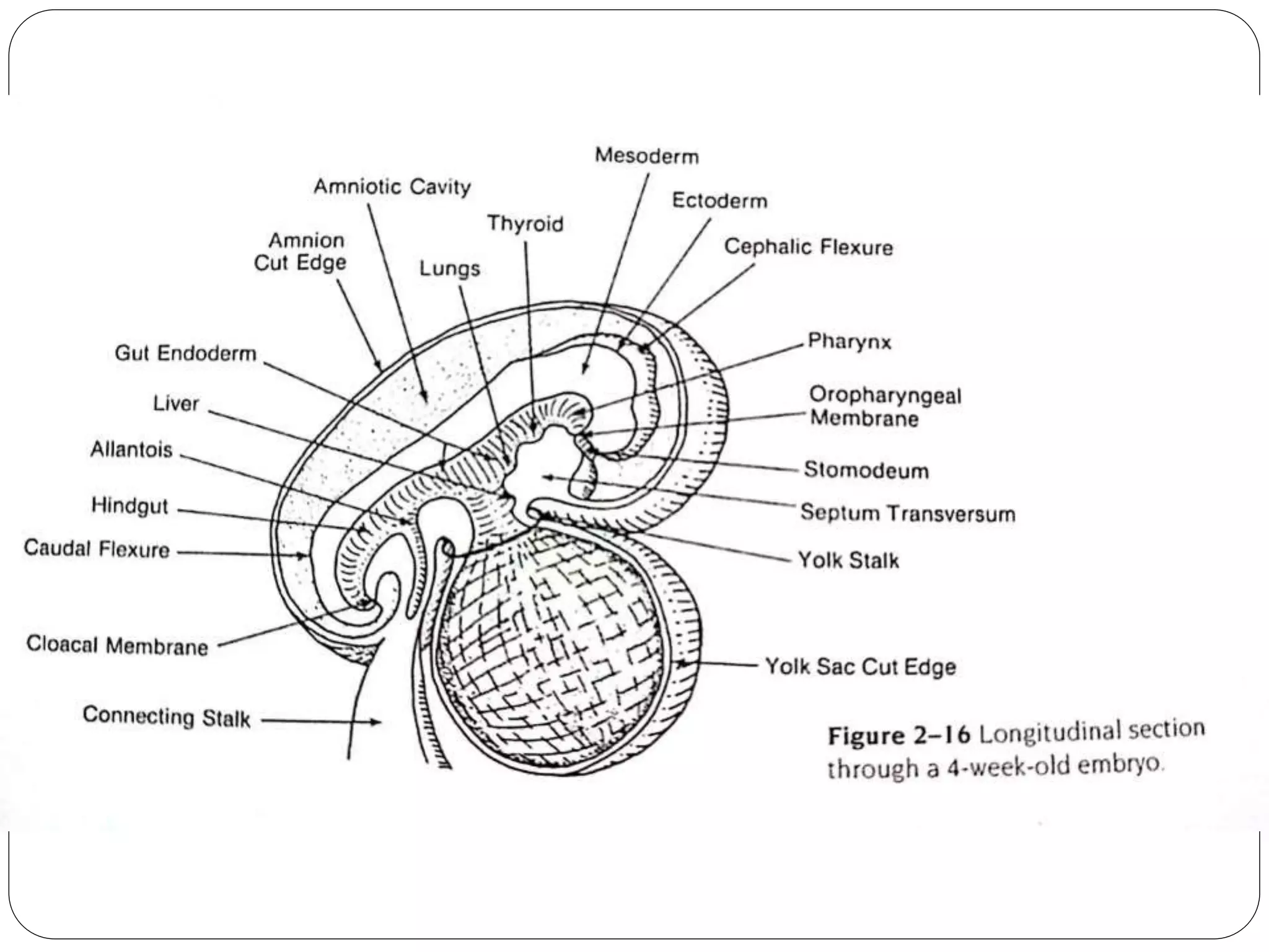
![PRESOMITE PERIOD
Development of Endoderm and Ectoderm
The primordial embryonic germ disk is composed of
two primary germ layers:
• The Ectoderm floor of the amniotic cavity
• Endoderm roof of the yolk sac
14th day endodermal thickening, the prechordal plate,
appears in the future midcephalic region [head-
organizing function]](https://image.slidesharecdn.com/sem3growthanddevelopmentoffaceparti-180520051643/75/growth-and-development-of-face-16-2048.jpg)
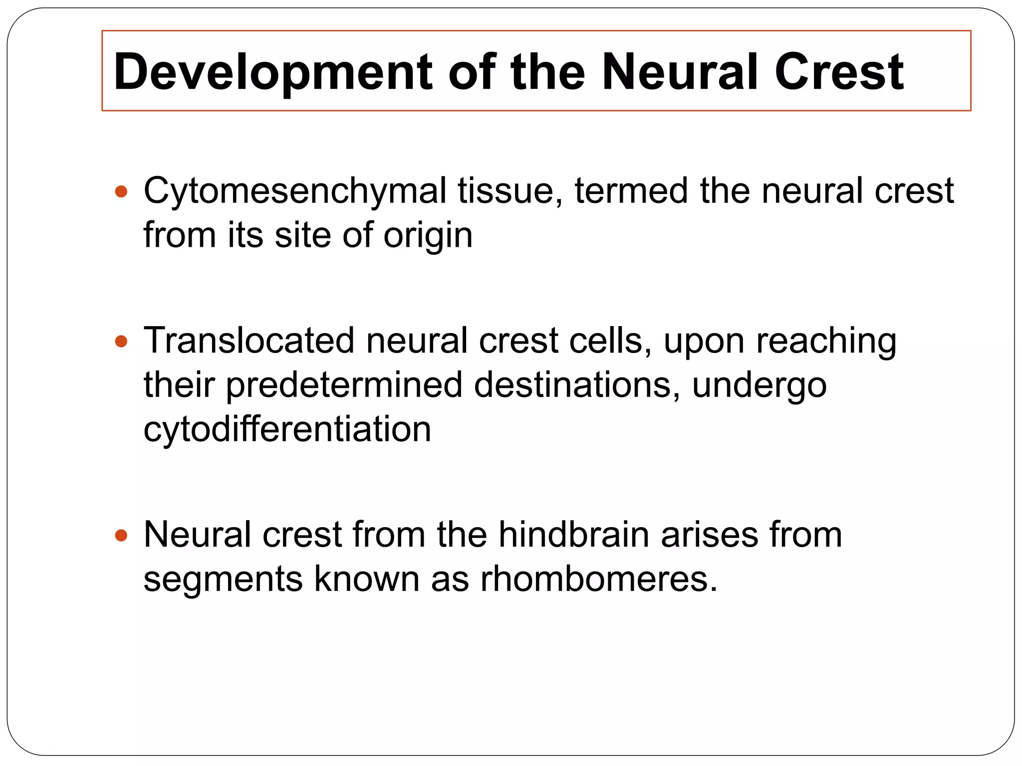
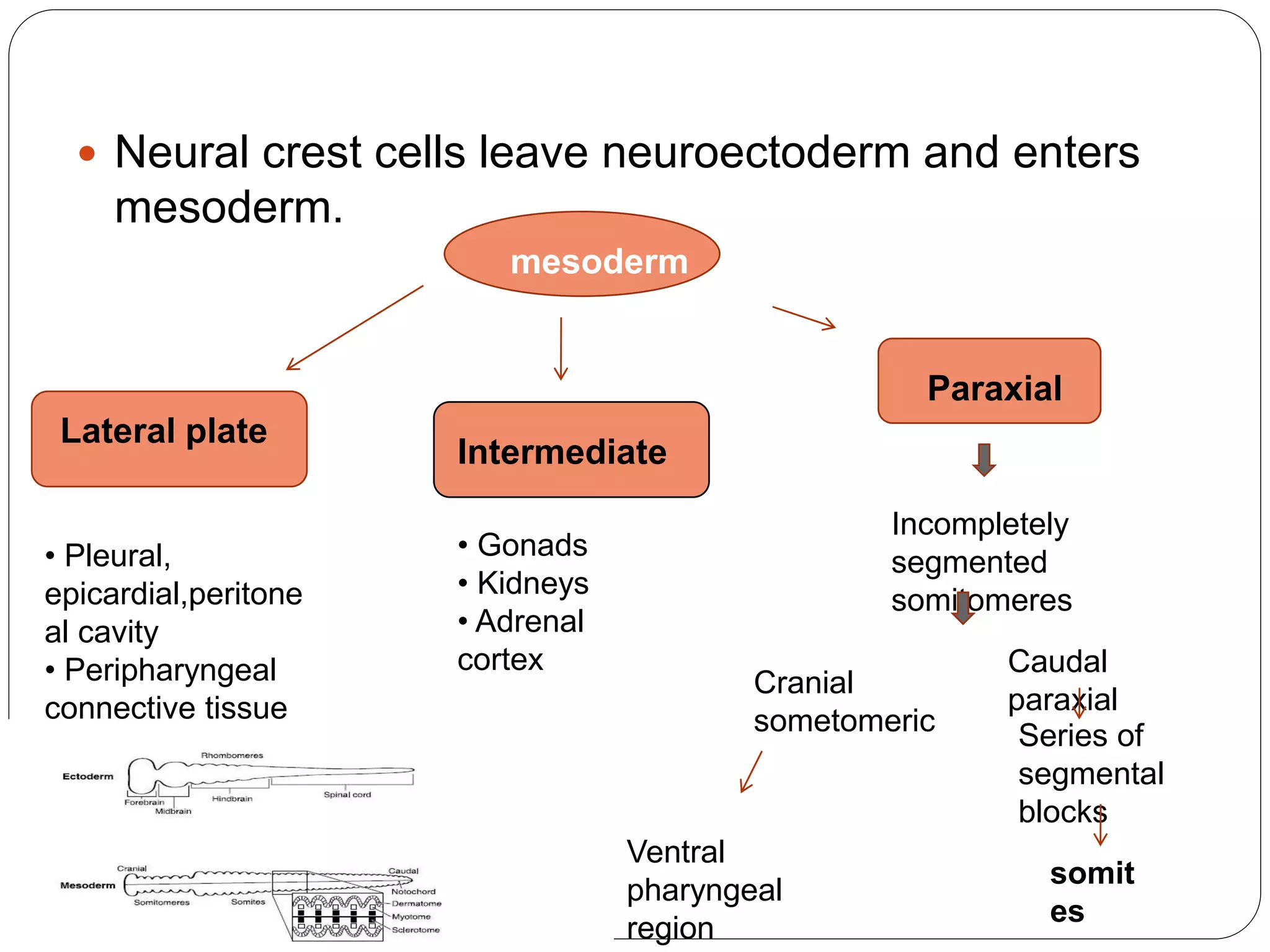
![SOMITE
Ventromedial part
[Sclerotome]
Lateral
aspect
[Dermatome]
Intermediate
portion
[Myotome]
Vertebral
column
(segmental
nature),
occipital bone
(fusion)
Dermis of
skin
Muscles
orofacial
region,
trunks,limb
s](https://image.slidesharecdn.com/sem3growthanddevelopmentoffaceparti-180520051643/75/growth-and-development-of-face-26-2048.jpg)
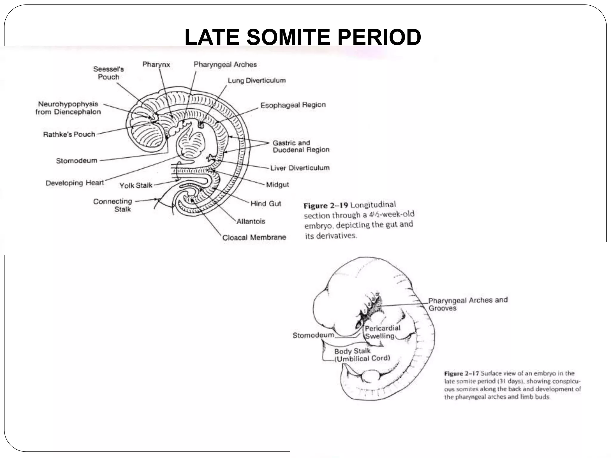


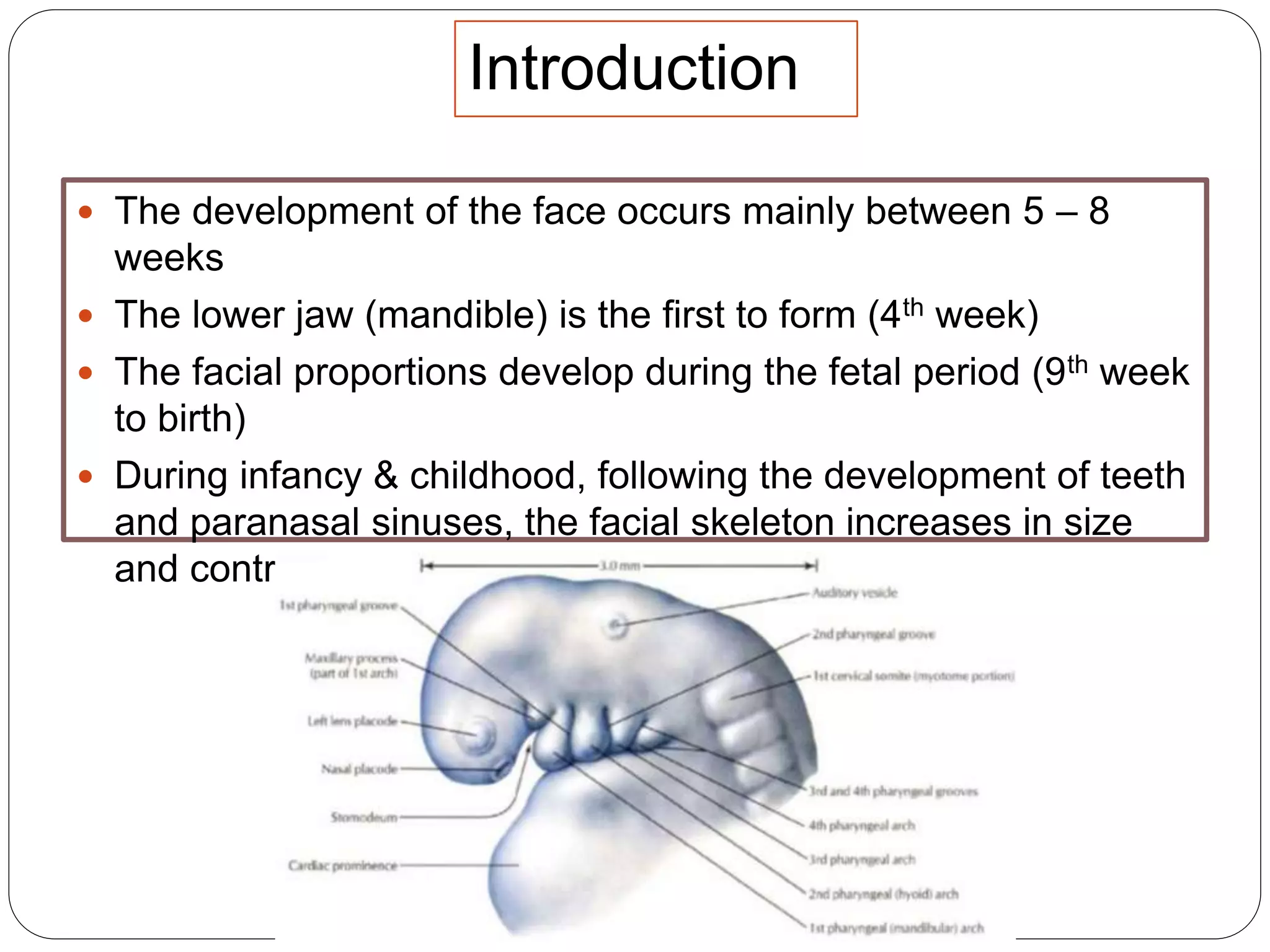
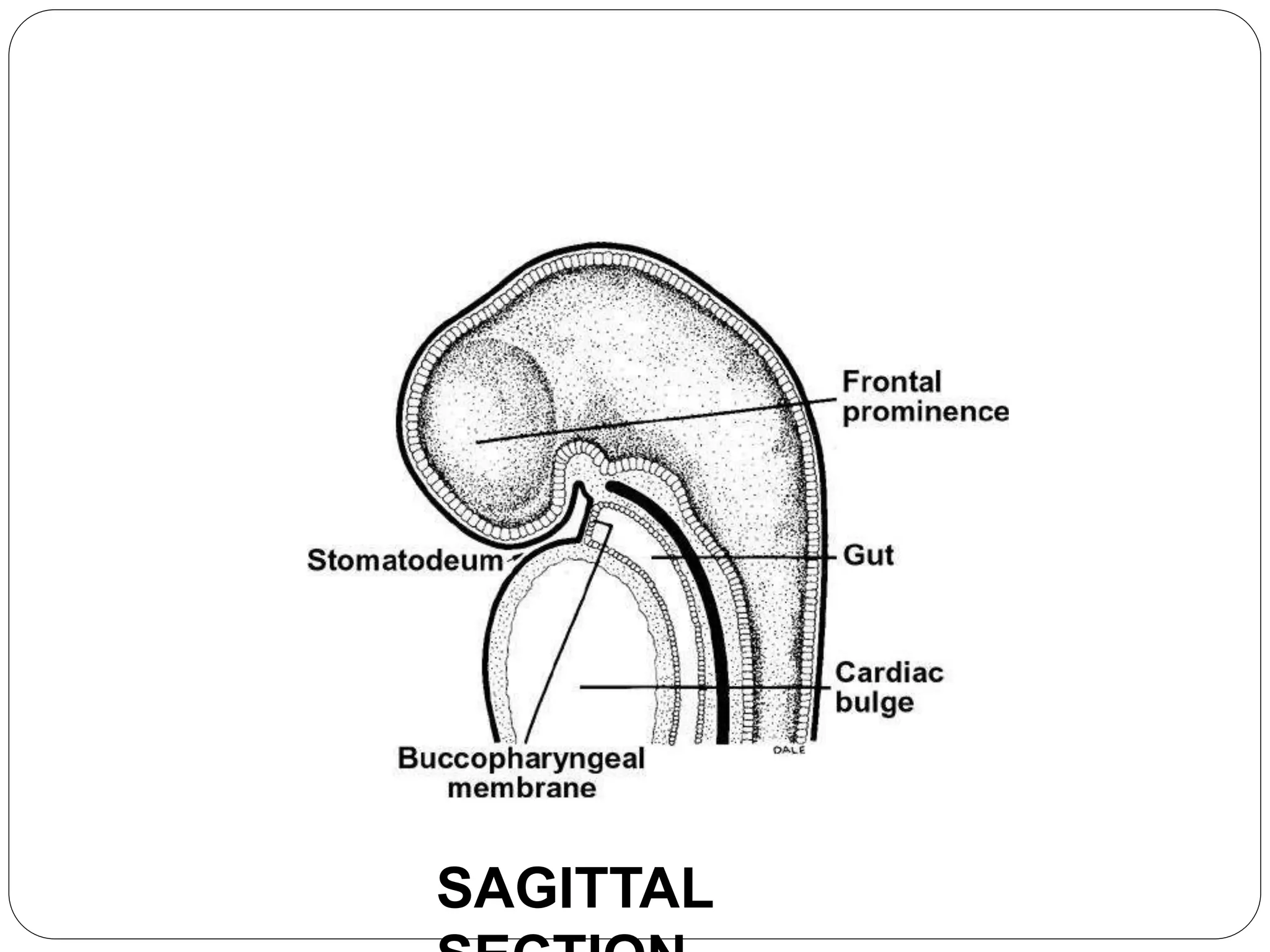


This document discusses embryogenesis and prenatal face formation. It covers early embryonic development from fertilization through the fetal period. Key points include: - Embryogenesis occurs in three main periods: preimplantation, embryonic, and fetal. - The pharyngeal arches play an important role in face development, with each arch contributing muscles, nerves, and blood vessels. - Organ systems like the cardiovascular, digestive, and nervous systems undergo differentiation during the somite period from 3-4 weeks. - The postsomite period from 5-8 weeks sees formation of external features and digit development. The embryo is now termed a fetus.


