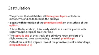
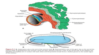
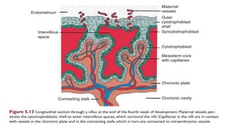
Third week of development
Themba Hospital FCOG(SA) Part 1 Tutorials
By Dr N.E Manana
Gastrulation
• The process that establishes all three germ layers (ectoderm,
mesoderm, and endoderm) in the embryo
• Begins with formation of the primitive streak on the surface of the
epiblast
• 15- to 16-day embryo, it is clearly visible as a narrow groove with
slightly bulging regions on either side
• The cephalic end of the streak, the primitive node, consists of a
slightly elevated area surrounding the small primitive pit
• Cells of the epiblast migrate toward the primitive streak and undergo
invagination (FGF8)
Gastrulation
• Once the cells have invaginated, some displace the hypoblast, creating the
embryonic endoderm
• Others come to lie between the epiblast and newly created endoderm to
form mesoderm
• Cells remaining in the epiblast then form ectoderm
• Thus, the epiblast, through the process of gastrulation, is the source of all
of the germ layers
• More cells move between the epiblast and hypoblast layers, they begin to
spread laterally and cranially
• They migrate beyond the margin of the disc and establish contact with the
extraembryonic mesoderm
Gastrulation
• In the cephalic direction, they pass on each side of the prechordal plate.
• Which is between the tip of the notochord and the oropharyngeal
membrane
• Later, the prechordal plate will be important for induction of the forebrain
• The oropharyngeal membrane at the cranial end of the disc consists of a
small region of tightly adherent ectoderm and endoderm cells that
represents the future opening of the oral cavity
Notochord
• Prenotochordal cells invaginating in the primitive node move forward
cranially in the midline until they reach the prechordal plate
• As the hypoblast is replaced by endoderm cells moving in at the streak,
cells of the notochordal plate proliferate and detach from the endoderm
• They then form a solid cord of cells, the definitive notochord.
• The cranial end forms first, and caudal regions are added as the primitive
streak assumes a more caudal position
• The cloacal membrane is formed at the caudal end of the embryonic disc
• When the cloacal membrane appears, the posterior wall of the yolk sac
forms a small diverticulum that extends into the connecting stalk (allantois;
appears around the 16th day of development)
Body axes
• Establishment of the body axes, anteroposterior, dorsoventral, and left–
right, takes place before and during the period of gastrulation
• The anteroposterior axis is signalled by cells at the anterior (cranial) margin
of the embryonic disc,
• Anterior visceral endoderm (AVE) expresses genes essential for head
formation
• Once the streak is formed, Nodal upregulates a number of genes
responsible for formation of dorsal and ventral mesoderm and head and
tail structures (TGF-b)
• Without HNF-3b, embryos fail to gastrulate properly and lack forebrain and
midbrain structures
Trophoblast development
• At the beginning of the 3rd week the trophoblast is characterised by
the primary villi.
• During further development , mesodermal cells penetrate the core of
the primitive villi and grow towards the decidua (Secondary villi)
• By the end of the third week, mesodermal cells in the core of the
villus begin to differentiate into blood cells and small blood vessels,
forming the villous capillary system (tertiary villus)
• These vessels, in turn, establish contact with the intraembryonic
circulatory system, connecting the placenta and the embryo
chorionic cavity
• Becomes larger, and by the 19th or the 20th day, the embryo is
attached to its trophoblastic shell by a narrow connecting stalk
• The connecting stalk later develops into the umbilical cord
Thank you

3rd week

  • 1.
    Third week ofdevelopment Themba Hospital FCOG(SA) Part 1 Tutorials By Dr N.E Manana
  • 2.
    Gastrulation • The processthat establishes all three germ layers (ectoderm, mesoderm, and endoderm) in the embryo • Begins with formation of the primitive streak on the surface of the epiblast • 15- to 16-day embryo, it is clearly visible as a narrow groove with slightly bulging regions on either side • The cephalic end of the streak, the primitive node, consists of a slightly elevated area surrounding the small primitive pit • Cells of the epiblast migrate toward the primitive streak and undergo invagination (FGF8)
  • 4.
    Gastrulation • Once thecells have invaginated, some displace the hypoblast, creating the embryonic endoderm • Others come to lie between the epiblast and newly created endoderm to form mesoderm • Cells remaining in the epiblast then form ectoderm • Thus, the epiblast, through the process of gastrulation, is the source of all of the germ layers • More cells move between the epiblast and hypoblast layers, they begin to spread laterally and cranially • They migrate beyond the margin of the disc and establish contact with the extraembryonic mesoderm
  • 6.
    Gastrulation • In thecephalic direction, they pass on each side of the prechordal plate. • Which is between the tip of the notochord and the oropharyngeal membrane • Later, the prechordal plate will be important for induction of the forebrain • The oropharyngeal membrane at the cranial end of the disc consists of a small region of tightly adherent ectoderm and endoderm cells that represents the future opening of the oral cavity
  • 8.
    Notochord • Prenotochordal cellsinvaginating in the primitive node move forward cranially in the midline until they reach the prechordal plate • As the hypoblast is replaced by endoderm cells moving in at the streak, cells of the notochordal plate proliferate and detach from the endoderm • They then form a solid cord of cells, the definitive notochord. • The cranial end forms first, and caudal regions are added as the primitive streak assumes a more caudal position • The cloacal membrane is formed at the caudal end of the embryonic disc • When the cloacal membrane appears, the posterior wall of the yolk sac forms a small diverticulum that extends into the connecting stalk (allantois; appears around the 16th day of development)
  • 9.
    Body axes • Establishmentof the body axes, anteroposterior, dorsoventral, and left– right, takes place before and during the period of gastrulation • The anteroposterior axis is signalled by cells at the anterior (cranial) margin of the embryonic disc, • Anterior visceral endoderm (AVE) expresses genes essential for head formation • Once the streak is formed, Nodal upregulates a number of genes responsible for formation of dorsal and ventral mesoderm and head and tail structures (TGF-b) • Without HNF-3b, embryos fail to gastrulate properly and lack forebrain and midbrain structures
  • 10.
    Trophoblast development • Atthe beginning of the 3rd week the trophoblast is characterised by the primary villi. • During further development , mesodermal cells penetrate the core of the primitive villi and grow towards the decidua (Secondary villi) • By the end of the third week, mesodermal cells in the core of the villus begin to differentiate into blood cells and small blood vessels, forming the villous capillary system (tertiary villus) • These vessels, in turn, establish contact with the intraembryonic circulatory system, connecting the placenta and the embryo
  • 13.
    chorionic cavity • Becomeslarger, and by the 19th or the 20th day, the embryo is attached to its trophoblastic shell by a narrow connecting stalk • The connecting stalk later develops into the umbilical cord
  • 14.

Editor's Notes

  • #3 Cell migration and specification are controlled by fi broblast growth factor 8 (FGF8), which is synthesized by streak cells themselves.
  • #5  In the cephalic direction, they pass on each side of the prechordal plate. Which is between the tip of the notochord and the oropharyngeal membrane and is derived from some of the fi rst cells that migrate through the node in the midline and move in a cephalic direction
  • #9  Notochord which underlies the neural tube and serves as the basis for the axial skeleton
  • #10  Thus, mesoderm formation in these regions depends on this gene product, and its absence results in shortening of the embryonic axis (caudal dysgenesis).
  • #11  Hence, when the heart begins to beat in the fourth week of development, the villous system is ready to supply the embryo proper with essential nutrients and oxygen. cytotrophoblast shell , Villi that extend from the chorionic plate to the decidua basalis (decidual plate: the part of the endometrium where the placenta will form; see Chapter 8) are called stem or anchoring villi Those that branch from the sides of stem villi are free (terminal) villi, through which exchange of nutrients and other factors will occur