Downloaded 152 times
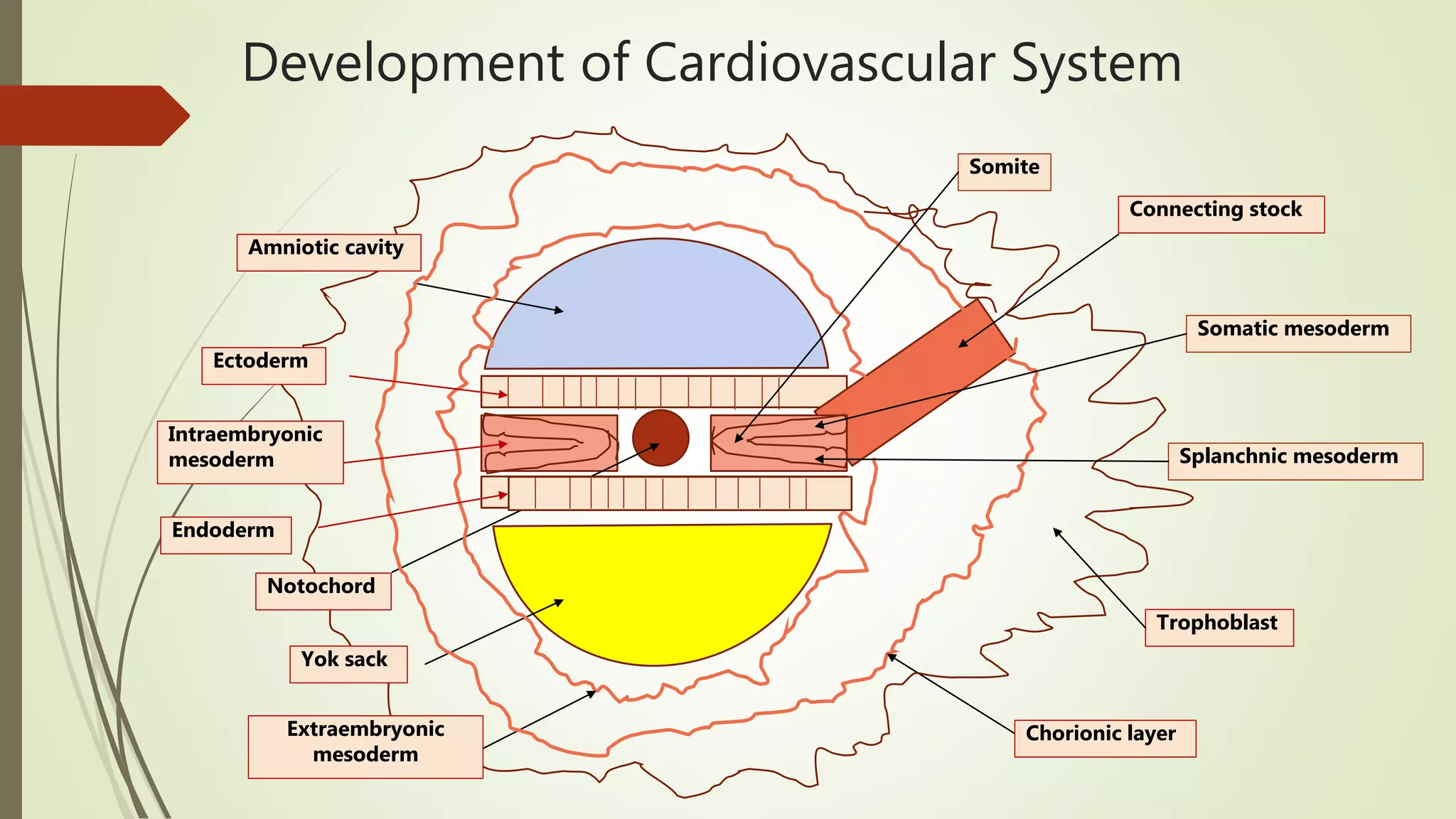

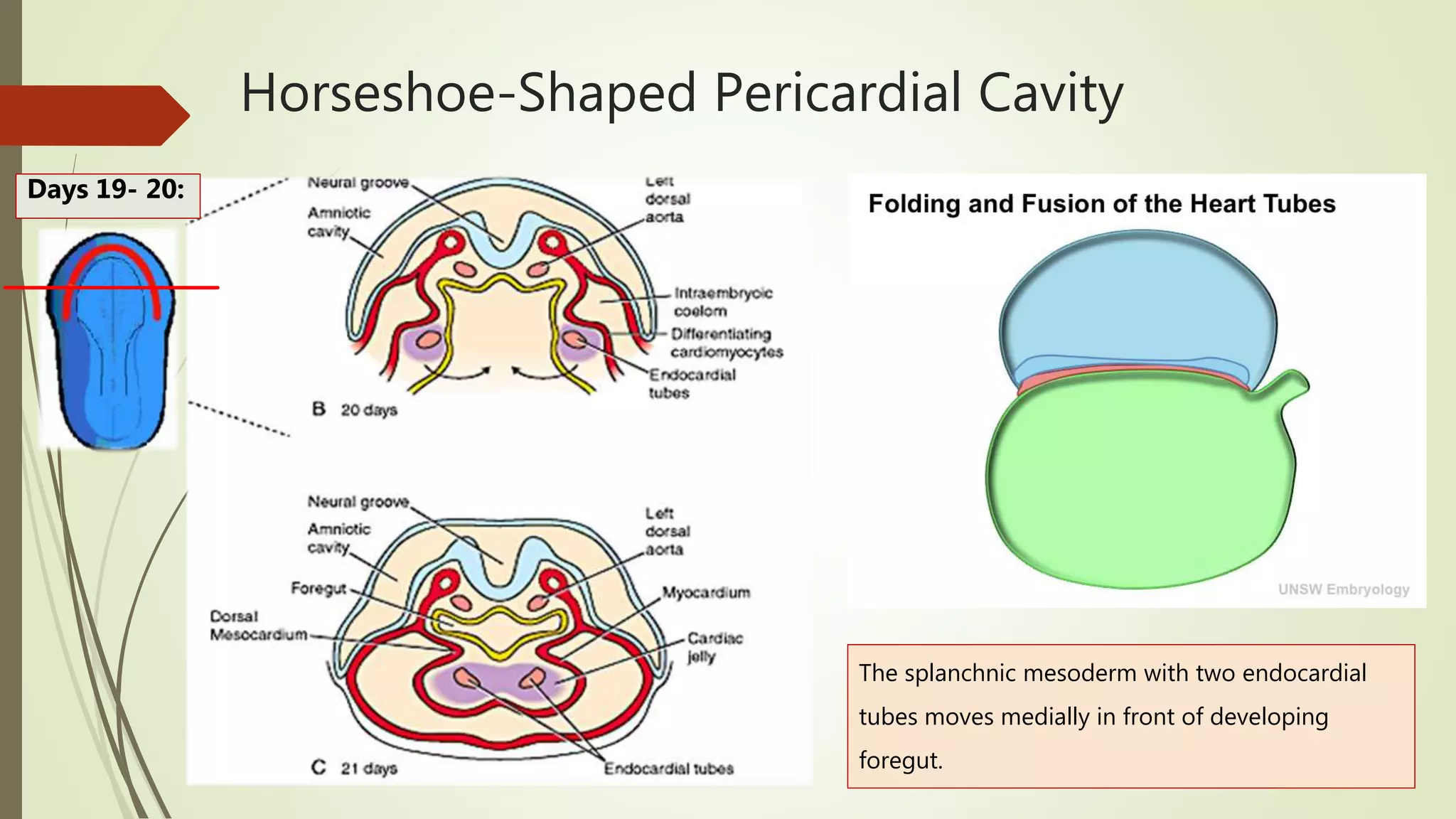
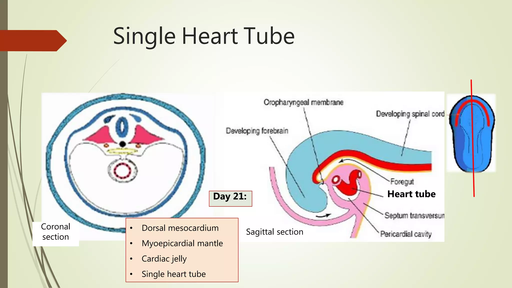
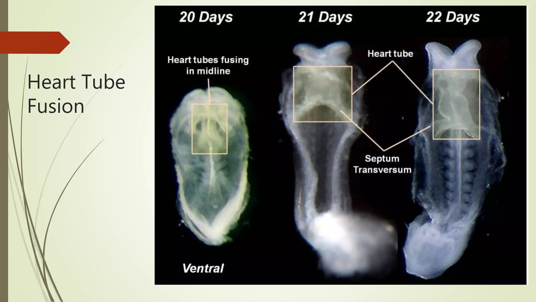
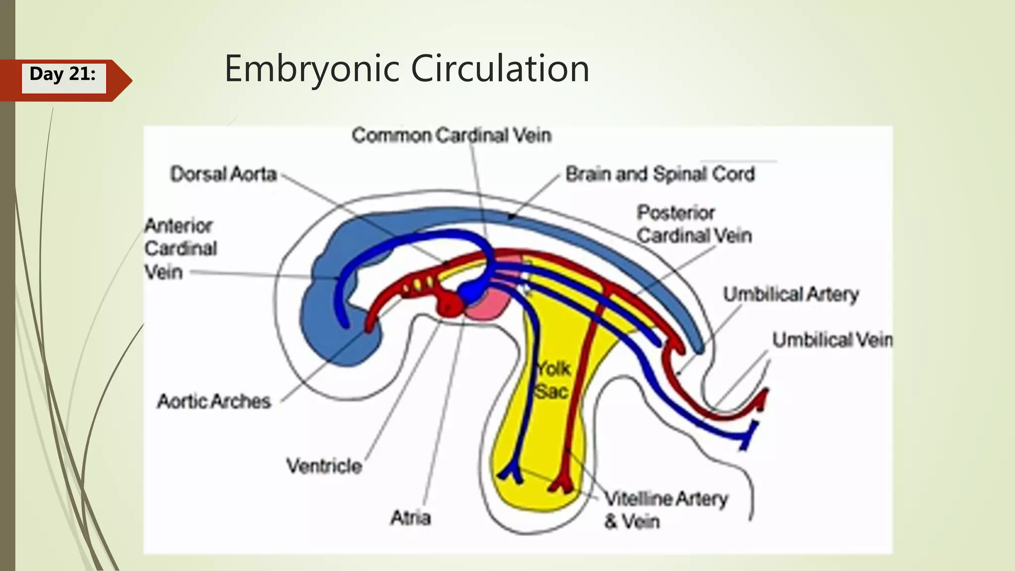

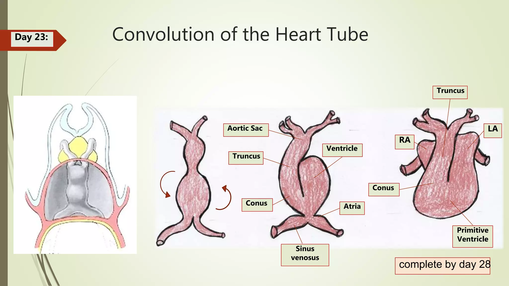

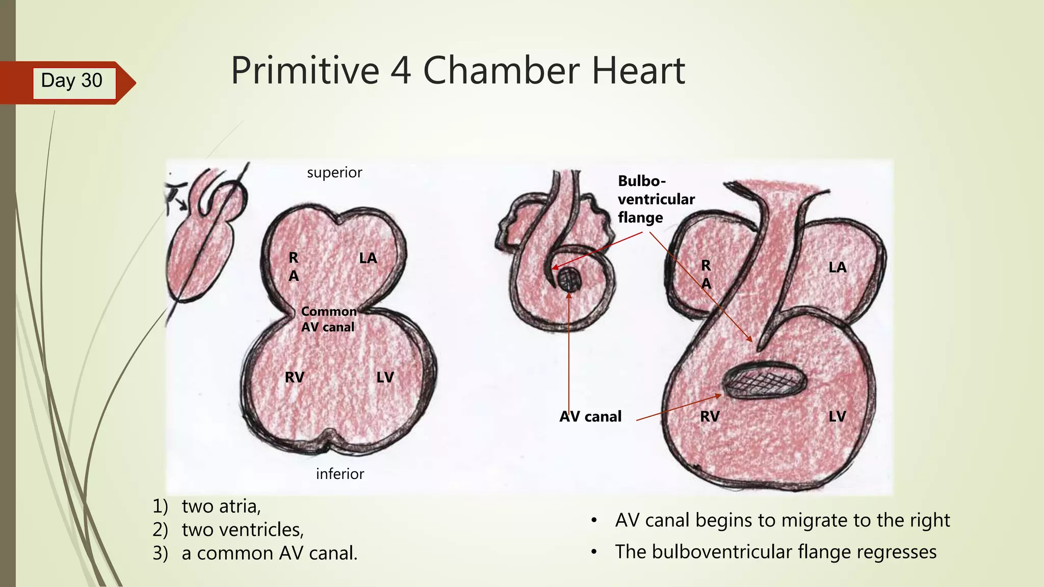
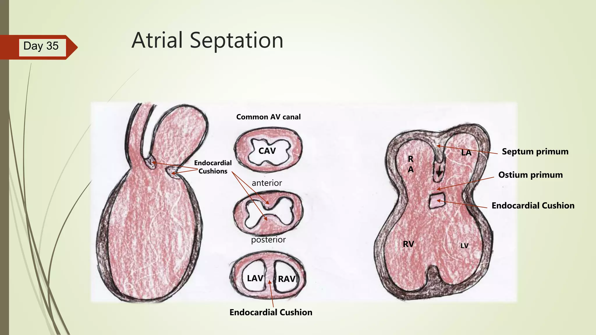
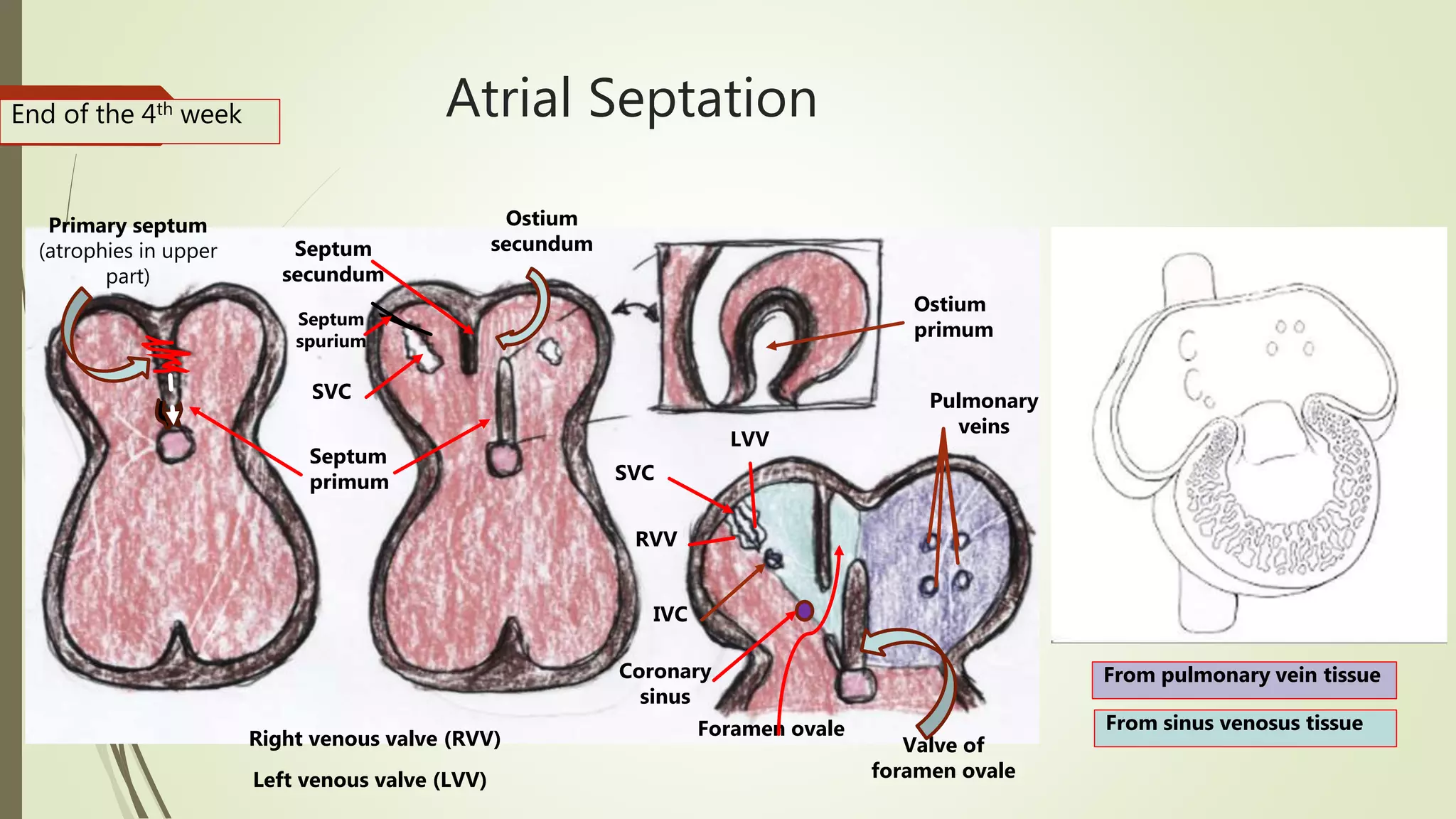
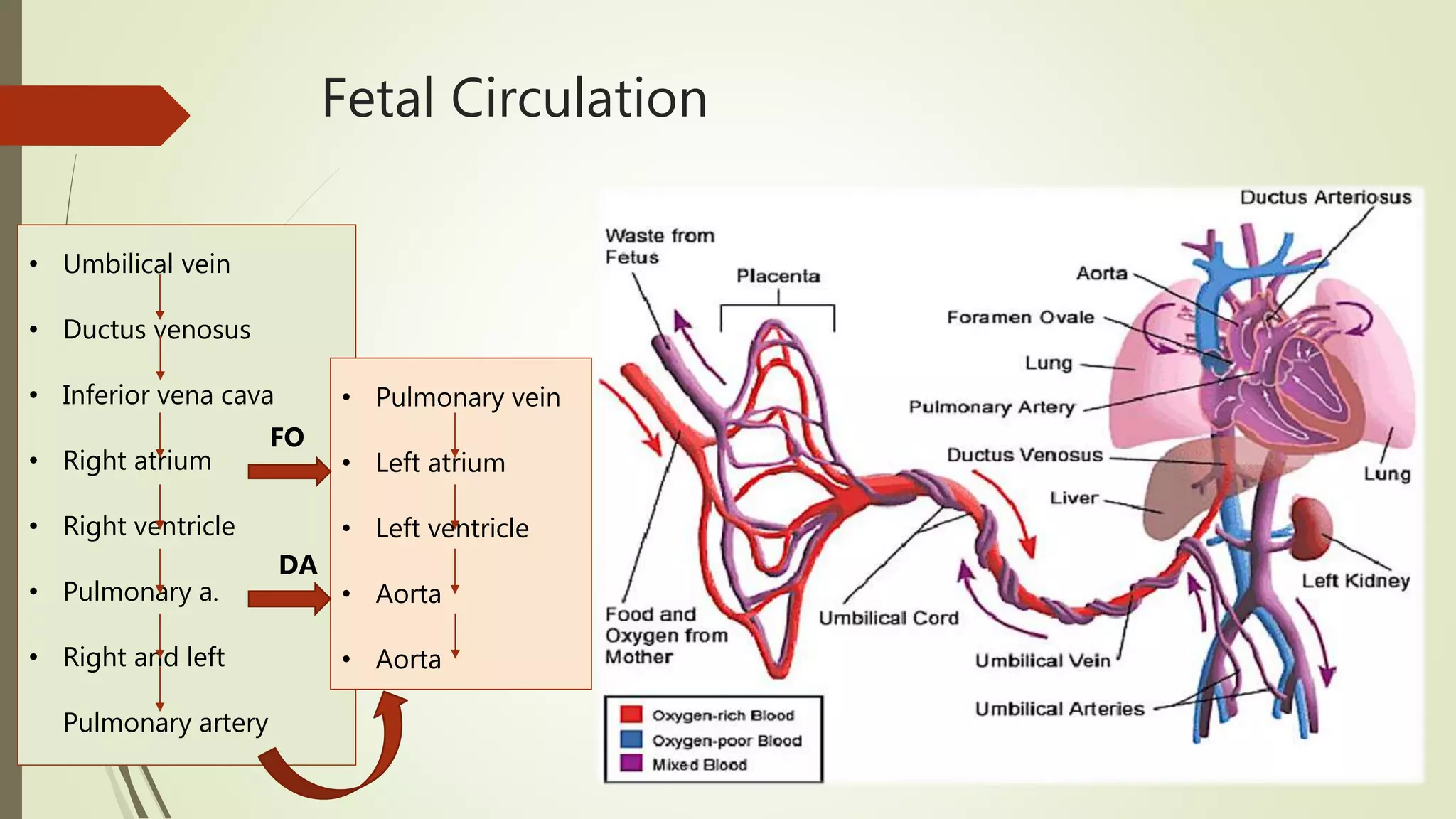

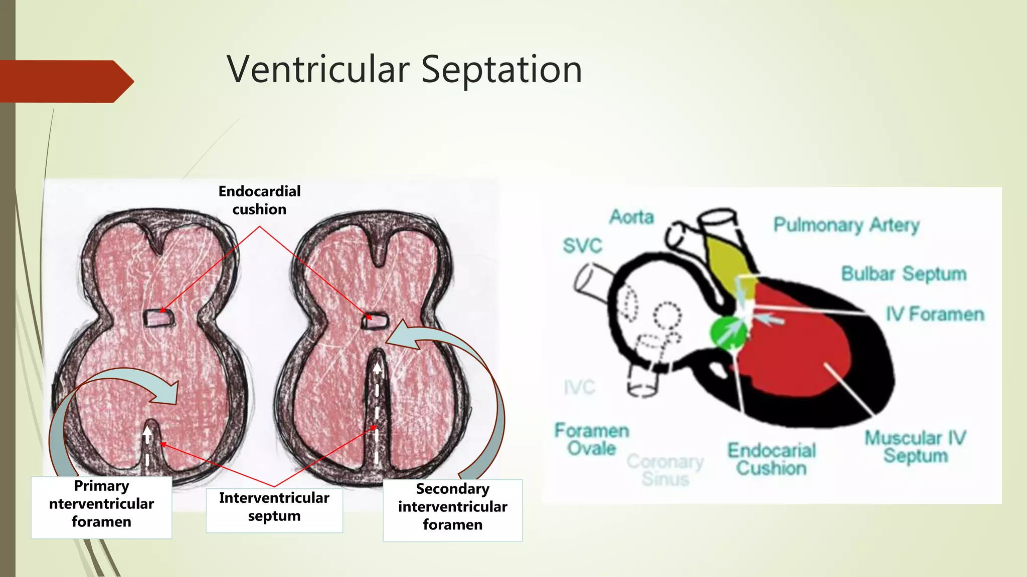


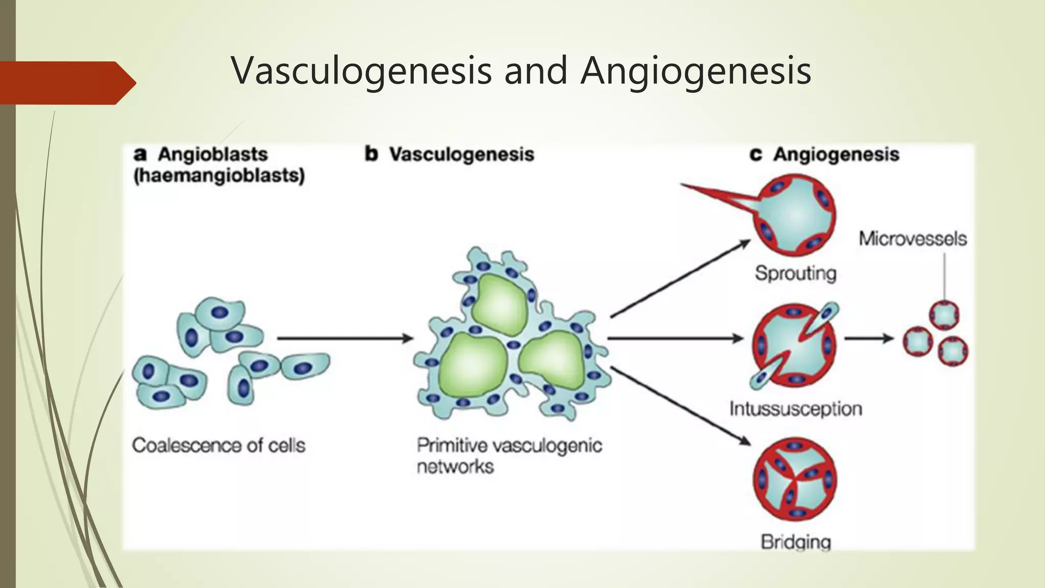
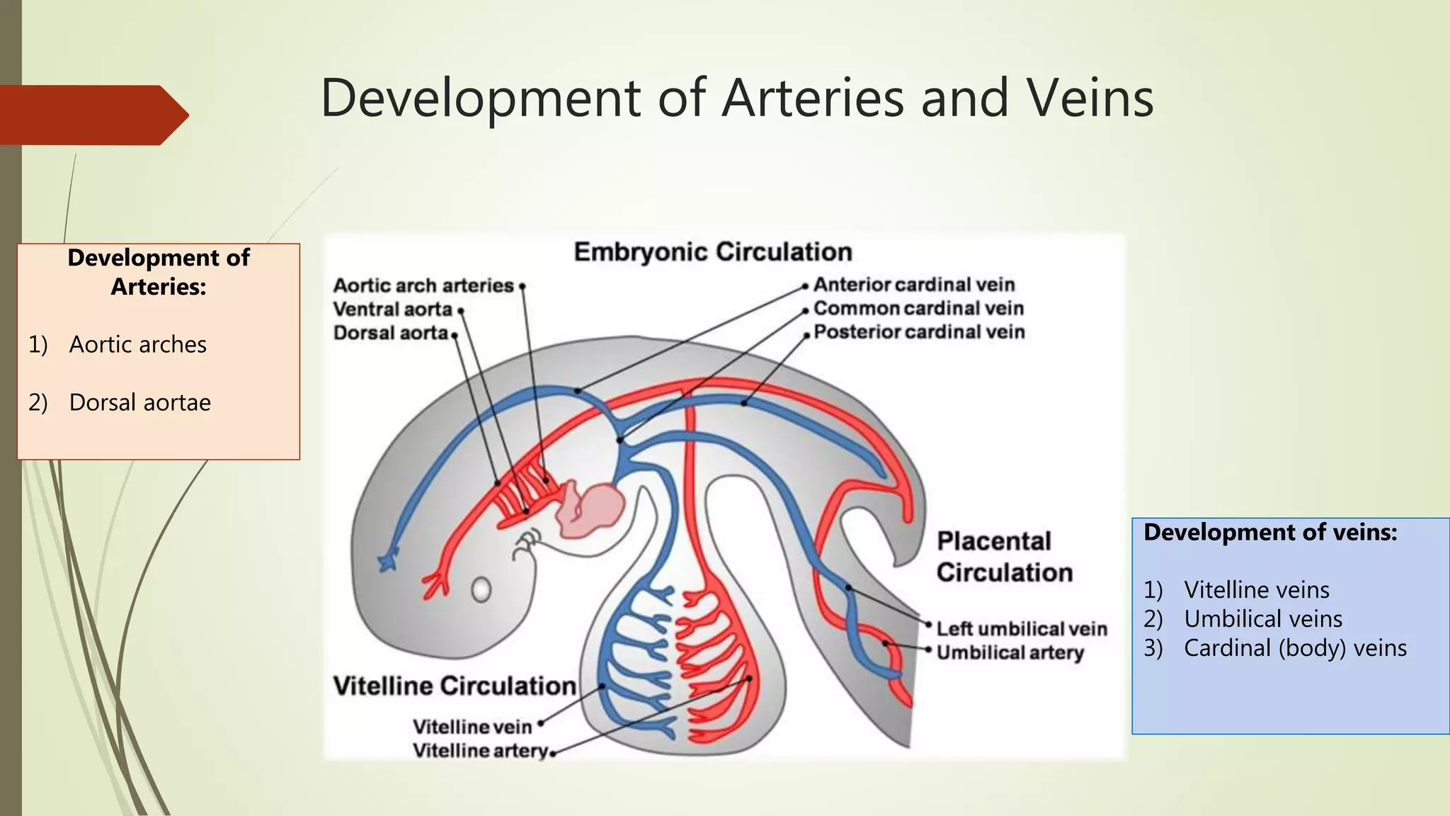

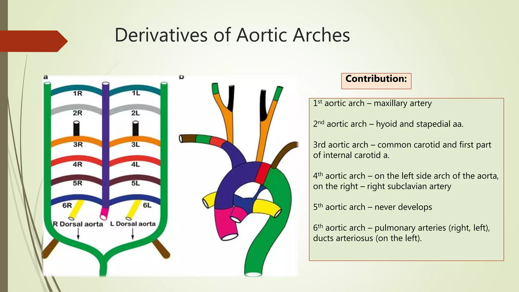

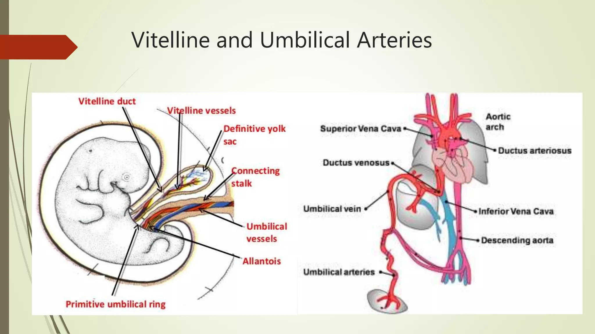
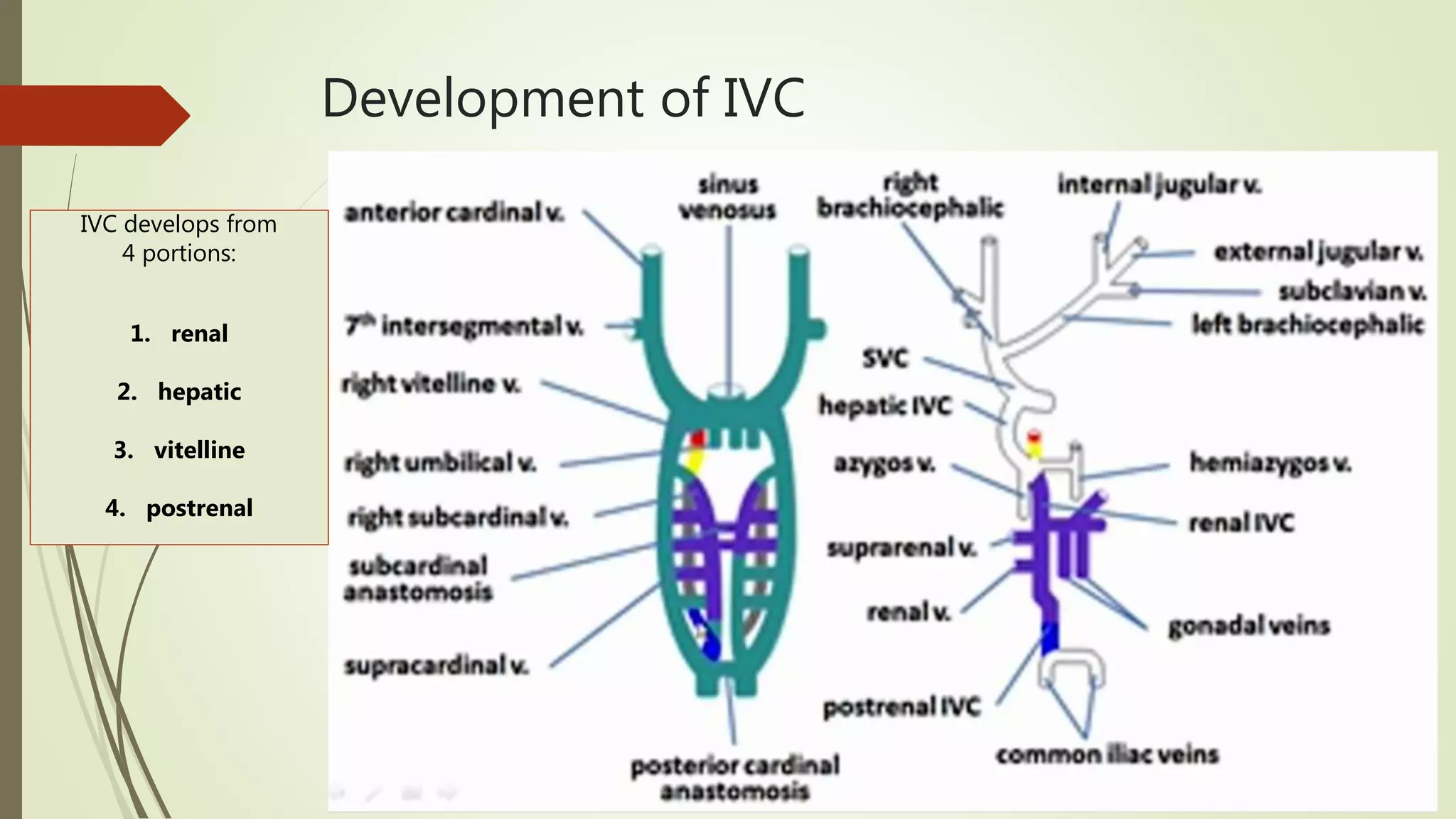
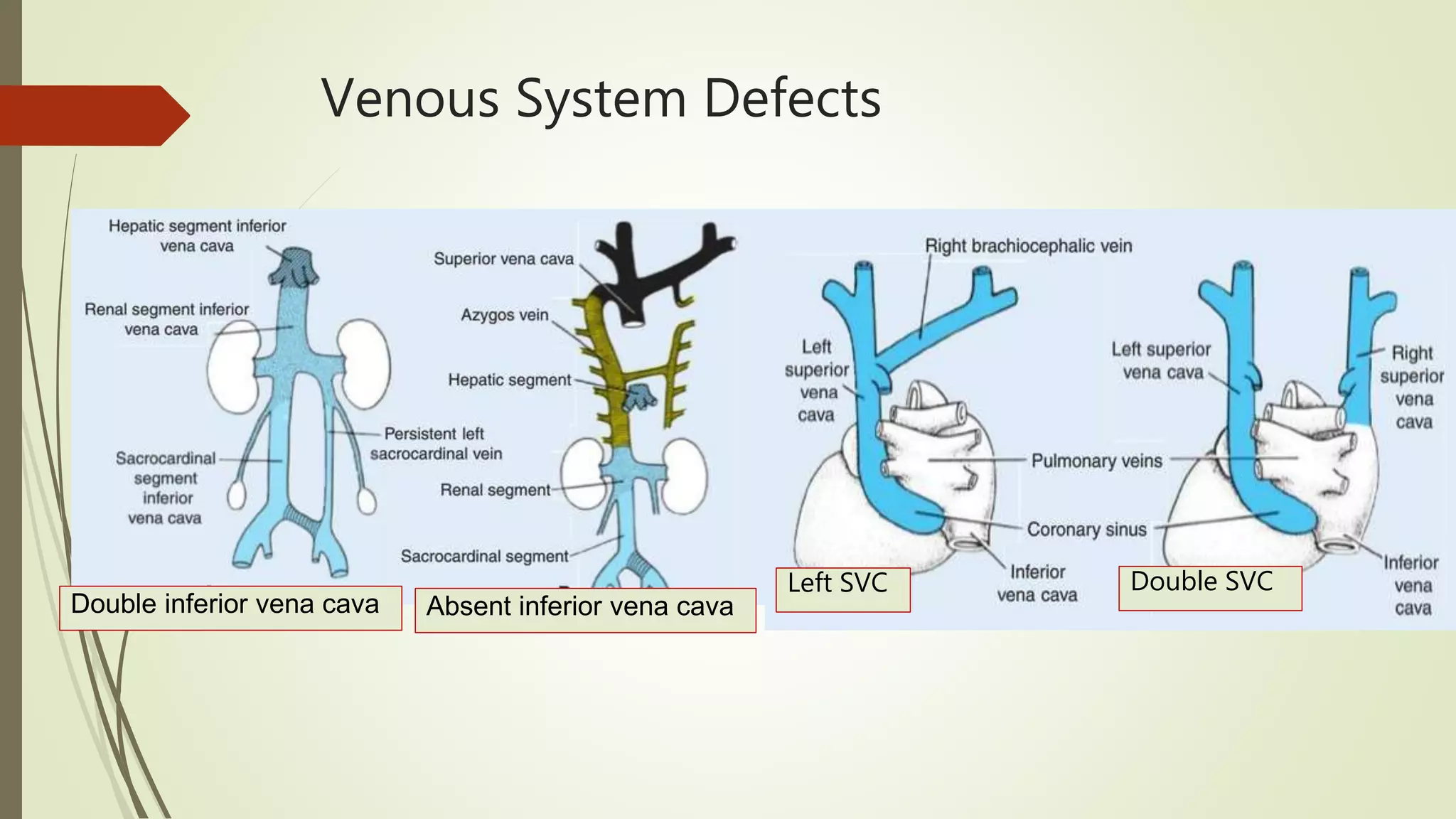
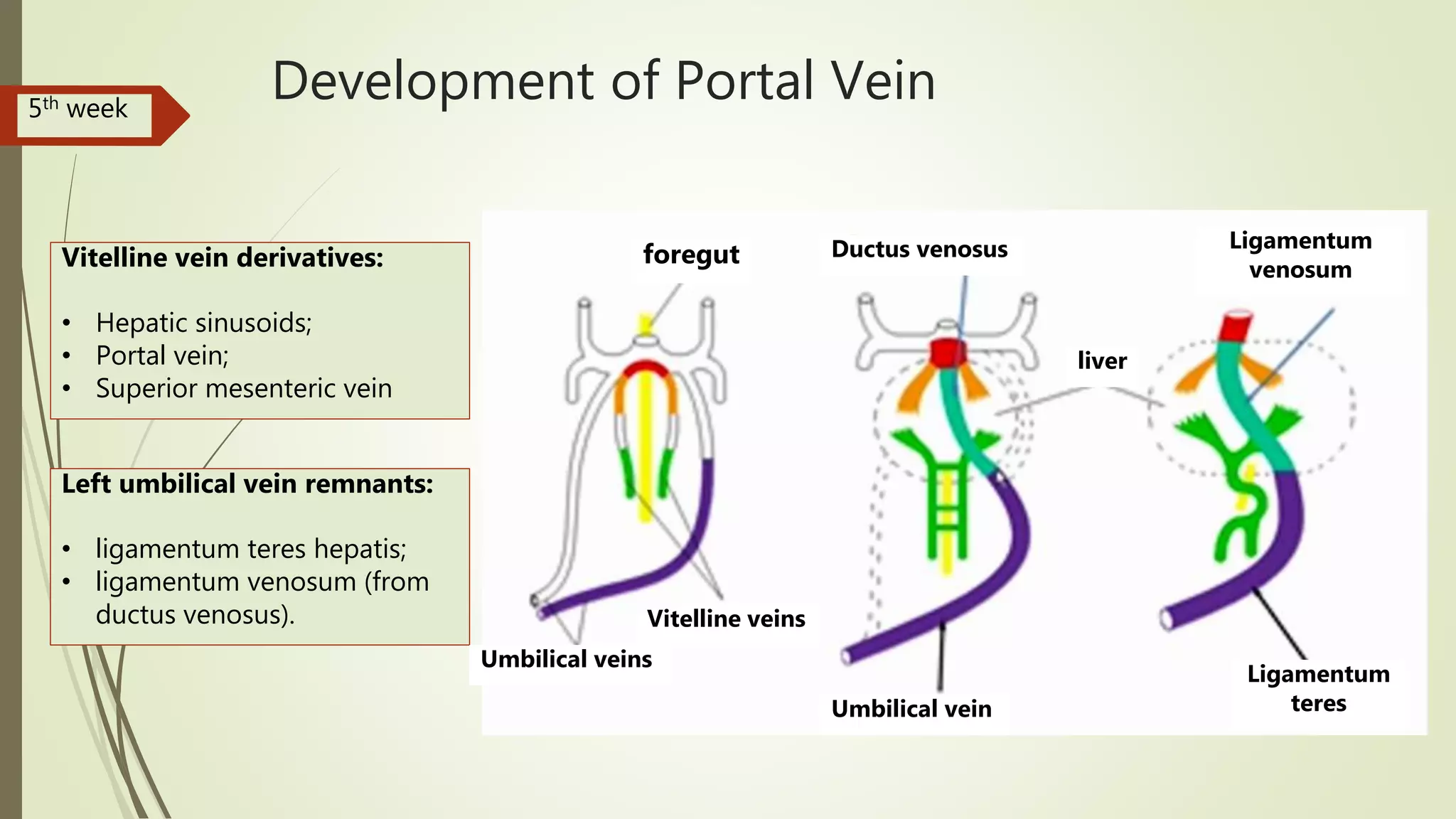
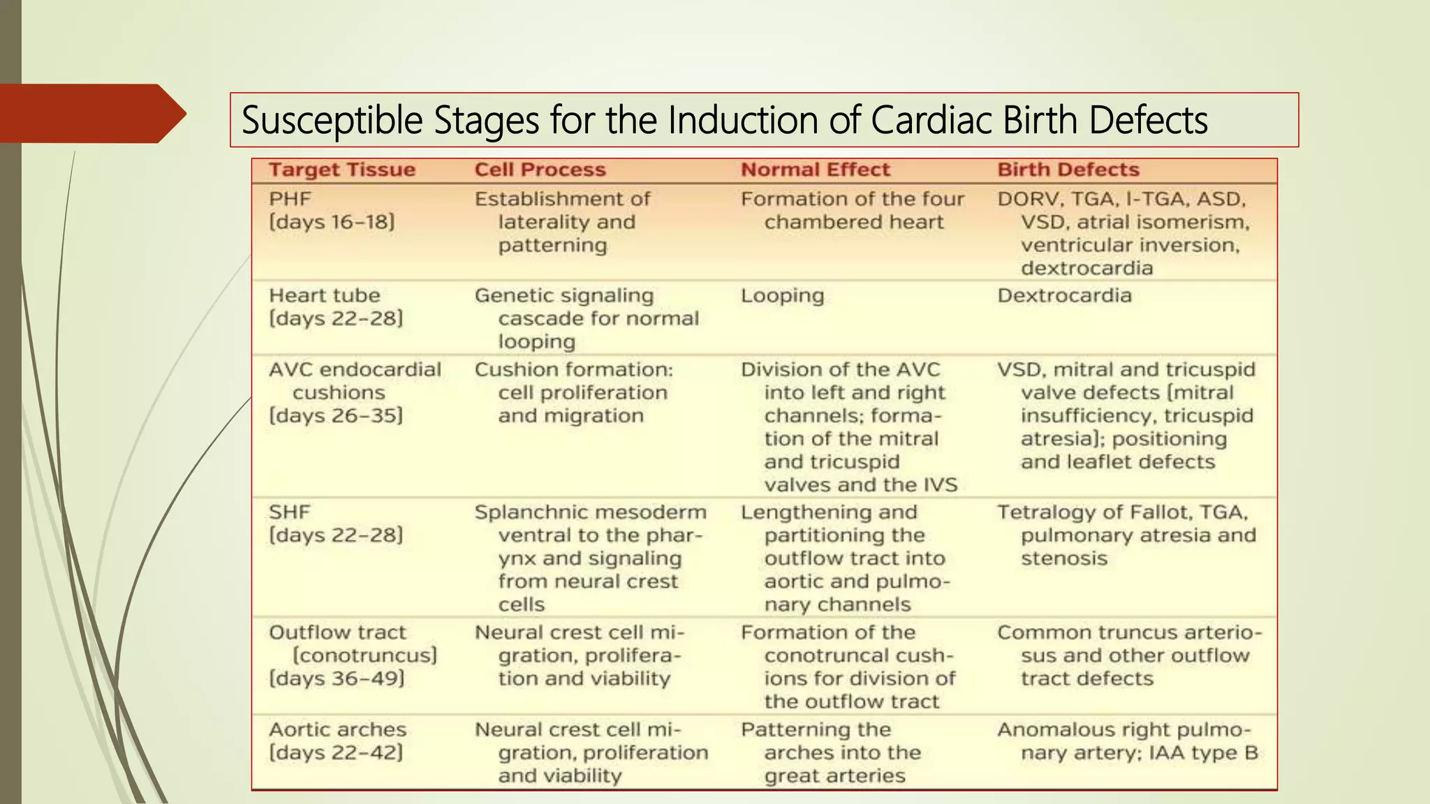
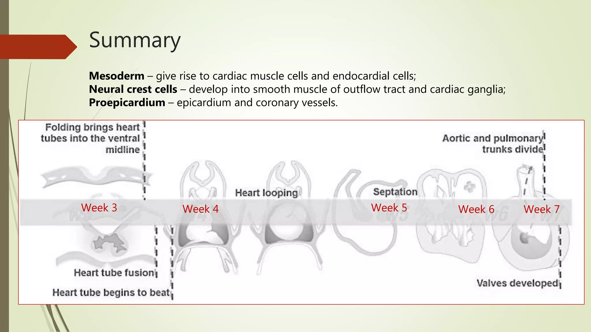

The document summarizes cardiovascular embryology, including: 1. Heart development begins with the formation of the horseshoe-shaped pericardial cavity and single heart tube, which undergoes convolution to form the primitive 4-chambered heart. 2. Septation occurs through atrial septation, ventricular septation, and aorticopulmonary septation to separate the chambers. 3. Arteries develop from the aortic arches and dorsal aortae, while veins form from the vitelline veins, umbilical veins, and cardinal veins. 4. The fetal circulation differs from the adult circulation, with blood bypassing the lungs via the ductus arteriosus and


