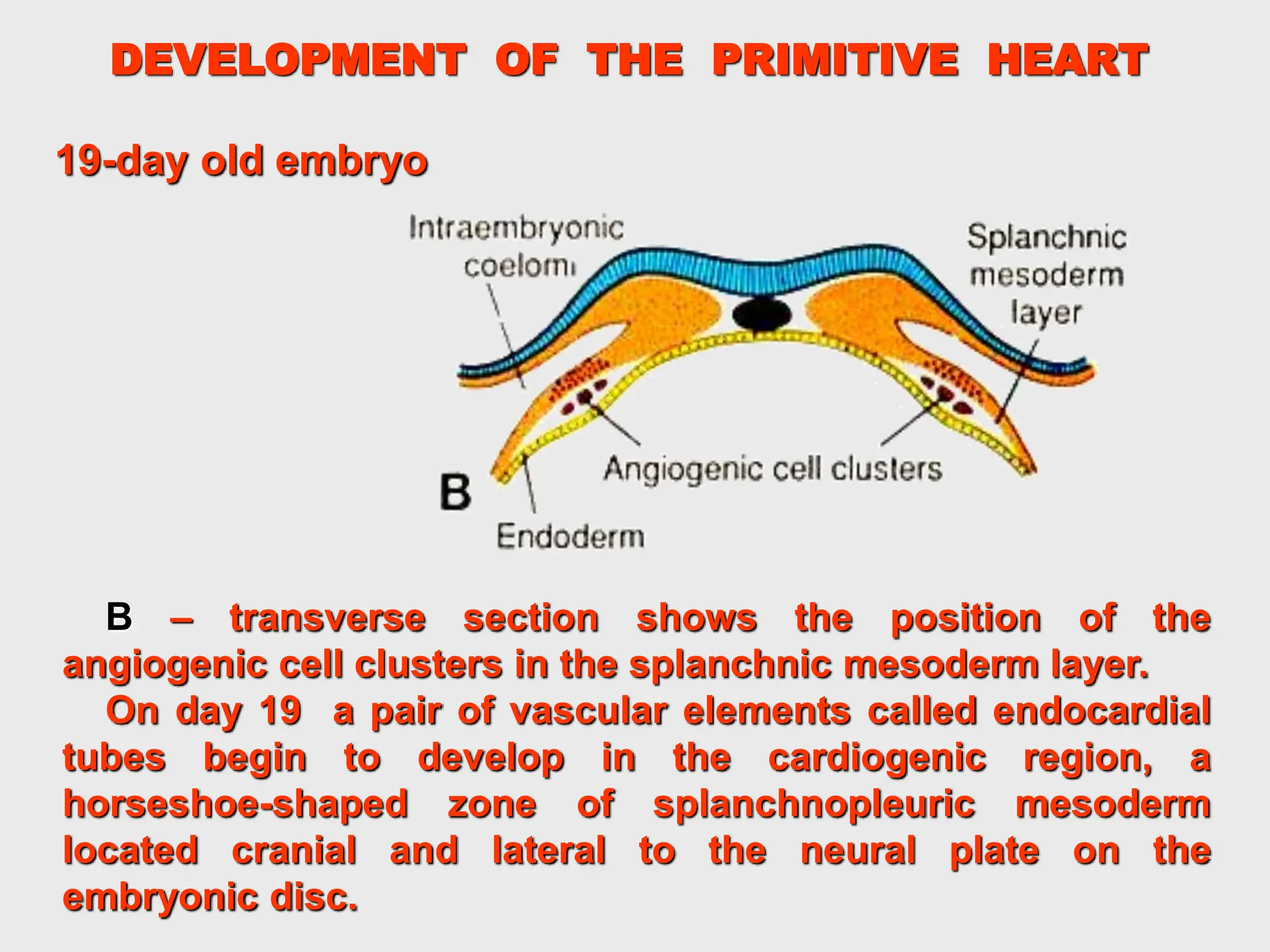
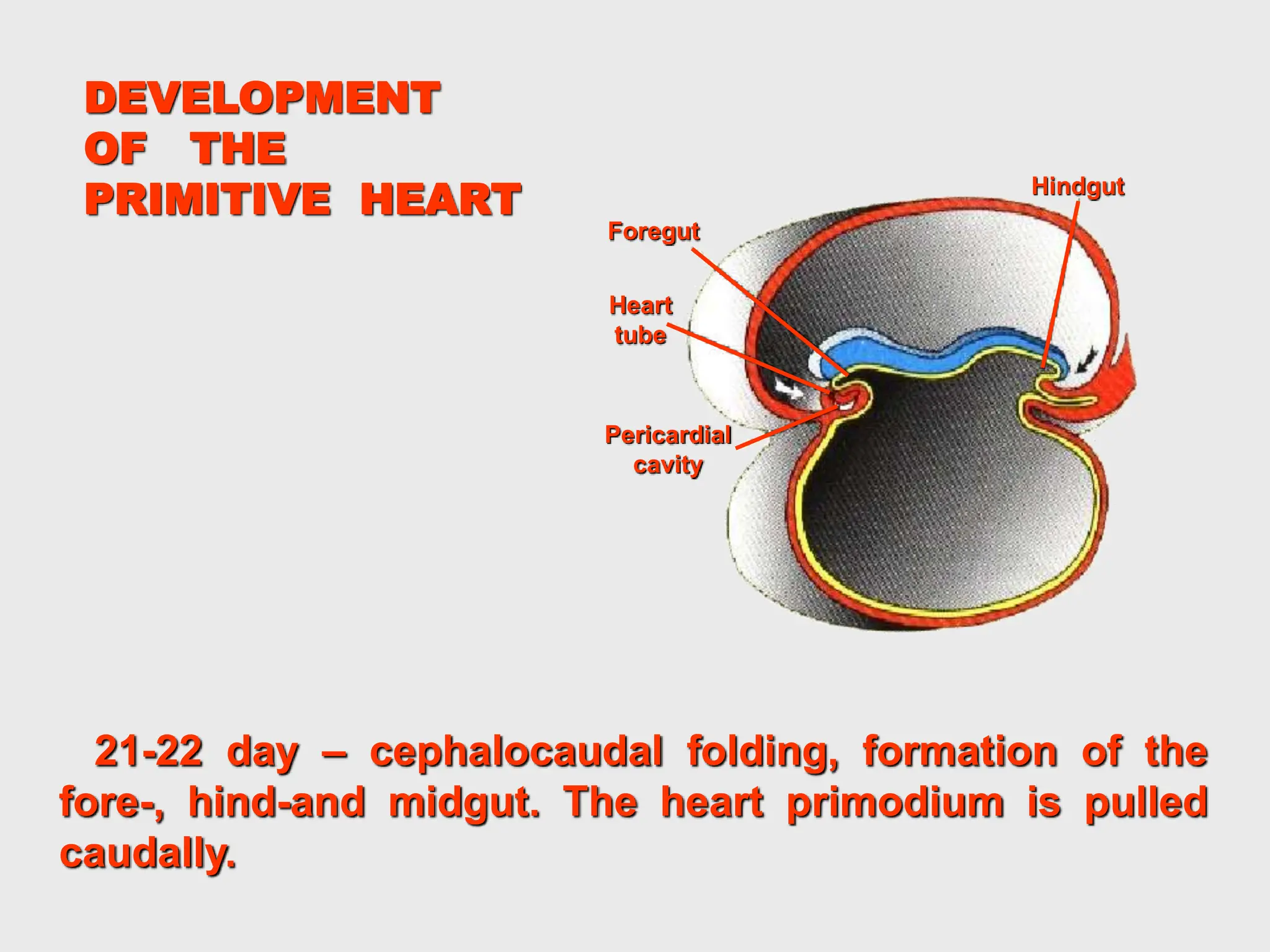
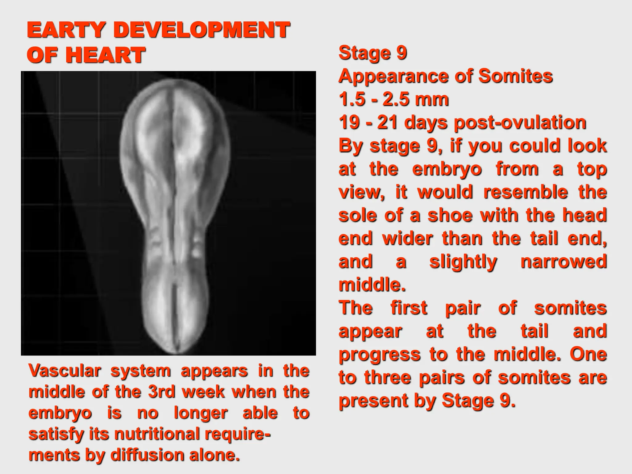
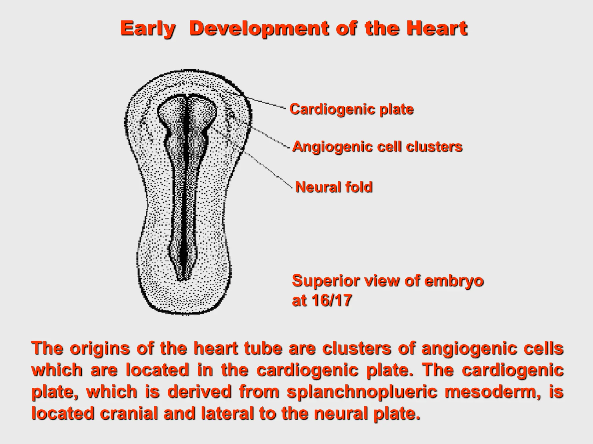
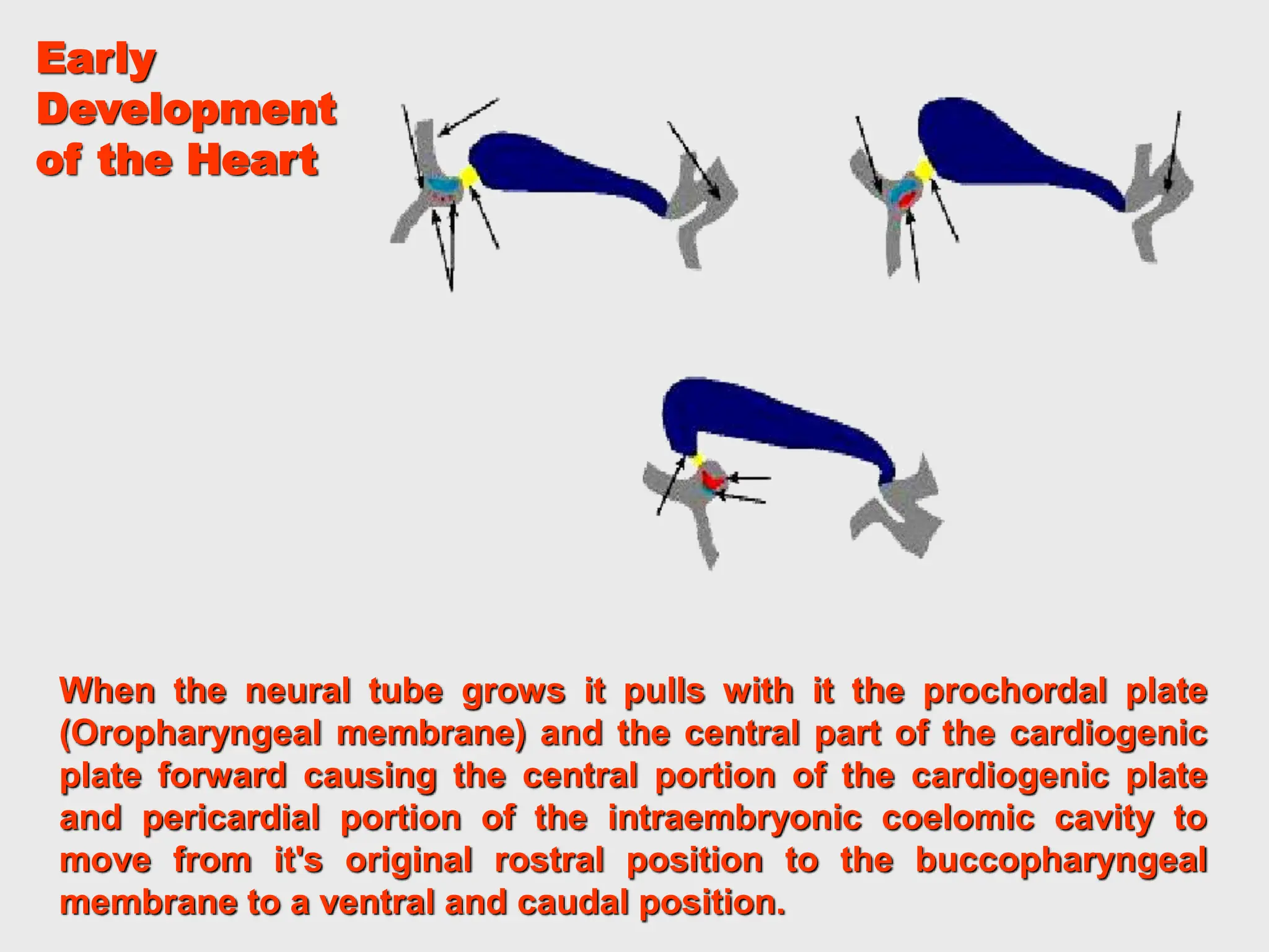
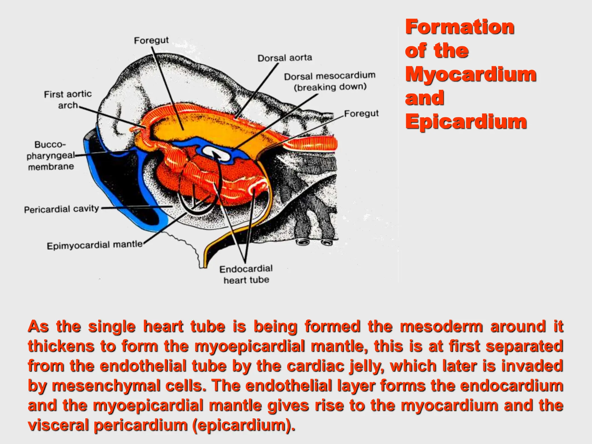
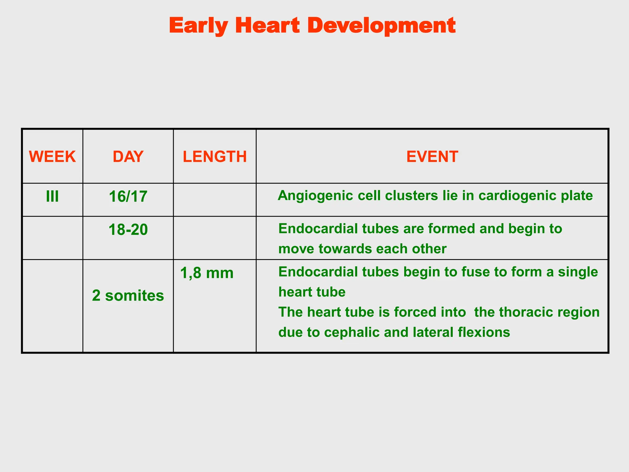
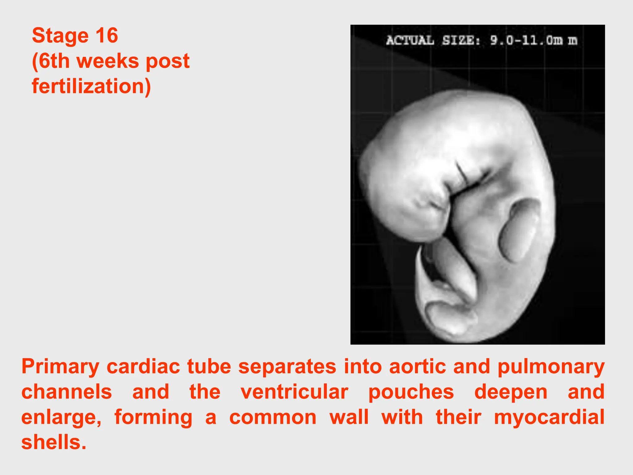
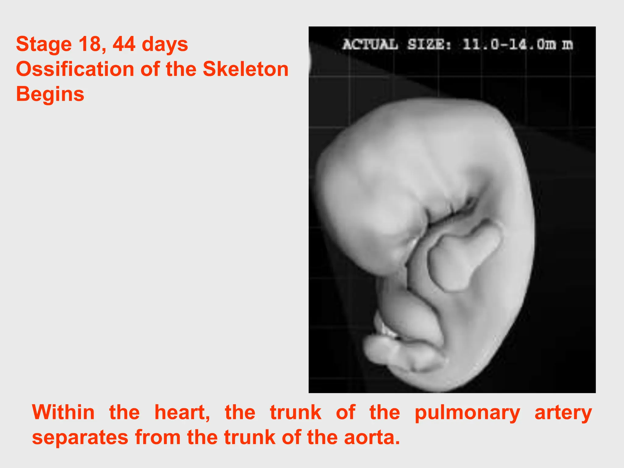
SlideShare a Scribd company logo
1 of 73
Lecture for the general medicine IInd course
english medium students
VOLGOGRAD STATE MEDICAL UNIVERSITY
Department of histology, embryology, cytology
Volgograd, 2015
THE OBJECTIVES:
Describe the main features of heart development to the four-
chambered system.
Describe the development of the pericardium
Describe the development of primary and secondary atrial
septa and the ventricular septum.
Explain the changes occurring in the bulbis cordis and
truncus arteriosus in its transformation from a single to a
double tube.
Describe the developmental aberrations responsible for the
following malformations: patent ductus arteriosus (P.D.A.);
atrial septal defects (A.S.D.) and ventricular septal defects
(V.S.D.); tetralogy of Fallot.
GENERAL PROVISIONS:
the CVS is the first system to function in the
embryo,
vascular system appears in the middle of the 3rd
week when the embryo is no longer able to satisfy
its nutritional requirements by diffusion alone,
blood begins to circulate by the end of the 3rd
week.
Endoderm
Ectoderm
Angiogenic
cell cluster
Prechor
dal
plate
Amniotic cavity
Connecting
stalk
Allantois
Cloacal
membrane
EARLY
DEVELOPMENT OF
THE EMBRYO
Middle of the 3rd week – presomite stage, pan-cake appearance of
embryonic disc, intraembryonic endoderm constitutes the roof of
the spherical yolk sac.
Formation of the angiogenic cell clusters: splanchnic mesoderm
gives rise to angioblasts – cells of the mesenchymal origin
condensing into interconnecting cords of cells.
DEVELOPMENT OF THE MESODERM
Notochord
Amniotic
cavity
Mesoderm
Ectoderm
Dorsal
aorta
Paraxial
mesoderm
Intermediate
mesoderm
Intercellular
cavities
in lateral
plate
A. day 17, initially cells of the mesodermal germ layer form a thin sheet
of loosely woven tissue on each side of the midline, by 17th day cells
close to the midline proliferate and form a thickened plate of tissue -
paraxial mesoderm.
A
B. day 19, more laterally, the mesoderm layer remains thin (lateral plate).
Intermediate mesoderm connects paraxial and lateral plate
mesoderm. Intercellular cavities in the lateral plate then appear.
B
Intra-
embryonic
coelomic
cavity
Endoderm
Somite
Intermediate
mesoderm
DEVELOPMENT OF THE MESODERM
C. day 20 - intercellular cavities in the lateral plate coalesce, it is divided
into two layers: the one continuous with mesoderm covering amnion
(somatic or parietal mesodermal layer); and the other continuous with
mesoderm covering the yolk sac (splanchnic or visceral layer).
C
D. day 21 - space bordered by these two layers forms a newly formed
cavity, the intraembryonic coelomic cavity or body cavity, which, on
each side of the embryo, is continuous with the extraembryonic
coelom.
D
Amnion
Parietal
mesoderm
layer
Endoderm
Neural groove
Visceral
mesoderm
layer
DEVELOPMENT OF THE HEART PRIMODIUM
At first the angiogenic clusters are located on the lateral
sides of the embryo, but they rapidly spread in a cephalic
direction. The anterior central portion of these clusters is
known as the cardiogenic area (region).
18-day old embryo
Angiogenic
cell clusters
Neural
plate
Cut edge
of amnion
Primitive
node
Primitive
streak
19-day old embryo
DEVELOPMENT OF THE PRIMITIVE HEART
B – transverse section shows the position of the
angiogenic cell clusters in the splanchnic mesoderm layer.
On day 19 a pair of vasсular elements called endocardial
tubes begin to develop in the cardiogenic region, a
horseshoe-shaped zone of splanchnopleuric mesoderm
located cranial and lateral to the neural plate on the
embryonic disc.
DEVELOPMENT OF THE PRIMITIVE HEART
Cephalocaudal section showing the position of the
cardiogenic area and pericardial cavity (part of the
intraembryonic coelom) in the cranial end of the embryo.
Cardiogenic
area
19-day old embryo
Pericardial
cavity
Heart
tube
Foregut
Hindgut
DEVELOPMENT
OF THE
PRIMITIVE HEART
21-22 day – cephalocaudal folding, formation of the
fore-, hind-and midgut. The heart primodium is pulled
caudally.
DEVELOPMENT OF THE HEART
C. 23-24 day, as a result of brain growth and cephalo-
caudal folding the prechordal plate (future bucco-
pharyngeal membrane) is pulled forward, while
primodium of the heart becomes located first in the
cervical region,
Buccopharyngeal
membrane
Cloacal
membrane
Heart
tube
C
Lung bud
Remnant
of the
buccopharyngeal
membrane
Vitelline duct
Yolk sac
Allantois
Midgut
Liver
bud
Heart tube
D
D. End of the 1st month – and then in the thoracic
region.
Cells of splanchnic mesodermal layer form the visceral layer of the serous
membranes covering the abdominal organs, lungs and heart. Visceral and
parietal layers are continuous with each other as the dorsal mesentery, which
suspends the gut tube in the peritoneal cavity. Ventral mesentery is a result of
thinning of septum transversum.
DEVELOPMENT OF THE SEROUS MEMBRANES
Ectoderm
Parietal
mesoderm
layer
Mesonephros
Body wall
Intraembryonic
coelomic cavity
Endoderm of
yolk sac
Wall of gut Serous membrane
(peritoneum)
Parietal
mesoderm
layer
Visceral
mesoderm
layer
Dorsal
mesentery
Amniotic cavity
A
B. end of the 4th week, cells of somatic mesoderm lining the intraembryonic
coelom, become mesothelial and form parietal layer of the serous
membrane (lining the outside of the future peritoneal, pleural and
pericardial cavities).
B
Stage 9
Appearance of Somites
1.5 - 2.5 mm
19 - 21 days post-ovulation
By stage 9, if you could look
at the embryo from a top
view, it would resemble the
sole of a shoe with the head
end wider than the tail end,
and a slightly narrowed
middle.
The first pair of somites
appear at the tail and
progress to the middle. One
to three pairs of somites are
present by Stage 9.
EARTY DEVELOPMENT
OF HEART
Vascular system appears in the
middle of the 3rd week when the
embryo is no longer able to
satisfy its nutritional require-
ments by diffusion alone.
Weeks Days Somites Length in mm Cardiac Events
1-2 0-20 1 1,5 No heart or great vessels
3 20 2 1,5 Cardiogenic plate
3 21 5 1,5 Endocardial tubes
4 22 10 2 Fusion of endocardial tubes
4 23 12 2
Single median cardiac tube, first contraction
(ineffective)
4 25 17 2,5 Cardiogenic loop
4 26 20 3 Single atrium
5 29 25 4 Bilobed atrium
5 31 26 4 Beginning of circulation
5 31 28 4,8 Septum primum
5 35 7,5 A-V orifice, 3 chamber heart
6 36 8,5 Septum secundum
6 39 10 Complete inferior septum
6 40 10,5 Septation of bulbus and ventricle
6 42 13 Divided truncus arteriosus
7 49 20
4-chambered heart, Absorption of pulmonary
veins
The origins of the heart tube are clusters of angiogenic cells
which are located in the cardiogenic plate. The cardiogenic
plate, which is derived from splanchnoplueric mesoderm, is
located cranial and lateral to the neural plate.
Early Development of the Heart
Cardiogenic plate
Angiogenic cell clusters
Neural fold
Superior view of embryo
at 16/17
When the neural tube grows it pulls with it the prochordal plate
(Oropharyngeal membrane) and the central part of the cardiogenic
plate forward causing the central portion of the cardiogenic plate
and pericardial portion of the intraembryonic coelomic cavity to
move from it's original rostral position to the buccopharyngeal
membrane to a ventral and caudal position.
Early
Development
of the Heart
Angiogenic cell clusters which lie in a horse-shoe shape
configuration in the plate coalesce to form two endocardial tubes.
These tubes are then forced into the thoracic region due to cephalic
and lateral foldings where they fuse together forming a single
endocardial tube.
EARLY DEVELOPMENT OF THE HEART
Early presomite embryo –
17 days
Late presomite embryo –
18 days
8 somite embryo-
22 days
5 somite stage
21 days
These angiogenic cell clusters coalesce to form right and left
endocardial tubes.
Each tube is continuous cranially with a dorsal aorta, its outflow
tract, and caudally with a vitelloumbilical vein, its inflow tract.
Early Development
of the Heart
Day 20
Dorsal aorta
Left endocardial tube
Vitelloumbilical vein
The lateral and cranial folding of the embryo forces the tubes
into the thoracic cavity. As a result, these tubes come to lie
closer to each other and begin to fuse in a cranial to caudal
direction.
Early Development of the Heart
Neural fold
L.Dorsal aorta
Foregut
Endocardial heart tube
Pericardial cavity
Angiogenic
cell clusters
As the single heart tube is being formed the mesoderm around it
thickens to form the myoepicardial mantle, this is at first separated
from the endothelial tube by the cardiac jelly, which later is invaded
by mesenchymal cells. The endothelial layer forms the endocardium
and the myoepicardial mantle gives rise to the myocardium and the
visceral pericardium (epicardium).
Formation
of the
Myocardium
and
Epicardium
At approximately day 21 the endocardial tubes are completely
fused.
The heart starts to beat at day 22, but the circulation does not
start until days 27 to 29.
Early Heart Development, SEM
Central veiw of fusing
endocardial tubes within
pericardial cavity
The single tubular heart develops
many constrictions outlining future
structures. The cranial most area is
the bulbus cordis which extends
cranially into the truncus arteriosus,
which in turn is connected to the aortic
sac and through the aortic arches to
the dorsal aorta. The primitive ventricle
is caudal to the bulbus cordis and the
primitive atrium is the caudal most
structure of the tubular heart. The
atrium which is paired connects to the
sinus venosus which receives the
viteline (from yolk sac), common
cardinal (from embryo) and umbilical
(from primitive placenta) veins. The
primitive atrium and sinus venosus lay
outside the caudal end of the
pericardial sac, and the truncus
arteriosus is outside the cranial end of
the pericardial sac.
Early Development
of the Heart
Dorsal aorta
Aortic sac
Bulbus cordis
Primitive ventricle
Atrioventricular
sulcus
Primitive atria
Sinus venosus
The newly formed heart tube may
be divided into regions. Starting
caudally:
 sinus venosus - consisting of right
and left horns,
 paired primitive atria. These
structures will later fuse together
to form common atrium,
 atrioventricular sulcus divides the
atria and the primitive ventricle.
 primitive ventricle expands to
become the left ventricle.
 interventricular sulcus divides the
primitive ventricle and the bulbus
cordis,
 bulbus cordis which may be
divided as follows:
1. conus cordis
2. truncus arteriosus aortic sac.
3. bulbus cordis - the proximal
portion forms the right
ventricle
Early Heart Development
WEEK DAY LENGTH EVENT
III 16/17 Angiogenic cell clusters lie in cardiogenic plate
18-20 Endocardial tubes are formed and begin to
move towards each other
2 somites
1,8 mm Endocardial tubes begin to fuse to form a single
heart tube
The heart tube is forced into the thoracic region
due to cephalic and lateral flexions
The original paired
cardiac tubes fuse, with the
"ventricular" primordia
initially lying above the
"atria".
Growth of the cardiac
tube flexes it into an "S-
shape" tube, rotating the
"ventricles“.
Early Development of the Heart
The bulboventricular portion of the heart grow faster than the
pericardial sac and the rest of the embryo. Since the
bulboventricular portion is fixed at the cranial end by the aortic arch
arteries and at it's caudal end by the septum transversum, the
bulboiventricular portion will fold as it elongates.
The cephalic end of the heart tube will bend ventrally, caudally and
slightly to the right.
Early Development of the Heart
The bulboventricular sulcus will become visible from the
outside, and from the inside there will be a primitive interventricular
foramen. The internal fold formed by the bulboventricular sulcus is
known as the bulboventricular fold. The bulboventricular segment
of the heart is now U-shaped, bulbus cordis occupies the right arm
of the U-shape and the primitive ventricle occupy the left arm of the
U-shaped bulbo-ventricular segment. The looping of the
bulboventricular segment of the heart will cause the atrium and
sinus venosus to become dorsal to the heart loop.
LOOPENING
By the time the heart tube has formed the bulboventricular
loop, the two primitive right and left atria have fused to form a
common atrium. Note that it now lies cranial to the primitive
ventricle and dorsal to the bulbus cordis. The truncus arteriosus
lies on the roof of the common atrium causing a depression and
indicates where septation of the atrium will occur. AS = Aortic sac,
BC = Bulbus cordis, CC = Conus cordis, LA = Left atrium, LV = Left
ventricle, RA = Right atrium
•SV = Sinus venosus, TA = Truncus arteriosus.
LOOPENING
The newly formed heart tube bulges into the pericardial cavity
and is attached to the dorsal wall by a fold of tissue, the dorsal
mesoderm. This is a derivative of foregut splanchnoplueric mesoderm.
Eventually this will rupture leaving the heart tube suspended in the
pericardial cavity anchored cranially by the dorsal aortae and caudally
by the vitelloumbilical veins.
As it bulges into the cavity it becomes invested in a layer of
myocardium. A layer of acellular matrix, the cardiac jelly, separates the
myocardium and the endothelial heart tube.
The primitive heart tube can be subdivided into
primordial heart chambers starting caudally at the inflow
end: the sinus venosus, primitive atria, ventricle, and
bulbus cordis (conus).
Early Development of the Heart
WEEK DAY LENGTH EVENT
IV 22 2,0 mm
4 somites
Endocardial heart tubes have completely fused to
form a single heart tube.
The heart begins to beat.
Splanchnopleuric mesoderm invests the heart tube
and forms the pericardial cavity.
23 2,2-2,5 mm
7/8 som.
The heart tube begins to grow rapidly, forcing it to fold
upon itself.
24 14 somites The centrally located sinoatrial junction begins to shift
to the right.
Perforations appear in the dorsal mesocardium.
25-28 3,2-5 mm The bulboventricular loop is formed.
Septum primum appears.
28 5 mm
16 somites
The ventricular septum appears as a small ridge on
the floor of the common ventricle.
The ventricules begin to dilate.
A single pulmonary artery grows from the outer dorsal
wall of the left atrium.
The endocardial cushions appear.
The partitioning of the atrium begins with the appearance of septum
primum at about the 28th day. This is a crest of tissue that grows
from the dorsal wall of the atrium towards the endocardial cushions
- the ostium (opening) formed by the free edge of septum primum is
the ostium primum.
ATRIAL PARTITIONING
Before the septum primum fuses with the endocardial cushions,
perforations appear in the upper portion of the septum primum. These
perforations will coalesce to form the ostium secundum.
• SAO = Sinoatrial orifice
• SS = Septum spurium
• S1 = Septum primum
• Perf = Perforations
• O1 = Ostium secundum
• EC = Endocardial cushions
ATRIAL PARTITIONING
Unlike the septum primum, septum secundum does not fuse with
the endocardial cushions. Its free edge forms the foramen ovale.
The left venous valve and the septum spurium located on the dorsal
wall of the right atrium, fuse with the septum secundum as it grows.
EC = Endocardial cushions, LVV = Left venous valve, O1 = Ostium
secundum, SS = Septum spurium, S1 = Septum primum,
S2 = Septum secundum.
ATRIAL PARTITIONING
At the end of the 7th week the human heart has reached its final stage of
development. Because the fetus does not use its lungs, most of the blood
is diverted to the systemic circulation. This is accomplished by a right to
left shunting of blood that occurs between the two atria.
The foramen ovale and the septum primum control this right and left
communication. The septum primum acts as a valve over the foramen
ovale. At birth the child will use its lungs for the first time and
consequently more blood will flow into the pulmonary circulation. The
pressure increase in the left atrium (where the pulmonary veins empty) will
force septum primum to be pushed up against septum secundum. Shortly
thereafter the two septa fuse to form a common atrial septum.
FO - foramen ovale
ATRIAL
PARTITIONING
DEVELOPMENT OF THE HEART
WEEK DAY LENGTH EVENT
V 29 6-7 mm Truncal swellings appear
30/31 Perforations appear in septum primum appear
32/33 9 mm The bulboventricular flange begins to recede
The atrioventricular canal gains a “dog’s bone”
appearance
The ostium secundum is formed by the free edge of
septum primum
35 The sinoatrial junction has shifted completely to the
right
VI 37 14 mm The septum secundum ceases to grow; its free edge
forms the foramen ovale.
42 Superior and inferior endocardial cushions fuse.
Early Development of the Heart
WEEK DAY LENGTH EVENT
VII 46 The ventricular septum ceases to grow.
The coronary sinus is formed.
end of
7th week
The interventricular canal is completely obliterated.
VIII early
In wk
The outflow tracts (the aorta and the pulmonary
trunk) are completely separated.
Fate of the Sinus Venosus
(Formation of the Right Atrium)
Communication between the sinus venosus and the primitive
atrium, the sinoatrial orifice, is centrally located.
• VV = Vitelline vein
• UV = Umbilical vein
• CC = Common cardinal vein
• SA = Sinoatrial orifice
• RSH, LSH = right, left sinus
horn
• RA, LA = right, left atrium
Unlike the atria, the sinus vinosus remains a paired structure with
right and left horns. Each horn receives venous blood from three
vessels:
1. Vitelline vein
2. Umbilical vein
3. Commom cardinal vein
Conversely, the left vein counterparts are obliterated and the left
sinus horn diminishes in size and forms the coronary sinus and the
oblique vein of the left ventricle.
• SVC = Superior vena cava
• IVC = Inferior vena cava
• SA = Sinoatrial junction
• CS/OV = Coronary sinus/ oblique
vein of left ventricle
• LA, RA = Left, right atrium
• LV, RV = Left, right ventricle
DEVELOPMENT
OF THE ATRIA
Gradually the sinoatrial oriface shifts to the right, due to the shunting of
blood to the right, until the sinus venosus communicates with only the
right atrium. The fate of each structure is as follows:
 the right sinus horn becomes enlarged
 the right anterior cardinal vein becomes the superior vena cava
 the right vitelline vein becomes the inferior vena cava
 the right umbilical vein is obliterated
Internally, the sinoatrial orifice is flanked by two valves,
the right and left venous valves. Superiorly these two
valves meet to form the septum spurium. Note that the left
horn opens up underneath the orifice of the right horn
(sinoatrial oriface). This is the orifice of the coronary
sinus.
• LVV, RVV = left, right venous
valve
• SS = Septum spurium
• SA = Sinoatrial oriface
• OCS = orifice of the coronary
sinus
SEPTATION
Further into development the right sinus horn is incorporated into
the expanding right atium. As the atrium expands the smooth tissue
of the sinus venosus displaces the trabeculated tissue of the
primitive right atrium anteriorly and laterally where it becomes the
adult right auricle. The smooth tissue forms part of the atrium called
the sinus venarum. Crista Terminalis, a ridge of tissue located to the
right of the sinoatrial orifice, forms the boundary between the
auricle and the sinus venarum.
• RAu = Right auricle
• SV = Sinus venarum
• CT = Crista terminalis
• OCS = Orifice of the
coronary sinus
Formation of the Right Auricle
The final morphological change in the heart is the partitioning of the
outflow tract - - the truncus arteriosus and the conus cordis - - into
the aorta and the pulmonary trunk. This is accomplished by the
development of a septum that forms in the outflow tract and the
emergence of the two great vessels.
The septum forms from two pairs of swellings which grow from the
walls of the outflow tract. These are the truncus swellings and the
conus swellings.
Formation of the Aorta and
Pulmonary Tract
• RSTS/ LITS = Right superior/
Left inferior truncal swelling,
• RDCS/ LVCS = Right dorsal/
Left ventral conus swelling
Truncal swellings: Right superior which grows distally and to the left. Left
inferior which grows distally and to the right. Both develop at the proximal
part of the truncus and proceed to grow in two directions; 1) distally
towards the aortic sac and 2) into the lumen of the outflow tract where they
will eventually fuse together.
Conus swellings: Right dorsal which is continuous with the right superior
Left ventral which is continuous with the left inferior Like the truncal
swellings, the conal swellings grow distally and towards each other,
however they appear after the first pair. These conus swellings eventually
fuse with the truncal swellings.
Formation of the Aorta and
Pulmonary Tract
• RSTS/ LITS = Right superior/
Left inferior truncal swelling,
• RDCS/ LVCS = Right dorsal/
Left ventral conus swelling
Pulmonary Veins (Formation of the Left Atrium)
Development of the left atrium occurs concurrently with that of the
right atrium. During the early part of the fourth week an outgrowth of
the pulmonary veins appear from the left atrium. This "sprout" will
bifurcate until there are four veins. These vessels will then grow
towards the lung buds.
• LA = Left atrium
• OPV = orifice of
pulmonary vein
• PV = pulmonary vein
The left atrium begins to expand gradually accepting the four
branches. As the atrial wall expands, the smooth tissue of the
pulmonary veins is incorporated into the wall of the atrium and
displaces the trabeculated tissue anteriorly and laterally which will
then form the adult auricles. Compare this process to the formation
of the adult right auricle.
• LAu = Left auricle
• OPV = Orifice of
the four pulmo-
nary veins
• PV = four pulmo-
nary veins
Pulmonary Veins (Formation of the Left Atrium)
Recall that the proximal bulbus cordis gives rise to the right ventricle. Thus,
blood flows from the primitive atrium to the left ventricle then to the right
ventricle. There is no direct communication between the atria and the right
ventricle even after the formation of the bulboventriclular loop. The
atrioventricular canal must shift to the right in order to acheive
communication to the right ventricle in addition to the left ventricle. During
this shift the proximal bulbus widens and the bulboventricular flange
begins to recede. Swellings of mesenchymal tissue, the endocardial
cushions, appear on the borders of the atrioventricular canal. There are four
cushions: inferior and superior (ventral and dorsal), left and right. The first
appear before the latter. These swellings give the atrioventricular canal a
"dog's bone" shape.
• AVC = Atrioventricular
canal
• BC = Bulbus Cordis
• LV = Left ventricle
Atrioventricular
Canals
At approximately day 42 the superior and inferior cushions fuse
forming a right and a left atrioventricular canal. The left atrium
communicates with the left ventricle and the right atrium
communicates with the right ventricle. The shifting process brings
the conus cordis to lie superior to the interventricular foramen,
which at this point, has not yet been obliterated. The fused
endocardial cushions are also responsible for the closure of the
ostium primum by fusing with the free edge of the septum primum.
• R/LAVC = Right/left
atrioventricular canal
Atrioventricular Canals
The final morphological change in the heart is the partitioning of the
outflow tract - - the truncus arteriosus and the conus cordis - - into
the aorta and the pulmonary trunk. This is accomplished by the
development of a septum that forms in the outflow tract and the
emergence of the two great vessels.
The septum forms from two pairs of swellings which grow from the
walls of the outflow tract. These are the truncus swellings and the
conus swellings.
Formation of the Aorta and
Pulmonary Tract
• RSTS/ LITS = Right superior/
Left inferior truncal swelling,
• RDCS/ LVCS = Right dorsal/
Left ventral conus swelling
In the newly formed bulboventricular loop the primitive right and left
ventricles appear as expansions in the heart tube. Externally the
interventricular sulcus separates the right and left ventricles and
internally they are separated by the bulboventricular flange.
Remember that the right ventricle arises from the proximal bulbus
cordis.
• AVC = Atrioventricular
canal
• BC = Bulbus Cordis
• BVF = Bulboventricular
flange
• IVS = Interventricular
sulcus
• RV = Right ventricle
• LV = Left ventricle
Formation of the Ventricles
During the shifting of the atrioventriclar canal the proximal bulbus
cordis expands forming the right ventricle. Both ventricles will
continue to expand until the late 7th/early 8th week. The growth of
the ventricles is due to the centrifugal growth of the myocardium
and the diverticulation of the internal walls. (This is what gives the
ventricle its trabeculated appearance). The muscular interventricular
septum forms as a result of the expanding ventricles. The walls of
the right and left ventricles grow in opposition to each other to form
the muscluar septum. Thus, the septum will cease to grow when the
ventriclar walls are no longer expanding.
• BC = Bulbus cordis
• IVS = Interventricular
septum
• MC = Myocardium
• RV = Right ventricle
Formation of the
Ventricles
 Secundum type involves septum primum or septum
secundum.
Atrial Septal Defect
In a heart with an Atrial Septal Defect (ASD) there is
communication between the right and left atria which
causes a left to right shunting of blood due to the lower
pressure in the pulmonary circulatory system.
Consequently there is a mixing of oxygenated (systemic)
and deoxygenated (pulmonary) blood.
There are two types of ASD:
 Primum type involves the endocardial cushions.
In this case, the ostium primum is patent because the septum primum
does not fuse with the endocardial cushions. Recall that the cushions
are responsible in forming a portion of septum primum, thus
obliterating ostium primum.
• FO = Foramen ovale
• IEC = Inferior endocardial
cushion
• SEC = Superior endocar-
dial cushion
• O1 = ostium primum
Primum Type ASD
Truncal swellings: Right superior which grows distally and to the left.
Left inferior which grows distally and to the right. Both develop at the
proximal part of the truncus and proceed to grow in two directions;
1) distally towards the aortic sac and 2) into the lumen of the outflow
tract where they will eventually fuse together.
Conus swellings: Right dorsal which is continuous with the right
superior Left ventral which is continuous with the left inferior Like the
truncal swellings, the conal swellings grow distally and towards each
other, however they appear after the first pair. These conus swellings
eventually fuse with the truncal swellings.
Formation of the Aorta and
Pulmonary Tract
• RSTS/ LITS = Right superior/
Left inferior truncal swelling,
• RDCS/ LVCS = Right dorsal/
Left ventral conus swelling
Secundum Atrial Septal Defect
This type involves septum primum and/or septum secundum. In both
cases the result is a patent foramen ovale.
There may be excessive resorbtion of septum primum where very
little septum remains or it has been completely resorbed. The short
septum primum does not overlap the foramen ovale leaving a
communication between right and left atrium. If the septum secundum
is involved it is because it did not reach its full growth and thus
results in a large foramen ovale.
• FO = Foramen
ovale
• S1 = Septum
primum
• S2 = Septum
secundum
Persistent Atrio-
ventricular Canal
The persistent atrioventricular canal results from the failure of the
superior and inferior cushions to fuse. Thus there is a single
atrioventricular canal in which all four chambers may freely
communicate. Because the cushions do not fuse the atrial and
ventricular septa cannot fully form as they rely on the cushions to form
the membranous portions of these septa.
• IEC = Inferior endocardial
cushion
• SEC = Superior endocardial
cushion
• PAVC = Persistent
atrioventricular canal
• S1 = Septum primum
• S2 = Septum secundum
In the case of a VSD there is a massive left to right shunting of blood and
pulmonary hypertension. The absence of the interventricular septum results
in a Common Ventricle.
Ventricular
Septal
Defect
• Mem = Membra-
nous septum
• Musc = Muscu-
lar septum
The ventricular septal defect is the most common of all congenital heart
anomalies. It may be caused by any of the four malformations:
1) Deficient development of the proximal conus swellings.
2) Failure of the muscular portion of the interventricular septum to
fuse with the free edge of the conus septum. (Membranous VSD)
3) Failure of the endocardial cushions to fuse.
4) Excessive diverticulation of the muscular septum- perforations in
the muscular interventricular septum. (Muscular VSD)
Transposition of
the Great
Vessels
Transposition is a condition in which the aorta arises from the
right ventricle and the pulmonary trunk from the left. This anomally is
due to the failure of the truncoconal swellings to grow in the normal
spiral direction. There is also a ventricular septal defect and a patent
ductus arteriosus. However, these secondary defects make life
possible as they provide a way for oxygenated blood to reach the
entire body.
• AO = Aorta
• PT = Pulmonary
trunk
• PDA = Persistent
Dunctus Arteriosus
• RV/LV = right and
left ventricles
Persistent Truncus Arteriosus
A persistent truncus arteriosus results when the truncoconal
swellings fail to grow. The single artery, the truncus arteriosus, arises
from both ventricles above the ventricular septal defect, allowing
pulmonary and systemic blood to mix. Distally, the single artery is
divided into the aorta and pulmonary trunk by an incomplete septum.
• AO = Aorta
• PT = Pulmonary
trunk
• PTA = Persistent
truncus arterio-
sus
• RV/LV = Right
and left ventricles
Tetralogy of Fallot
4) right ventricular hypertrophy due to the shunting of blood from left
to right. (The pressure in the right ventricle is increased causing
the walls of the right ventricle to expand.)
• AO = Aorta
• PT = Pulmonary trunk
(stenotic)
• RV/LV = Right and left
ventricles
This condition results from a single error: the conus septum develops
too far anteriorly giving rise to two unequally proportioned vessels- - a
large aorta and a smaller stenotic pulmonary trunk. The four main
characteristics of Tetralogy of Fallot are:
1) pulmonary stenosis
2) ventricular septal defect (VSD) of the membranous portion (the
septum is displaced too far anteriorly to contribute to the septum)
3) overriding aorta (the aorta straddles the VSD)
Dextrocardia
Dextrocardia is an anomaly in which the primitive heart tube folds to
the left in a mirror image of a normal bulboventricular loop. This
usually occurs when all the organ systems are reversed, a condition
called situs inversus.
• RA/LA = Right and left atrium
• RV = Right ventricle
• BC = Bulbus cordis
The embryo is shaped in a modified S curve. The embryo has a bulb-
like tail and a connecting stalk to the developing placenta.
A primitive S-shaped tubal heart is beating and peristalsis, the
rhythmic flow propelling fluids throughout the body, begins.
However, this is not true circulation because blood vessel
development is still incomplete.
Stage 11
13-20 Somite Pairs, Rostral
Neuropore Closes, Optic Vesicle
Appears,
Two Pharyngeal Arches Appear
2.5 - 3.0 mm
23 - 25 days post-ovulation
Time-Line Schedule of Heart Development
The heart tube begins to grow rapidly forcing it to bend upon
itself.
The result is the bulboventricular loop.
By the end of the eighth week partitioning is completed and
the fetal heart has formed.
Septa begin to grow in the atria, ventricle and bulbus cordis to
form right and left atria, right and left ventricles and two great
vessels- the pulmonary artery and the aorta.
MAIN STAGES OF THE ERALY
HEART DEVELOPMENT
Heart chambers are filled with plasma and blood cells
making the heart seem distended and prominent. The heart
and liver combined are equal in volume to the head by this
stage. Blood circulation is well established, though true
valves are not yet present.
Stage 13
(approximately 27-
29 postovulatory
days)
Four Limb Buds,
Lens Disc and
Optic Vesicle, the
first thin surface
layer of skin
appears covering
the embryo. 30-40
somite pairs.
The embryo curves into a C shape. The arches that
form the face and neck are now becoming evident under
the enlarging forebrain. A blood system continues to
develop. Blood cells follow the surface of yolk sac where
they originate, move along the central nervous system, and
move in the chorionic villi, the maternal blood system.
Valves and septa may appear in the heart in Stage 12.
Stage 12
21-29 Somite Pairs,
Caudal Neuropore
Closes, Three to Four
Pharyngeal Arches
Appear, Upper Limb
Buds Appear
3.0 - 5.0 mm
25 - 27 days post-
ovulation
Septum primum fuses with septum intermedium in the
heart.
Stage 19,
(approximately
47-48 post ovulatory days)
Stage 14, 5th week
Lens Pit and
Optic Cup
Appear,
Endolymphatic
Appendage
Distinct
Semilunar valves begin to form in the heart. Four major
subdivisions of the heart (the trabeculated left and right
ventricles, the conus cords and the truncus arteriosus) are
clearly defined. Two sprouts, a ventral one from the aortic
sac and a dorsal one from the aorta, form the pulmonary
(sixth aortic) arch.
Blood flow through the arioventricular canal is divided into
left and right streams, which continue through the outflow
tract and aortic sac. The left ventricle is larger than the right
and has a thicker wall.
Stage 15
(6 to 8 weeks post
fertilization)
Lens Vesicle,
Nasal Pit, Hand
Plate; Trunk
Widens, Future
Cerebral
Hemispheres
Distinct
Stage 16
(6th weeks post
fertilization)
Primary cardiac tube separates into aortic and pulmonary
channels and the ventricular pouches deepen and
enlarge, forming a common wall with their myocardial
shells.
Stage 17
(approximately
41 postovulatory
days)
A Four
Chambered Heart
and a Sense of
Smell
The heart begins to separate into four chambers.
Within the heart, the trunk of the pulmonary artery
separates from the trunk of the aorta.
Stage 18, 44 days
Ossification of the Skeleton
Begins
CONGENITAL MALFORMATIONS OF THE HEART AND
GREAT VESSELS.
1. They are common.
2. The overall incidence is 0.7% of live births and 2.7% of
stillbirths.
- Atrial Septal Defects (ASD) – are among the most common
(6.4/10,000births).
- One of the most significant defects is ostium secundum
defect. This anomaly is characterized by a large opening
between the left and the right atria and is caused either by
excessive cell death or resoption of the septum primum. Or
by inadequate development of the septum secundum.
Depending on the size of the opening considerable
intercardiac shunting may occur from left to the right.
Ventricular Septal Defects involve the membranous
portion of the septum. The occur in 12/10,000.
Depending on the size of the opening, blood carried by the
pulmonary artery may be 1.5 times more abundant than that
carried by aorta. The defect may be not restricted to the
membranous part and involve the muscular part of the septum.
Tetralogy of Fallot is the most frequently occured
abnormality of the conotruncal region. The defect is due to an
unequal division of the conus, resulting from anterior
displacement of the conotruncal septum. Displacement of the
septum produces four CV-alterations:
-a narrow right ventricular outflow region,i.e. Pulmonary
infundibular stenosis,
-a large defect of the interventricular septum,
-an overriding aorta that arrises directly above the septal
defect,
-hypertrophy of the right ventricular wall due to the hight
pressure on the right side. The rate is 9.6/10,000.

More Related Content

Similar to Development of Heart 83453453456939987.ppt

Cardiac embryology
Cardiac embryologyCardiac embryology
Cardiac embryologyRezaul hayat
 
The_embryonic_(Organogenetic)_Period(3th_to_8th_weeks_).pptx
The_embryonic_(Organogenetic)_Period(3th_to_8th_weeks_).pptxThe_embryonic_(Organogenetic)_Period(3th_to_8th_weeks_).pptx
The_embryonic_(Organogenetic)_Period(3th_to_8th_weeks_).pptxAhmedYousseryBatan
 
Trilaminar germ disc (week 3 embryology)
Trilaminar germ disc (week 3 embryology)Trilaminar germ disc (week 3 embryology)
Trilaminar germ disc (week 3 embryology)Abdoulwahab Mahde
 
Embryology cardiovascular system (heart development)
Embryology   cardiovascular system (heart development)Embryology   cardiovascular system (heart development)
Embryology cardiovascular system (heart development)MBBS IMS MSU
 
Lecture11 development of the heart and blood vessels
Lecture11   development  of the heart and blood vesselsLecture11   development  of the heart and blood vessels
Lecture11 development of the heart and blood vesselsMUBOSScz
 
Ch 5. CVS.pptxgjjhnmkikuygfvbnkjikkuytfv
Ch 5. CVS.pptxgjjhnmkikuygfvbnkjikkuytfvCh 5. CVS.pptxgjjhnmkikuygfvbnkjikkuytfv
Ch 5. CVS.pptxgjjhnmkikuygfvbnkjikkuytfvprettyfahim20
 
lec 5- week 4-8 weeks of development.pdf
lec 5- week 4-8 weeks of development.pdflec 5- week 4-8 weeks of development.pdf
lec 5- week 4-8 weeks of development.pdfSriRam071
 
Ventricle anatomy and CSF physiology.pptx
Ventricle anatomy and CSF physiology.pptxVentricle anatomy and CSF physiology.pptx
Ventricle anatomy and CSF physiology.pptxMichael Sintayehu
 
Cardiac Embryology basics.pptx
Cardiac Embryology basics.pptxCardiac Embryology basics.pptx
Cardiac Embryology basics.pptxShivani Rao
 
Development of the Heart
Development of the HeartDevelopment of the Heart
Development of the HeartJean Vitor
 
General embryology growth & development of mandible
General embryology growth & development of mandibleGeneral embryology growth & development of mandible
General embryology growth & development of mandibleIman Zubair
 
Embryology Course II - 2nd and 3rd Weeks of Development
Embryology Course II - 2nd and 3rd Weeks of DevelopmentEmbryology Course II - 2nd and 3rd Weeks of Development
Embryology Course II - 2nd and 3rd Weeks of DevelopmentRawa Muhsin
 
Development of face and oral cavity 4 /certified fixed orthodontic courses by...
Development of face and oral cavity 4 /certified fixed orthodontic courses by...Development of face and oral cavity 4 /certified fixed orthodontic courses by...
Development of face and oral cavity 4 /certified fixed orthodontic courses by...Indian dental academy
 
Develop sy
Develop syDevelop sy
Develop syMUBOSScz
 
Embryology of head & neck
Embryology of head & neckEmbryology of head & neck
Embryology of head & neckSonu Pandit
 
DEVELOPMENT OF FACE/ Development of face, palate and jaw
DEVELOPMENT OF FACE/ Development of face, palate and jawDEVELOPMENT OF FACE/ Development of face, palate and jaw
DEVELOPMENT OF FACE/ Development of face, palate and jawDishikaBhagwani27
 

Similar to Development of Heart 83453453456939987.ppt (20)

Cardiac embryology
Cardiac embryologyCardiac embryology
Cardiac embryology
 
Embryology of the heart
Embryology of the heartEmbryology of the heart
Embryology of the heart
 
The_embryonic_(Organogenetic)_Period(3th_to_8th_weeks_).pptx
The_embryonic_(Organogenetic)_Period(3th_to_8th_weeks_).pptxThe_embryonic_(Organogenetic)_Period(3th_to_8th_weeks_).pptx
The_embryonic_(Organogenetic)_Period(3th_to_8th_weeks_).pptx
 
Development of heart
Development of heartDevelopment of heart
Development of heart
 
Trilaminar germ disc (week 3 embryology)
Trilaminar germ disc (week 3 embryology)Trilaminar germ disc (week 3 embryology)
Trilaminar germ disc (week 3 embryology)
 
Embryology cardiovascular system (heart development)
Embryology   cardiovascular system (heart development)Embryology   cardiovascular system (heart development)
Embryology cardiovascular system (heart development)
 
Lecture11 development of the heart and blood vessels
Lecture11   development  of the heart and blood vesselsLecture11   development  of the heart and blood vessels
Lecture11 development of the heart and blood vessels
 
Ch 5. CVS.pptxgjjhnmkikuygfvbnkjikkuytfv
Ch 5. CVS.pptxgjjhnmkikuygfvbnkjikkuytfvCh 5. CVS.pptxgjjhnmkikuygfvbnkjikkuytfv
Ch 5. CVS.pptxgjjhnmkikuygfvbnkjikkuytfv
 
lec 5- week 4-8 weeks of development.pdf
lec 5- week 4-8 weeks of development.pdflec 5- week 4-8 weeks of development.pdf
lec 5- week 4-8 weeks of development.pdf
 
Human Embryology II
Human Embryology IIHuman Embryology II
Human Embryology II
 
Ventricle anatomy and CSF physiology.pptx
Ventricle anatomy and CSF physiology.pptxVentricle anatomy and CSF physiology.pptx
Ventricle anatomy and CSF physiology.pptx
 
Cardiac Embryology basics.pptx
Cardiac Embryology basics.pptxCardiac Embryology basics.pptx
Cardiac Embryology basics.pptx
 
Development of the Heart
Development of the HeartDevelopment of the Heart
Development of the Heart
 
General embryology growth & development of mandible
General embryology growth & development of mandibleGeneral embryology growth & development of mandible
General embryology growth & development of mandible
 
4embryonic period...
4embryonic period...4embryonic period...
4embryonic period...
 
Embryology Course II - 2nd and 3rd Weeks of Development
Embryology Course II - 2nd and 3rd Weeks of DevelopmentEmbryology Course II - 2nd and 3rd Weeks of Development
Embryology Course II - 2nd and 3rd Weeks of Development
 
Development of face and oral cavity 4 /certified fixed orthodontic courses by...
Development of face and oral cavity 4 /certified fixed orthodontic courses by...Development of face and oral cavity 4 /certified fixed orthodontic courses by...
Development of face and oral cavity 4 /certified fixed orthodontic courses by...
 
Develop sy
Develop syDevelop sy
Develop sy
 
Embryology of head & neck
Embryology of head & neckEmbryology of head & neck
Embryology of head & neck
 
DEVELOPMENT OF FACE/ Development of face, palate and jaw
DEVELOPMENT OF FACE/ Development of face, palate and jawDEVELOPMENT OF FACE/ Development of face, palate and jaw
DEVELOPMENT OF FACE/ Development of face, palate and jaw
 

More from Abdelrhman abooda

VOLCANO ERUPTION VOLCANO ERUPTIO VOLCANO ERUPTIO.ppsx
VOLCANO ERUPTION VOLCANO ERUPTIO VOLCANO ERUPTIO.ppsxVOLCANO ERUPTION VOLCANO ERUPTIO VOLCANO ERUPTIO.ppsx
VOLCANO ERUPTION VOLCANO ERUPTIO VOLCANO ERUPTIO.ppsxAbdelrhman abooda
 
W_2_parasympathetic_agonists_antagonist_New_Nur_144360_Read_Only.pptx
W_2_parasympathetic_agonists_antagonist_New_Nur_144360_Read_Only.pptxW_2_parasympathetic_agonists_antagonist_New_Nur_144360_Read_Only.pptx
W_2_parasympathetic_agonists_antagonist_New_Nur_144360_Read_Only.pptxAbdelrhman abooda
 
Amazon TQM (2) Amazon TQM (2)Amazon TQM (2).pptx
Amazon TQM (2) Amazon TQM (2)Amazon TQM (2).pptxAmazon TQM (2) Amazon TQM (2)Amazon TQM (2).pptx
Amazon TQM (2) Amazon TQM (2)Amazon TQM (2).pptxAbdelrhman abooda
 
6Experiment164Experiment8319-Experiment-3.ppt
6Experiment164Experiment8319-Experiment-3.ppt6Experiment164Experiment8319-Experiment-3.ppt
6Experiment164Experiment8319-Experiment-3.pptAbdelrhman abooda
 
333333333333333333333333333333333333333333333RP.pptx
333333333333333333333333333333333333333333333RP.pptx333333333333333333333333333333333333333333333RP.pptx
333333333333333333333333333333333333333333333RP.pptxAbdelrhman abooda
 
Reduction - Oxidation Titrations Theory and Application.ppt
Reduction - Oxidation Titrations Theory and Application.pptReduction - Oxidation Titrations Theory and Application.ppt
Reduction - Oxidation Titrations Theory and Application.pptAbdelrhman abooda
 
GMP Module 123123- Laboratory Controls.pptx
GMP Module 123123- Laboratory Controls.pptxGMP Module 123123- Laboratory Controls.pptx
GMP Module 123123- Laboratory Controls.pptxAbdelrhman abooda
 
general principle of cell energy12312.ppt
general principle of cell energy12312.pptgeneral principle of cell energy12312.ppt
general principle of cell energy12312.pptAbdelrhman abooda
 
Insulin and oral hypoglycemic drugs short.ppt
Insulin and oral hypoglycemic drugs short.pptInsulin and oral hypoglycemic drugs short.ppt
Insulin and oral hypoglycemic drugs short.pptAbdelrhman abooda
 
Fetal381503888-Fetal-Growth-Disorders.ppt
Fetal381503888-Fetal-Growth-Disorders.pptFetal381503888-Fetal-Growth-Disorders.ppt
Fetal381503888-Fetal-Growth-Disorders.pptAbdelrhman abooda
 
German-Measles 355588092-German-Measles.ppt
German-Measles 355588092-German-Measles.pptGerman-Measles 355588092-German-Measles.ppt
German-Measles 355588092-German-Measles.pptAbdelrhman abooda
 
Communcation 233652412-1-Communcation-Skills.ppt
Communcation 233652412-1-Communcation-Skills.pptCommuncation 233652412-1-Communcation-Skills.ppt
Communcation 233652412-1-Communcation-Skills.pptAbdelrhman abooda
 
4214730 voltammetry and polarography .ppt
4214730 voltammetry and polarography .ppt4214730 voltammetry and polarography .ppt
4214730 voltammetry and polarography .pptAbdelrhman abooda
 
4 ANS Intro + Cholinergics 123456-41.ppt
4 ANS Intro + Cholinergics  123456-41.ppt4 ANS Intro + Cholinergics  123456-41.ppt
4 ANS Intro + Cholinergics 123456-41.pptAbdelrhman abooda
 
4 ANS Intro + Cholinergics -41 ff(1).ppt
4 ANS Intro + Cholinergics  -41 ff(1).ppt4 ANS Intro + Cholinergics  -41 ff(1).ppt
4 ANS Intro + Cholinergics -41 ff(1).pptAbdelrhman abooda
 
123 Work related MSD Work related MSD.ppt
123 Work related MSD Work related MSD.ppt123 Work related MSD Work related MSD.ppt
123 Work related MSD Work related MSD.pptAbdelrhman abooda
 
percutaneous absorption 4355447679450.ppt
percutaneous absorption 4355447679450.pptpercutaneous absorption 4355447679450.ppt
percutaneous absorption 4355447679450.pptAbdelrhman abooda
 
784465834343 Effect of Stress 7844658.ppt
784465834343 Effect of Stress 7844658.ppt784465834343 Effect of Stress 7844658.ppt
784465834343 Effect of Stress 7844658.pptAbdelrhman abooda
 
46037073323-1-Chemical-Reactions-ppt.ppt
46037073323-1-Chemical-Reactions-ppt.ppt46037073323-1-Chemical-Reactions-ppt.ppt
46037073323-1-Chemical-Reactions-ppt.pptAbdelrhman abooda
 
002_Food chemicals (1) 002_Food chemicals .ppt
002_Food chemicals (1) 002_Food chemicals .ppt002_Food chemicals (1) 002_Food chemicals .ppt
002_Food chemicals (1) 002_Food chemicals .pptAbdelrhman abooda
 

More from Abdelrhman abooda (20)

VOLCANO ERUPTION VOLCANO ERUPTIO VOLCANO ERUPTIO.ppsx
VOLCANO ERUPTION VOLCANO ERUPTIO VOLCANO ERUPTIO.ppsxVOLCANO ERUPTION VOLCANO ERUPTIO VOLCANO ERUPTIO.ppsx
VOLCANO ERUPTION VOLCANO ERUPTIO VOLCANO ERUPTIO.ppsx
 
W_2_parasympathetic_agonists_antagonist_New_Nur_144360_Read_Only.pptx
W_2_parasympathetic_agonists_antagonist_New_Nur_144360_Read_Only.pptxW_2_parasympathetic_agonists_antagonist_New_Nur_144360_Read_Only.pptx
W_2_parasympathetic_agonists_antagonist_New_Nur_144360_Read_Only.pptx
 
Amazon TQM (2) Amazon TQM (2)Amazon TQM (2).pptx
Amazon TQM (2) Amazon TQM (2)Amazon TQM (2).pptxAmazon TQM (2) Amazon TQM (2)Amazon TQM (2).pptx
Amazon TQM (2) Amazon TQM (2)Amazon TQM (2).pptx
 
6Experiment164Experiment8319-Experiment-3.ppt
6Experiment164Experiment8319-Experiment-3.ppt6Experiment164Experiment8319-Experiment-3.ppt
6Experiment164Experiment8319-Experiment-3.ppt
 
333333333333333333333333333333333333333333333RP.pptx
333333333333333333333333333333333333333333333RP.pptx333333333333333333333333333333333333333333333RP.pptx
333333333333333333333333333333333333333333333RP.pptx
 
Reduction - Oxidation Titrations Theory and Application.ppt
Reduction - Oxidation Titrations Theory and Application.pptReduction - Oxidation Titrations Theory and Application.ppt
Reduction - Oxidation Titrations Theory and Application.ppt
 
GMP Module 123123- Laboratory Controls.pptx
GMP Module 123123- Laboratory Controls.pptxGMP Module 123123- Laboratory Controls.pptx
GMP Module 123123- Laboratory Controls.pptx
 
general principle of cell energy12312.ppt
general principle of cell energy12312.pptgeneral principle of cell energy12312.ppt
general principle of cell energy12312.ppt
 
Insulin and oral hypoglycemic drugs short.ppt
Insulin and oral hypoglycemic drugs short.pptInsulin and oral hypoglycemic drugs short.ppt
Insulin and oral hypoglycemic drugs short.ppt
 
Fetal381503888-Fetal-Growth-Disorders.ppt
Fetal381503888-Fetal-Growth-Disorders.pptFetal381503888-Fetal-Growth-Disorders.ppt
Fetal381503888-Fetal-Growth-Disorders.ppt
 
German-Measles 355588092-German-Measles.ppt
German-Measles 355588092-German-Measles.pptGerman-Measles 355588092-German-Measles.ppt
German-Measles 355588092-German-Measles.ppt
 
Communcation 233652412-1-Communcation-Skills.ppt
Communcation 233652412-1-Communcation-Skills.pptCommuncation 233652412-1-Communcation-Skills.ppt
Communcation 233652412-1-Communcation-Skills.ppt
 
4214730 voltammetry and polarography .ppt
4214730 voltammetry and polarography .ppt4214730 voltammetry and polarography .ppt
4214730 voltammetry and polarography .ppt
 
4 ANS Intro + Cholinergics 123456-41.ppt
4 ANS Intro + Cholinergics  123456-41.ppt4 ANS Intro + Cholinergics  123456-41.ppt
4 ANS Intro + Cholinergics 123456-41.ppt
 
4 ANS Intro + Cholinergics -41 ff(1).ppt
4 ANS Intro + Cholinergics  -41 ff(1).ppt4 ANS Intro + Cholinergics  -41 ff(1).ppt
4 ANS Intro + Cholinergics -41 ff(1).ppt
 
123 Work related MSD Work related MSD.ppt
123 Work related MSD Work related MSD.ppt123 Work related MSD Work related MSD.ppt
123 Work related MSD Work related MSD.ppt
 
percutaneous absorption 4355447679450.ppt
percutaneous absorption 4355447679450.pptpercutaneous absorption 4355447679450.ppt
percutaneous absorption 4355447679450.ppt
 
784465834343 Effect of Stress 7844658.ppt
784465834343 Effect of Stress 7844658.ppt784465834343 Effect of Stress 7844658.ppt
784465834343 Effect of Stress 7844658.ppt
 
46037073323-1-Chemical-Reactions-ppt.ppt
46037073323-1-Chemical-Reactions-ppt.ppt46037073323-1-Chemical-Reactions-ppt.ppt
46037073323-1-Chemical-Reactions-ppt.ppt
 
002_Food chemicals (1) 002_Food chemicals .ppt
002_Food chemicals (1) 002_Food chemicals .ppt002_Food chemicals (1) 002_Food chemicals .ppt
002_Food chemicals (1) 002_Food chemicals .ppt
 

Recently uploaded

Call Girls Ahmedabad Just Call 9630942363 Top Class Call Girl Service Available
Call Girls Ahmedabad Just Call 9630942363 Top Class Call Girl Service AvailableCall Girls Ahmedabad Just Call 9630942363 Top Class Call Girl Service Available
Call Girls Ahmedabad Just Call 9630942363 Top Class Call Girl Service AvailableGENUINE ESCORT AGENCY
 
Call Girls Service Jaipur {8445551418} ❤️VVIP BHAWNA Call Girl in Jaipur Raja...
Call Girls Service Jaipur {8445551418} ❤️VVIP BHAWNA Call Girl in Jaipur Raja...Call Girls Service Jaipur {8445551418} ❤️VVIP BHAWNA Call Girl in Jaipur Raja...
Call Girls Service Jaipur {8445551418} ❤️VVIP BHAWNA Call Girl in Jaipur Raja...parulsinha
 
Night 7k to 12k Chennai City Center Call Girls 👉👉 7427069034⭐⭐ 100% Genuine E...
Night 7k to 12k Chennai City Center Call Girls 👉👉 7427069034⭐⭐ 100% Genuine E...Night 7k to 12k Chennai City Center Call Girls 👉👉 7427069034⭐⭐ 100% Genuine E...
Night 7k to 12k Chennai City Center Call Girls 👉👉 7427069034⭐⭐ 100% Genuine E...hotbabesbook
 
8980367676 Call Girls In Ahmedabad Escort Service Available 24×7 In Ahmedabad
8980367676 Call Girls In Ahmedabad Escort Service Available 24×7 In Ahmedabad8980367676 Call Girls In Ahmedabad Escort Service Available 24×7 In Ahmedabad
8980367676 Call Girls In Ahmedabad Escort Service Available 24×7 In AhmedabadGENUINE ESCORT AGENCY
 
All Time Service Available Call Girls Marine Drive 📳 9820252231 For 18+ VIP C...
All Time Service Available Call Girls Marine Drive 📳 9820252231 For 18+ VIP C...All Time Service Available Call Girls Marine Drive 📳 9820252231 For 18+ VIP C...
All Time Service Available Call Girls Marine Drive 📳 9820252231 For 18+ VIP C...Arohi Goyal
 
Call Girls Hyderabad Just Call 8250077686 Top Class Call Girl Service Available
Call Girls Hyderabad Just Call 8250077686 Top Class Call Girl Service AvailableCall Girls Hyderabad Just Call 8250077686 Top Class Call Girl Service Available
Call Girls Hyderabad Just Call 8250077686 Top Class Call Girl Service AvailableDipal Arora
 
The Most Attractive Hyderabad Call Girls Kothapet 𖠋 9332606886 𖠋 Will You Mis...
The Most Attractive Hyderabad Call Girls Kothapet 𖠋 9332606886 𖠋 Will You Mis...The Most Attractive Hyderabad Call Girls Kothapet 𖠋 9332606886 𖠋 Will You Mis...
The Most Attractive Hyderabad Call Girls Kothapet 𖠋 9332606886 𖠋 Will You Mis...chandars293
 
Call Girls Rishikesh Just Call 9667172968 Top Class Call Girl Service Available
Call Girls Rishikesh Just Call 9667172968 Top Class Call Girl Service AvailableCall Girls Rishikesh Just Call 9667172968 Top Class Call Girl Service Available
Call Girls Rishikesh Just Call 9667172968 Top Class Call Girl Service Availableperfect solution
 
Jogeshwari ! Call Girls Service Mumbai - 450+ Call Girl Cash Payment 90042684...
Jogeshwari ! Call Girls Service Mumbai - 450+ Call Girl Cash Payment 90042684...Jogeshwari ! Call Girls Service Mumbai - 450+ Call Girl Cash Payment 90042684...
Jogeshwari ! Call Girls Service Mumbai - 450+ Call Girl Cash Payment 90042684...Anamika Rawat
 
Russian Call Girls Service Jaipur {8445551418} ❤️PALLAVI VIP Jaipur Call Gir...
Russian Call Girls Service  Jaipur {8445551418} ❤️PALLAVI VIP Jaipur Call Gir...Russian Call Girls Service  Jaipur {8445551418} ❤️PALLAVI VIP Jaipur Call Gir...
Russian Call Girls Service Jaipur {8445551418} ❤️PALLAVI VIP Jaipur Call Gir...parulsinha
 
9630942363 Genuine Call Girls In Ahmedabad Gujarat Call Girls Service
9630942363 Genuine Call Girls In Ahmedabad Gujarat Call Girls Service9630942363 Genuine Call Girls In Ahmedabad Gujarat Call Girls Service
9630942363 Genuine Call Girls In Ahmedabad Gujarat Call Girls ServiceGENUINE ESCORT AGENCY
 
Models Call Girls In Hyderabad 9630942363 Hyderabad Call Girl & Hyderabad Esc...
Models Call Girls In Hyderabad 9630942363 Hyderabad Call Girl & Hyderabad Esc...Models Call Girls In Hyderabad 9630942363 Hyderabad Call Girl & Hyderabad Esc...
Models Call Girls In Hyderabad 9630942363 Hyderabad Call Girl & Hyderabad Esc...GENUINE ESCORT AGENCY
 
Call Girl in Indore 8827247818 {LowPrice} ❤️ (ahana) Indore Call Girls * UPA...
Call Girl in Indore 8827247818 {LowPrice} ❤️ (ahana) Indore Call Girls  * UPA...Call Girl in Indore 8827247818 {LowPrice} ❤️ (ahana) Indore Call Girls  * UPA...
Call Girl in Indore 8827247818 {LowPrice} ❤️ (ahana) Indore Call Girls * UPA...mahaiklolahd
 
Coimbatore Call Girls in Thudiyalur : 7427069034 High Profile Model Escorts |...
Coimbatore Call Girls in Thudiyalur : 7427069034 High Profile Model Escorts |...Coimbatore Call Girls in Thudiyalur : 7427069034 High Profile Model Escorts |...
Coimbatore Call Girls in Thudiyalur : 7427069034 High Profile Model Escorts |...chennailover
 
Top Rated Hyderabad Call Girls Erragadda ⟟ 9332606886 ⟟ Call Me For Genuine ...
Top Rated  Hyderabad Call Girls Erragadda ⟟ 9332606886 ⟟ Call Me For Genuine ...Top Rated  Hyderabad Call Girls Erragadda ⟟ 9332606886 ⟟ Call Me For Genuine ...
Top Rated Hyderabad Call Girls Erragadda ⟟ 9332606886 ⟟ Call Me For Genuine ...chandars293
 
Call Girls Jaipur Just Call 9521753030 Top Class Call Girl Service Available
Call Girls Jaipur Just Call 9521753030 Top Class Call Girl Service AvailableCall Girls Jaipur Just Call 9521753030 Top Class Call Girl Service Available
Call Girls Jaipur Just Call 9521753030 Top Class Call Girl Service AvailableJanvi Singh
 
Andheri East ) Call Girls in Mumbai Phone No 9004268417 Elite Escort Service ...
Andheri East ) Call Girls in Mumbai Phone No 9004268417 Elite Escort Service ...Andheri East ) Call Girls in Mumbai Phone No 9004268417 Elite Escort Service ...
Andheri East ) Call Girls in Mumbai Phone No 9004268417 Elite Escort Service ...Anamika Rawat
 
Coimbatore Call Girls in Coimbatore 7427069034 genuine Escort Service Girl 10...
Coimbatore Call Girls in Coimbatore 7427069034 genuine Escort Service Girl 10...Coimbatore Call Girls in Coimbatore 7427069034 genuine Escort Service Girl 10...
Coimbatore Call Girls in Coimbatore 7427069034 genuine Escort Service Girl 10...chennailover
 
Most Beautiful Call Girl in Bangalore Contact on Whatsapp
Most Beautiful Call Girl in Bangalore Contact on WhatsappMost Beautiful Call Girl in Bangalore Contact on Whatsapp
Most Beautiful Call Girl in Bangalore Contact on WhatsappInaaya Sharma
 

Recently uploaded (20)

Call Girls Ahmedabad Just Call 9630942363 Top Class Call Girl Service Available
Call Girls Ahmedabad Just Call 9630942363 Top Class Call Girl Service AvailableCall Girls Ahmedabad Just Call 9630942363 Top Class Call Girl Service Available
Call Girls Ahmedabad Just Call 9630942363 Top Class Call Girl Service Available
 
Call Girls Service Jaipur {8445551418} ❤️VVIP BHAWNA Call Girl in Jaipur Raja...
Call Girls Service Jaipur {8445551418} ❤️VVIP BHAWNA Call Girl in Jaipur Raja...Call Girls Service Jaipur {8445551418} ❤️VVIP BHAWNA Call Girl in Jaipur Raja...
Call Girls Service Jaipur {8445551418} ❤️VVIP BHAWNA Call Girl in Jaipur Raja...
 
Night 7k to 12k Chennai City Center Call Girls 👉👉 7427069034⭐⭐ 100% Genuine E...
Night 7k to 12k Chennai City Center Call Girls 👉👉 7427069034⭐⭐ 100% Genuine E...Night 7k to 12k Chennai City Center Call Girls 👉👉 7427069034⭐⭐ 100% Genuine E...
Night 7k to 12k Chennai City Center Call Girls 👉👉 7427069034⭐⭐ 100% Genuine E...
 
8980367676 Call Girls In Ahmedabad Escort Service Available 24×7 In Ahmedabad
8980367676 Call Girls In Ahmedabad Escort Service Available 24×7 In Ahmedabad8980367676 Call Girls In Ahmedabad Escort Service Available 24×7 In Ahmedabad
8980367676 Call Girls In Ahmedabad Escort Service Available 24×7 In Ahmedabad
 
All Time Service Available Call Girls Marine Drive 📳 9820252231 For 18+ VIP C...
All Time Service Available Call Girls Marine Drive 📳 9820252231 For 18+ VIP C...All Time Service Available Call Girls Marine Drive 📳 9820252231 For 18+ VIP C...
All Time Service Available Call Girls Marine Drive 📳 9820252231 For 18+ VIP C...
 
Call Girls Hyderabad Just Call 8250077686 Top Class Call Girl Service Available
Call Girls Hyderabad Just Call 8250077686 Top Class Call Girl Service AvailableCall Girls Hyderabad Just Call 8250077686 Top Class Call Girl Service Available
Call Girls Hyderabad Just Call 8250077686 Top Class Call Girl Service Available
 
The Most Attractive Hyderabad Call Girls Kothapet 𖠋 9332606886 𖠋 Will You Mis...
The Most Attractive Hyderabad Call Girls Kothapet 𖠋 9332606886 𖠋 Will You Mis...The Most Attractive Hyderabad Call Girls Kothapet 𖠋 9332606886 𖠋 Will You Mis...
The Most Attractive Hyderabad Call Girls Kothapet 𖠋 9332606886 𖠋 Will You Mis...
 
Call Girls Rishikesh Just Call 9667172968 Top Class Call Girl Service Available
Call Girls Rishikesh Just Call 9667172968 Top Class Call Girl Service AvailableCall Girls Rishikesh Just Call 9667172968 Top Class Call Girl Service Available
Call Girls Rishikesh Just Call 9667172968 Top Class Call Girl Service Available
 
Jogeshwari ! Call Girls Service Mumbai - 450+ Call Girl Cash Payment 90042684...
Jogeshwari ! Call Girls Service Mumbai - 450+ Call Girl Cash Payment 90042684...Jogeshwari ! Call Girls Service Mumbai - 450+ Call Girl Cash Payment 90042684...
Jogeshwari ! Call Girls Service Mumbai - 450+ Call Girl Cash Payment 90042684...
 
Russian Call Girls Service Jaipur {8445551418} ❤️PALLAVI VIP Jaipur Call Gir...
Russian Call Girls Service  Jaipur {8445551418} ❤️PALLAVI VIP Jaipur Call Gir...Russian Call Girls Service  Jaipur {8445551418} ❤️PALLAVI VIP Jaipur Call Gir...
Russian Call Girls Service Jaipur {8445551418} ❤️PALLAVI VIP Jaipur Call Gir...
 
9630942363 Genuine Call Girls In Ahmedabad Gujarat Call Girls Service
9630942363 Genuine Call Girls In Ahmedabad Gujarat Call Girls Service9630942363 Genuine Call Girls In Ahmedabad Gujarat Call Girls Service
9630942363 Genuine Call Girls In Ahmedabad Gujarat Call Girls Service
 
Models Call Girls In Hyderabad 9630942363 Hyderabad Call Girl & Hyderabad Esc...
Models Call Girls In Hyderabad 9630942363 Hyderabad Call Girl & Hyderabad Esc...Models Call Girls In Hyderabad 9630942363 Hyderabad Call Girl & Hyderabad Esc...
Models Call Girls In Hyderabad 9630942363 Hyderabad Call Girl & Hyderabad Esc...
 
Call Girl in Indore 8827247818 {LowPrice} ❤️ (ahana) Indore Call Girls * UPA...
Call Girl in Indore 8827247818 {LowPrice} ❤️ (ahana) Indore Call Girls  * UPA...Call Girl in Indore 8827247818 {LowPrice} ❤️ (ahana) Indore Call Girls  * UPA...
Call Girl in Indore 8827247818 {LowPrice} ❤️ (ahana) Indore Call Girls * UPA...
 
Coimbatore Call Girls in Thudiyalur : 7427069034 High Profile Model Escorts |...
Coimbatore Call Girls in Thudiyalur : 7427069034 High Profile Model Escorts |...Coimbatore Call Girls in Thudiyalur : 7427069034 High Profile Model Escorts |...
Coimbatore Call Girls in Thudiyalur : 7427069034 High Profile Model Escorts |...
 
Top Rated Hyderabad Call Girls Erragadda ⟟ 9332606886 ⟟ Call Me For Genuine ...
Top Rated  Hyderabad Call Girls Erragadda ⟟ 9332606886 ⟟ Call Me For Genuine ...Top Rated  Hyderabad Call Girls Erragadda ⟟ 9332606886 ⟟ Call Me For Genuine ...
Top Rated Hyderabad Call Girls Erragadda ⟟ 9332606886 ⟟ Call Me For Genuine ...
 
Call Girls Jaipur Just Call 9521753030 Top Class Call Girl Service Available
Call Girls Jaipur Just Call 9521753030 Top Class Call Girl Service AvailableCall Girls Jaipur Just Call 9521753030 Top Class Call Girl Service Available
Call Girls Jaipur Just Call 9521753030 Top Class Call Girl Service Available
 
Andheri East ) Call Girls in Mumbai Phone No 9004268417 Elite Escort Service ...
Andheri East ) Call Girls in Mumbai Phone No 9004268417 Elite Escort Service ...Andheri East ) Call Girls in Mumbai Phone No 9004268417 Elite Escort Service ...
Andheri East ) Call Girls in Mumbai Phone No 9004268417 Elite Escort Service ...
 
Coimbatore Call Girls in Coimbatore 7427069034 genuine Escort Service Girl 10...
Coimbatore Call Girls in Coimbatore 7427069034 genuine Escort Service Girl 10...Coimbatore Call Girls in Coimbatore 7427069034 genuine Escort Service Girl 10...
Coimbatore Call Girls in Coimbatore 7427069034 genuine Escort Service Girl 10...
 
Most Beautiful Call Girl in Bangalore Contact on Whatsapp
Most Beautiful Call Girl in Bangalore Contact on WhatsappMost Beautiful Call Girl in Bangalore Contact on Whatsapp
Most Beautiful Call Girl in Bangalore Contact on Whatsapp
 
🌹Attapur⬅️ Vip Call Girls Hyderabad 📱9352852248 Book Well Trand Call Girls In...
🌹Attapur⬅️ Vip Call Girls Hyderabad 📱9352852248 Book Well Trand Call Girls In...🌹Attapur⬅️ Vip Call Girls Hyderabad 📱9352852248 Book Well Trand Call Girls In...
🌹Attapur⬅️ Vip Call Girls Hyderabad 📱9352852248 Book Well Trand Call Girls In...
 

Development of Heart 83453453456939987.ppt

  • 1. Lecture for the general medicine IInd course english medium students VOLGOGRAD STATE MEDICAL UNIVERSITY Department of histology, embryology, cytology Volgograd, 2015
  • 2. THE OBJECTIVES: Describe the main features of heart development to the four- chambered system. Describe the development of the pericardium Describe the development of primary and secondary atrial septa and the ventricular septum. Explain the changes occurring in the bulbis cordis and truncus arteriosus in its transformation from a single to a double tube. Describe the developmental aberrations responsible for the following malformations: patent ductus arteriosus (P.D.A.); atrial septal defects (A.S.D.) and ventricular septal defects (V.S.D.); tetralogy of Fallot.
  • 3. GENERAL PROVISIONS: the CVS is the first system to function in the embryo, vascular system appears in the middle of the 3rd week when the embryo is no longer able to satisfy its nutritional requirements by diffusion alone, blood begins to circulate by the end of the 3rd week.
  • 4. Endoderm Ectoderm Angiogenic cell cluster Prechor dal plate Amniotic cavity Connecting stalk Allantois Cloacal membrane EARLY DEVELOPMENT OF THE EMBRYO Middle of the 3rd week – presomite stage, pan-cake appearance of embryonic disc, intraembryonic endoderm constitutes the roof of the spherical yolk sac. Formation of the angiogenic cell clusters: splanchnic mesoderm gives rise to angioblasts – cells of the mesenchymal origin condensing into interconnecting cords of cells.
  • 5. DEVELOPMENT OF THE MESODERM Notochord Amniotic cavity Mesoderm Ectoderm Dorsal aorta Paraxial mesoderm Intermediate mesoderm Intercellular cavities in lateral plate A. day 17, initially cells of the mesodermal germ layer form a thin sheet of loosely woven tissue on each side of the midline, by 17th day cells close to the midline proliferate and form a thickened plate of tissue - paraxial mesoderm. A B. day 19, more laterally, the mesoderm layer remains thin (lateral plate). Intermediate mesoderm connects paraxial and lateral plate mesoderm. Intercellular cavities in the lateral plate then appear. B
  • 6. Intra- embryonic coelomic cavity Endoderm Somite Intermediate mesoderm DEVELOPMENT OF THE MESODERM C. day 20 - intercellular cavities in the lateral plate coalesce, it is divided into two layers: the one continuous with mesoderm covering amnion (somatic or parietal mesodermal layer); and the other continuous with mesoderm covering the yolk sac (splanchnic or visceral layer). C D. day 21 - space bordered by these two layers forms a newly formed cavity, the intraembryonic coelomic cavity or body cavity, which, on each side of the embryo, is continuous with the extraembryonic coelom. D Amnion Parietal mesoderm layer Endoderm Neural groove Visceral mesoderm layer
  • 7. DEVELOPMENT OF THE HEART PRIMODIUM At first the angiogenic clusters are located on the lateral sides of the embryo, but they rapidly spread in a cephalic direction. The anterior central portion of these clusters is known as the cardiogenic area (region). 18-day old embryo Angiogenic cell clusters Neural plate Cut edge of amnion Primitive node Primitive streak
  • 8. 19-day old embryo DEVELOPMENT OF THE PRIMITIVE HEART B – transverse section shows the position of the angiogenic cell clusters in the splanchnic mesoderm layer. On day 19 a pair of vasсular elements called endocardial tubes begin to develop in the cardiogenic region, a horseshoe-shaped zone of splanchnopleuric mesoderm located cranial and lateral to the neural plate on the embryonic disc.
  • 9. DEVELOPMENT OF THE PRIMITIVE HEART Cephalocaudal section showing the position of the cardiogenic area and pericardial cavity (part of the intraembryonic coelom) in the cranial end of the embryo. Cardiogenic area 19-day old embryo
  • 10. Pericardial cavity Heart tube Foregut Hindgut DEVELOPMENT OF THE PRIMITIVE HEART 21-22 day – cephalocaudal folding, formation of the fore-, hind-and midgut. The heart primodium is pulled caudally.
  • 11. DEVELOPMENT OF THE HEART C. 23-24 day, as a result of brain growth and cephalo- caudal folding the prechordal plate (future bucco- pharyngeal membrane) is pulled forward, while primodium of the heart becomes located first in the cervical region, Buccopharyngeal membrane Cloacal membrane Heart tube C Lung bud Remnant of the buccopharyngeal membrane Vitelline duct Yolk sac Allantois Midgut Liver bud Heart tube D D. End of the 1st month – and then in the thoracic region.
  • 12. Cells of splanchnic mesodermal layer form the visceral layer of the serous membranes covering the abdominal organs, lungs and heart. Visceral and parietal layers are continuous with each other as the dorsal mesentery, which suspends the gut tube in the peritoneal cavity. Ventral mesentery is a result of thinning of septum transversum. DEVELOPMENT OF THE SEROUS MEMBRANES Ectoderm Parietal mesoderm layer Mesonephros Body wall Intraembryonic coelomic cavity Endoderm of yolk sac Wall of gut Serous membrane (peritoneum) Parietal mesoderm layer Visceral mesoderm layer Dorsal mesentery Amniotic cavity A B. end of the 4th week, cells of somatic mesoderm lining the intraembryonic coelom, become mesothelial and form parietal layer of the serous membrane (lining the outside of the future peritoneal, pleural and pericardial cavities). B
  • 13. Stage 9 Appearance of Somites 1.5 - 2.5 mm 19 - 21 days post-ovulation By stage 9, if you could look at the embryo from a top view, it would resemble the sole of a shoe with the head end wider than the tail end, and a slightly narrowed middle. The first pair of somites appear at the tail and progress to the middle. One to three pairs of somites are present by Stage 9. EARTY DEVELOPMENT OF HEART Vascular system appears in the middle of the 3rd week when the embryo is no longer able to satisfy its nutritional require- ments by diffusion alone.
  • 14. Weeks Days Somites Length in mm Cardiac Events 1-2 0-20 1 1,5 No heart or great vessels 3 20 2 1,5 Cardiogenic plate 3 21 5 1,5 Endocardial tubes 4 22 10 2 Fusion of endocardial tubes 4 23 12 2 Single median cardiac tube, first contraction (ineffective) 4 25 17 2,5 Cardiogenic loop 4 26 20 3 Single atrium 5 29 25 4 Bilobed atrium 5 31 26 4 Beginning of circulation 5 31 28 4,8 Septum primum 5 35 7,5 A-V orifice, 3 chamber heart 6 36 8,5 Septum secundum 6 39 10 Complete inferior septum 6 40 10,5 Septation of bulbus and ventricle 6 42 13 Divided truncus arteriosus 7 49 20 4-chambered heart, Absorption of pulmonary veins
  • 15. The origins of the heart tube are clusters of angiogenic cells which are located in the cardiogenic plate. The cardiogenic plate, which is derived from splanchnoplueric mesoderm, is located cranial and lateral to the neural plate. Early Development of the Heart Cardiogenic plate Angiogenic cell clusters Neural fold Superior view of embryo at 16/17
  • 16. When the neural tube grows it pulls with it the prochordal plate (Oropharyngeal membrane) and the central part of the cardiogenic plate forward causing the central portion of the cardiogenic plate and pericardial portion of the intraembryonic coelomic cavity to move from it's original rostral position to the buccopharyngeal membrane to a ventral and caudal position. Early Development of the Heart
  • 17. Angiogenic cell clusters which lie in a horse-shoe shape configuration in the plate coalesce to form two endocardial tubes. These tubes are then forced into the thoracic region due to cephalic and lateral foldings where they fuse together forming a single endocardial tube. EARLY DEVELOPMENT OF THE HEART Early presomite embryo – 17 days Late presomite embryo – 18 days 8 somite embryo- 22 days 5 somite stage 21 days
  • 18. These angiogenic cell clusters coalesce to form right and left endocardial tubes. Each tube is continuous cranially with a dorsal aorta, its outflow tract, and caudally with a vitelloumbilical vein, its inflow tract. Early Development of the Heart Day 20 Dorsal aorta Left endocardial tube Vitelloumbilical vein
  • 19. The lateral and cranial folding of the embryo forces the tubes into the thoracic cavity. As a result, these tubes come to lie closer to each other and begin to fuse in a cranial to caudal direction. Early Development of the Heart Neural fold L.Dorsal aorta Foregut Endocardial heart tube Pericardial cavity Angiogenic cell clusters
  • 20. As the single heart tube is being formed the mesoderm around it thickens to form the myoepicardial mantle, this is at first separated from the endothelial tube by the cardiac jelly, which later is invaded by mesenchymal cells. The endothelial layer forms the endocardium and the myoepicardial mantle gives rise to the myocardium and the visceral pericardium (epicardium). Formation of the Myocardium and Epicardium
  • 21. At approximately day 21 the endocardial tubes are completely fused. The heart starts to beat at day 22, but the circulation does not start until days 27 to 29. Early Heart Development, SEM Central veiw of fusing endocardial tubes within pericardial cavity
  • 22.
  • 23. The single tubular heart develops many constrictions outlining future structures. The cranial most area is the bulbus cordis which extends cranially into the truncus arteriosus, which in turn is connected to the aortic sac and through the aortic arches to the dorsal aorta. The primitive ventricle is caudal to the bulbus cordis and the primitive atrium is the caudal most structure of the tubular heart. The atrium which is paired connects to the sinus venosus which receives the viteline (from yolk sac), common cardinal (from embryo) and umbilical (from primitive placenta) veins. The primitive atrium and sinus venosus lay outside the caudal end of the pericardial sac, and the truncus arteriosus is outside the cranial end of the pericardial sac.
  • 24. Early Development of the Heart Dorsal aorta Aortic sac Bulbus cordis Primitive ventricle Atrioventricular sulcus Primitive atria Sinus venosus The newly formed heart tube may be divided into regions. Starting caudally:  sinus venosus - consisting of right and left horns,  paired primitive atria. These structures will later fuse together to form common atrium,  atrioventricular sulcus divides the atria and the primitive ventricle.  primitive ventricle expands to become the left ventricle.  interventricular sulcus divides the primitive ventricle and the bulbus cordis,  bulbus cordis which may be divided as follows: 1. conus cordis 2. truncus arteriosus aortic sac. 3. bulbus cordis - the proximal portion forms the right ventricle
  • 25. Early Heart Development WEEK DAY LENGTH EVENT III 16/17 Angiogenic cell clusters lie in cardiogenic plate 18-20 Endocardial tubes are formed and begin to move towards each other 2 somites 1,8 mm Endocardial tubes begin to fuse to form a single heart tube The heart tube is forced into the thoracic region due to cephalic and lateral flexions
  • 26. The original paired cardiac tubes fuse, with the "ventricular" primordia initially lying above the "atria". Growth of the cardiac tube flexes it into an "S- shape" tube, rotating the "ventricles“. Early Development of the Heart
  • 27. The bulboventricular portion of the heart grow faster than the pericardial sac and the rest of the embryo. Since the bulboventricular portion is fixed at the cranial end by the aortic arch arteries and at it's caudal end by the septum transversum, the bulboiventricular portion will fold as it elongates. The cephalic end of the heart tube will bend ventrally, caudally and slightly to the right. Early Development of the Heart
  • 28. The bulboventricular sulcus will become visible from the outside, and from the inside there will be a primitive interventricular foramen. The internal fold formed by the bulboventricular sulcus is known as the bulboventricular fold. The bulboventricular segment of the heart is now U-shaped, bulbus cordis occupies the right arm of the U-shape and the primitive ventricle occupy the left arm of the U-shaped bulbo-ventricular segment. The looping of the bulboventricular segment of the heart will cause the atrium and sinus venosus to become dorsal to the heart loop. LOOPENING
  • 29. By the time the heart tube has formed the bulboventricular loop, the two primitive right and left atria have fused to form a common atrium. Note that it now lies cranial to the primitive ventricle and dorsal to the bulbus cordis. The truncus arteriosus lies on the roof of the common atrium causing a depression and indicates where septation of the atrium will occur. AS = Aortic sac, BC = Bulbus cordis, CC = Conus cordis, LA = Left atrium, LV = Left ventricle, RA = Right atrium •SV = Sinus venosus, TA = Truncus arteriosus. LOOPENING
  • 30. The newly formed heart tube bulges into the pericardial cavity and is attached to the dorsal wall by a fold of tissue, the dorsal mesoderm. This is a derivative of foregut splanchnoplueric mesoderm. Eventually this will rupture leaving the heart tube suspended in the pericardial cavity anchored cranially by the dorsal aortae and caudally by the vitelloumbilical veins. As it bulges into the cavity it becomes invested in a layer of myocardium. A layer of acellular matrix, the cardiac jelly, separates the myocardium and the endothelial heart tube.
  • 31. The primitive heart tube can be subdivided into primordial heart chambers starting caudally at the inflow end: the sinus venosus, primitive atria, ventricle, and bulbus cordis (conus).
  • 32. Early Development of the Heart WEEK DAY LENGTH EVENT IV 22 2,0 mm 4 somites Endocardial heart tubes have completely fused to form a single heart tube. The heart begins to beat. Splanchnopleuric mesoderm invests the heart tube and forms the pericardial cavity. 23 2,2-2,5 mm 7/8 som. The heart tube begins to grow rapidly, forcing it to fold upon itself. 24 14 somites The centrally located sinoatrial junction begins to shift to the right. Perforations appear in the dorsal mesocardium. 25-28 3,2-5 mm The bulboventricular loop is formed. Septum primum appears. 28 5 mm 16 somites The ventricular septum appears as a small ridge on the floor of the common ventricle. The ventricules begin to dilate. A single pulmonary artery grows from the outer dorsal wall of the left atrium. The endocardial cushions appear.
  • 33. The partitioning of the atrium begins with the appearance of septum primum at about the 28th day. This is a crest of tissue that grows from the dorsal wall of the atrium towards the endocardial cushions - the ostium (opening) formed by the free edge of septum primum is the ostium primum. ATRIAL PARTITIONING
  • 34. Before the septum primum fuses with the endocardial cushions, perforations appear in the upper portion of the septum primum. These perforations will coalesce to form the ostium secundum. • SAO = Sinoatrial orifice • SS = Septum spurium • S1 = Septum primum • Perf = Perforations • O1 = Ostium secundum • EC = Endocardial cushions ATRIAL PARTITIONING
  • 35. Unlike the septum primum, septum secundum does not fuse with the endocardial cushions. Its free edge forms the foramen ovale. The left venous valve and the septum spurium located on the dorsal wall of the right atrium, fuse with the septum secundum as it grows. EC = Endocardial cushions, LVV = Left venous valve, O1 = Ostium secundum, SS = Septum spurium, S1 = Septum primum, S2 = Septum secundum. ATRIAL PARTITIONING
  • 36. At the end of the 7th week the human heart has reached its final stage of development. Because the fetus does not use its lungs, most of the blood is diverted to the systemic circulation. This is accomplished by a right to left shunting of blood that occurs between the two atria. The foramen ovale and the septum primum control this right and left communication. The septum primum acts as a valve over the foramen ovale. At birth the child will use its lungs for the first time and consequently more blood will flow into the pulmonary circulation. The pressure increase in the left atrium (where the pulmonary veins empty) will force septum primum to be pushed up against septum secundum. Shortly thereafter the two septa fuse to form a common atrial septum. FO - foramen ovale ATRIAL PARTITIONING
  • 37. DEVELOPMENT OF THE HEART WEEK DAY LENGTH EVENT V 29 6-7 mm Truncal swellings appear 30/31 Perforations appear in septum primum appear 32/33 9 mm The bulboventricular flange begins to recede The atrioventricular canal gains a “dog’s bone” appearance The ostium secundum is formed by the free edge of septum primum 35 The sinoatrial junction has shifted completely to the right VI 37 14 mm The septum secundum ceases to grow; its free edge forms the foramen ovale. 42 Superior and inferior endocardial cushions fuse.
  • 38. Early Development of the Heart WEEK DAY LENGTH EVENT VII 46 The ventricular septum ceases to grow. The coronary sinus is formed. end of 7th week The interventricular canal is completely obliterated. VIII early In wk The outflow tracts (the aorta and the pulmonary trunk) are completely separated.
  • 39. Fate of the Sinus Venosus (Formation of the Right Atrium) Communication between the sinus venosus and the primitive atrium, the sinoatrial orifice, is centrally located. • VV = Vitelline vein • UV = Umbilical vein • CC = Common cardinal vein • SA = Sinoatrial orifice • RSH, LSH = right, left sinus horn • RA, LA = right, left atrium Unlike the atria, the sinus vinosus remains a paired structure with right and left horns. Each horn receives venous blood from three vessels: 1. Vitelline vein 2. Umbilical vein 3. Commom cardinal vein
  • 40. Conversely, the left vein counterparts are obliterated and the left sinus horn diminishes in size and forms the coronary sinus and the oblique vein of the left ventricle. • SVC = Superior vena cava • IVC = Inferior vena cava • SA = Sinoatrial junction • CS/OV = Coronary sinus/ oblique vein of left ventricle • LA, RA = Left, right atrium • LV, RV = Left, right ventricle DEVELOPMENT OF THE ATRIA Gradually the sinoatrial oriface shifts to the right, due to the shunting of blood to the right, until the sinus venosus communicates with only the right atrium. The fate of each structure is as follows:  the right sinus horn becomes enlarged  the right anterior cardinal vein becomes the superior vena cava  the right vitelline vein becomes the inferior vena cava  the right umbilical vein is obliterated
  • 41. Internally, the sinoatrial orifice is flanked by two valves, the right and left venous valves. Superiorly these two valves meet to form the septum spurium. Note that the left horn opens up underneath the orifice of the right horn (sinoatrial oriface). This is the orifice of the coronary sinus. • LVV, RVV = left, right venous valve • SS = Septum spurium • SA = Sinoatrial oriface • OCS = orifice of the coronary sinus SEPTATION
  • 42. Further into development the right sinus horn is incorporated into the expanding right atium. As the atrium expands the smooth tissue of the sinus venosus displaces the trabeculated tissue of the primitive right atrium anteriorly and laterally where it becomes the adult right auricle. The smooth tissue forms part of the atrium called the sinus venarum. Crista Terminalis, a ridge of tissue located to the right of the sinoatrial orifice, forms the boundary between the auricle and the sinus venarum. • RAu = Right auricle • SV = Sinus venarum • CT = Crista terminalis • OCS = Orifice of the coronary sinus Formation of the Right Auricle
  • 43. The final morphological change in the heart is the partitioning of the outflow tract - - the truncus arteriosus and the conus cordis - - into the aorta and the pulmonary trunk. This is accomplished by the development of a septum that forms in the outflow tract and the emergence of the two great vessels. The septum forms from two pairs of swellings which grow from the walls of the outflow tract. These are the truncus swellings and the conus swellings. Formation of the Aorta and Pulmonary Tract • RSTS/ LITS = Right superior/ Left inferior truncal swelling, • RDCS/ LVCS = Right dorsal/ Left ventral conus swelling
  • 44. Truncal swellings: Right superior which grows distally and to the left. Left inferior which grows distally and to the right. Both develop at the proximal part of the truncus and proceed to grow in two directions; 1) distally towards the aortic sac and 2) into the lumen of the outflow tract where they will eventually fuse together. Conus swellings: Right dorsal which is continuous with the right superior Left ventral which is continuous with the left inferior Like the truncal swellings, the conal swellings grow distally and towards each other, however they appear after the first pair. These conus swellings eventually fuse with the truncal swellings. Formation of the Aorta and Pulmonary Tract • RSTS/ LITS = Right superior/ Left inferior truncal swelling, • RDCS/ LVCS = Right dorsal/ Left ventral conus swelling
  • 45. Pulmonary Veins (Formation of the Left Atrium) Development of the left atrium occurs concurrently with that of the right atrium. During the early part of the fourth week an outgrowth of the pulmonary veins appear from the left atrium. This "sprout" will bifurcate until there are four veins. These vessels will then grow towards the lung buds. • LA = Left atrium • OPV = orifice of pulmonary vein • PV = pulmonary vein
  • 46. The left atrium begins to expand gradually accepting the four branches. As the atrial wall expands, the smooth tissue of the pulmonary veins is incorporated into the wall of the atrium and displaces the trabeculated tissue anteriorly and laterally which will then form the adult auricles. Compare this process to the formation of the adult right auricle. • LAu = Left auricle • OPV = Orifice of the four pulmo- nary veins • PV = four pulmo- nary veins Pulmonary Veins (Formation of the Left Atrium)
  • 47. Recall that the proximal bulbus cordis gives rise to the right ventricle. Thus, blood flows from the primitive atrium to the left ventricle then to the right ventricle. There is no direct communication between the atria and the right ventricle even after the formation of the bulboventriclular loop. The atrioventricular canal must shift to the right in order to acheive communication to the right ventricle in addition to the left ventricle. During this shift the proximal bulbus widens and the bulboventricular flange begins to recede. Swellings of mesenchymal tissue, the endocardial cushions, appear on the borders of the atrioventricular canal. There are four cushions: inferior and superior (ventral and dorsal), left and right. The first appear before the latter. These swellings give the atrioventricular canal a "dog's bone" shape. • AVC = Atrioventricular canal • BC = Bulbus Cordis • LV = Left ventricle Atrioventricular Canals
  • 48. At approximately day 42 the superior and inferior cushions fuse forming a right and a left atrioventricular canal. The left atrium communicates with the left ventricle and the right atrium communicates with the right ventricle. The shifting process brings the conus cordis to lie superior to the interventricular foramen, which at this point, has not yet been obliterated. The fused endocardial cushions are also responsible for the closure of the ostium primum by fusing with the free edge of the septum primum. • R/LAVC = Right/left atrioventricular canal Atrioventricular Canals
  • 49. The final morphological change in the heart is the partitioning of the outflow tract - - the truncus arteriosus and the conus cordis - - into the aorta and the pulmonary trunk. This is accomplished by the development of a septum that forms in the outflow tract and the emergence of the two great vessels. The septum forms from two pairs of swellings which grow from the walls of the outflow tract. These are the truncus swellings and the conus swellings. Formation of the Aorta and Pulmonary Tract • RSTS/ LITS = Right superior/ Left inferior truncal swelling, • RDCS/ LVCS = Right dorsal/ Left ventral conus swelling
  • 50. In the newly formed bulboventricular loop the primitive right and left ventricles appear as expansions in the heart tube. Externally the interventricular sulcus separates the right and left ventricles and internally they are separated by the bulboventricular flange. Remember that the right ventricle arises from the proximal bulbus cordis. • AVC = Atrioventricular canal • BC = Bulbus Cordis • BVF = Bulboventricular flange • IVS = Interventricular sulcus • RV = Right ventricle • LV = Left ventricle Formation of the Ventricles
  • 51. During the shifting of the atrioventriclar canal the proximal bulbus cordis expands forming the right ventricle. Both ventricles will continue to expand until the late 7th/early 8th week. The growth of the ventricles is due to the centrifugal growth of the myocardium and the diverticulation of the internal walls. (This is what gives the ventricle its trabeculated appearance). The muscular interventricular septum forms as a result of the expanding ventricles. The walls of the right and left ventricles grow in opposition to each other to form the muscluar septum. Thus, the septum will cease to grow when the ventriclar walls are no longer expanding. • BC = Bulbus cordis • IVS = Interventricular septum • MC = Myocardium • RV = Right ventricle Formation of the Ventricles
  • 52.  Secundum type involves septum primum or septum secundum. Atrial Septal Defect In a heart with an Atrial Septal Defect (ASD) there is communication between the right and left atria which causes a left to right shunting of blood due to the lower pressure in the pulmonary circulatory system. Consequently there is a mixing of oxygenated (systemic) and deoxygenated (pulmonary) blood. There are two types of ASD:  Primum type involves the endocardial cushions.
  • 53. In this case, the ostium primum is patent because the septum primum does not fuse with the endocardial cushions. Recall that the cushions are responsible in forming a portion of septum primum, thus obliterating ostium primum. • FO = Foramen ovale • IEC = Inferior endocardial cushion • SEC = Superior endocar- dial cushion • O1 = ostium primum Primum Type ASD
  • 54. Truncal swellings: Right superior which grows distally and to the left. Left inferior which grows distally and to the right. Both develop at the proximal part of the truncus and proceed to grow in two directions; 1) distally towards the aortic sac and 2) into the lumen of the outflow tract where they will eventually fuse together. Conus swellings: Right dorsal which is continuous with the right superior Left ventral which is continuous with the left inferior Like the truncal swellings, the conal swellings grow distally and towards each other, however they appear after the first pair. These conus swellings eventually fuse with the truncal swellings. Formation of the Aorta and Pulmonary Tract • RSTS/ LITS = Right superior/ Left inferior truncal swelling, • RDCS/ LVCS = Right dorsal/ Left ventral conus swelling
  • 55. Secundum Atrial Septal Defect This type involves septum primum and/or septum secundum. In both cases the result is a patent foramen ovale. There may be excessive resorbtion of septum primum where very little septum remains or it has been completely resorbed. The short septum primum does not overlap the foramen ovale leaving a communication between right and left atrium. If the septum secundum is involved it is because it did not reach its full growth and thus results in a large foramen ovale. • FO = Foramen ovale • S1 = Septum primum • S2 = Septum secundum
  • 56. Persistent Atrio- ventricular Canal The persistent atrioventricular canal results from the failure of the superior and inferior cushions to fuse. Thus there is a single atrioventricular canal in which all four chambers may freely communicate. Because the cushions do not fuse the atrial and ventricular septa cannot fully form as they rely on the cushions to form the membranous portions of these septa. • IEC = Inferior endocardial cushion • SEC = Superior endocardial cushion • PAVC = Persistent atrioventricular canal • S1 = Septum primum • S2 = Septum secundum
  • 57. In the case of a VSD there is a massive left to right shunting of blood and pulmonary hypertension. The absence of the interventricular septum results in a Common Ventricle. Ventricular Septal Defect • Mem = Membra- nous septum • Musc = Muscu- lar septum The ventricular septal defect is the most common of all congenital heart anomalies. It may be caused by any of the four malformations: 1) Deficient development of the proximal conus swellings. 2) Failure of the muscular portion of the interventricular septum to fuse with the free edge of the conus septum. (Membranous VSD) 3) Failure of the endocardial cushions to fuse. 4) Excessive diverticulation of the muscular septum- perforations in the muscular interventricular septum. (Muscular VSD)
  • 58. Transposition of the Great Vessels Transposition is a condition in which the aorta arises from the right ventricle and the pulmonary trunk from the left. This anomally is due to the failure of the truncoconal swellings to grow in the normal spiral direction. There is also a ventricular septal defect and a patent ductus arteriosus. However, these secondary defects make life possible as they provide a way for oxygenated blood to reach the entire body. • AO = Aorta • PT = Pulmonary trunk • PDA = Persistent Dunctus Arteriosus • RV/LV = right and left ventricles
  • 59. Persistent Truncus Arteriosus A persistent truncus arteriosus results when the truncoconal swellings fail to grow. The single artery, the truncus arteriosus, arises from both ventricles above the ventricular septal defect, allowing pulmonary and systemic blood to mix. Distally, the single artery is divided into the aorta and pulmonary trunk by an incomplete septum. • AO = Aorta • PT = Pulmonary trunk • PTA = Persistent truncus arterio- sus • RV/LV = Right and left ventricles
  • 60. Tetralogy of Fallot 4) right ventricular hypertrophy due to the shunting of blood from left to right. (The pressure in the right ventricle is increased causing the walls of the right ventricle to expand.) • AO = Aorta • PT = Pulmonary trunk (stenotic) • RV/LV = Right and left ventricles This condition results from a single error: the conus septum develops too far anteriorly giving rise to two unequally proportioned vessels- - a large aorta and a smaller stenotic pulmonary trunk. The four main characteristics of Tetralogy of Fallot are: 1) pulmonary stenosis 2) ventricular septal defect (VSD) of the membranous portion (the septum is displaced too far anteriorly to contribute to the septum) 3) overriding aorta (the aorta straddles the VSD)
  • 61. Dextrocardia Dextrocardia is an anomaly in which the primitive heart tube folds to the left in a mirror image of a normal bulboventricular loop. This usually occurs when all the organ systems are reversed, a condition called situs inversus. • RA/LA = Right and left atrium • RV = Right ventricle • BC = Bulbus cordis
  • 62. The embryo is shaped in a modified S curve. The embryo has a bulb- like tail and a connecting stalk to the developing placenta. A primitive S-shaped tubal heart is beating and peristalsis, the rhythmic flow propelling fluids throughout the body, begins. However, this is not true circulation because blood vessel development is still incomplete. Stage 11 13-20 Somite Pairs, Rostral Neuropore Closes, Optic Vesicle Appears, Two Pharyngeal Arches Appear 2.5 - 3.0 mm 23 - 25 days post-ovulation Time-Line Schedule of Heart Development
  • 63. The heart tube begins to grow rapidly forcing it to bend upon itself. The result is the bulboventricular loop. By the end of the eighth week partitioning is completed and the fetal heart has formed. Septa begin to grow in the atria, ventricle and bulbus cordis to form right and left atria, right and left ventricles and two great vessels- the pulmonary artery and the aorta. MAIN STAGES OF THE ERALY HEART DEVELOPMENT
  • 64. Heart chambers are filled with plasma and blood cells making the heart seem distended and prominent. The heart and liver combined are equal in volume to the head by this stage. Blood circulation is well established, though true valves are not yet present. Stage 13 (approximately 27- 29 postovulatory days) Four Limb Buds, Lens Disc and Optic Vesicle, the first thin surface layer of skin appears covering the embryo. 30-40 somite pairs.
  • 65. The embryo curves into a C shape. The arches that form the face and neck are now becoming evident under the enlarging forebrain. A blood system continues to develop. Blood cells follow the surface of yolk sac where they originate, move along the central nervous system, and move in the chorionic villi, the maternal blood system. Valves and septa may appear in the heart in Stage 12. Stage 12 21-29 Somite Pairs, Caudal Neuropore Closes, Three to Four Pharyngeal Arches Appear, Upper Limb Buds Appear 3.0 - 5.0 mm 25 - 27 days post- ovulation
  • 66. Septum primum fuses with septum intermedium in the heart. Stage 19, (approximately 47-48 post ovulatory days)
  • 67. Stage 14, 5th week Lens Pit and Optic Cup Appear, Endolymphatic Appendage Distinct Semilunar valves begin to form in the heart. Four major subdivisions of the heart (the trabeculated left and right ventricles, the conus cords and the truncus arteriosus) are clearly defined. Two sprouts, a ventral one from the aortic sac and a dorsal one from the aorta, form the pulmonary (sixth aortic) arch.
  • 68. Blood flow through the arioventricular canal is divided into left and right streams, which continue through the outflow tract and aortic sac. The left ventricle is larger than the right and has a thicker wall. Stage 15 (6 to 8 weeks post fertilization) Lens Vesicle, Nasal Pit, Hand Plate; Trunk Widens, Future Cerebral Hemispheres Distinct
  • 69. Stage 16 (6th weeks post fertilization) Primary cardiac tube separates into aortic and pulmonary channels and the ventricular pouches deepen and enlarge, forming a common wall with their myocardial shells.
  • 70. Stage 17 (approximately 41 postovulatory days) A Four Chambered Heart and a Sense of Smell The heart begins to separate into four chambers.
  • 71. Within the heart, the trunk of the pulmonary artery separates from the trunk of the aorta. Stage 18, 44 days Ossification of the Skeleton Begins
  • 72. CONGENITAL MALFORMATIONS OF THE HEART AND GREAT VESSELS. 1. They are common. 2. The overall incidence is 0.7% of live births and 2.7% of stillbirths. - Atrial Septal Defects (ASD) – are among the most common (6.4/10,000births). - One of the most significant defects is ostium secundum defect. This anomaly is characterized by a large opening between the left and the right atria and is caused either by excessive cell death or resoption of the septum primum. Or by inadequate development of the septum secundum. Depending on the size of the opening considerable intercardiac shunting may occur from left to the right.
  • 73. Ventricular Septal Defects involve the membranous portion of the septum. The occur in 12/10,000. Depending on the size of the opening, blood carried by the pulmonary artery may be 1.5 times more abundant than that carried by aorta. The defect may be not restricted to the membranous part and involve the muscular part of the septum. Tetralogy of Fallot is the most frequently occured abnormality of the conotruncal region. The defect is due to an unequal division of the conus, resulting from anterior displacement of the conotruncal septum. Displacement of the septum produces four CV-alterations: -a narrow right ventricular outflow region,i.e. Pulmonary infundibular stenosis, -a large defect of the interventricular septum, -an overriding aorta that arrises directly above the septal defect, -hypertrophy of the right ventricular wall due to the hight pressure on the right side. The rate is 9.6/10,000.