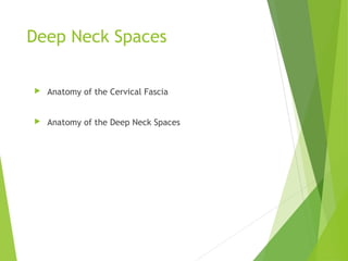
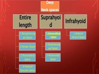
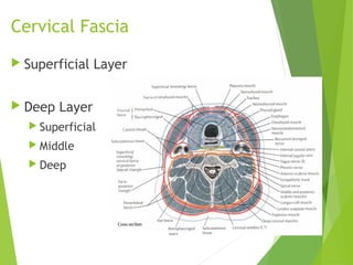
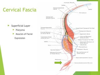
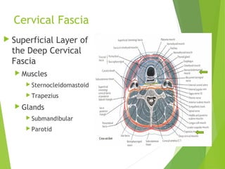
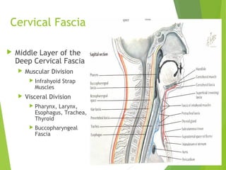
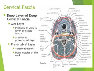
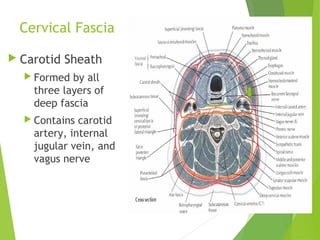
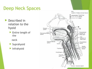
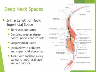
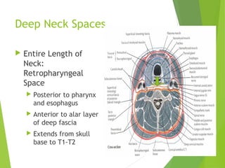
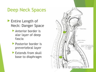
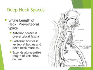
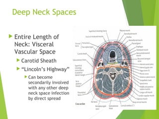
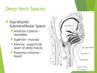
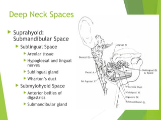
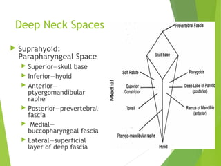
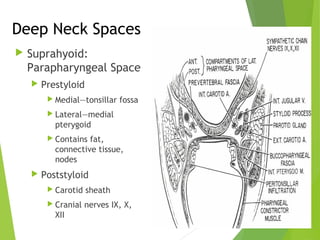
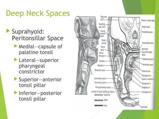
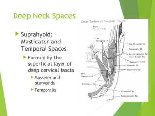
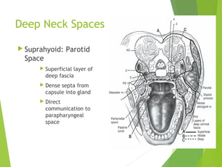
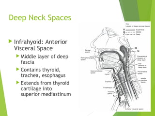
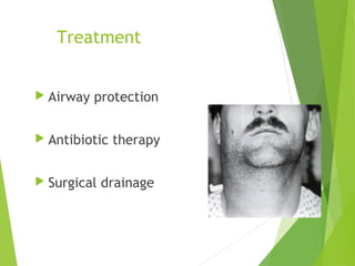
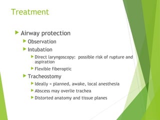
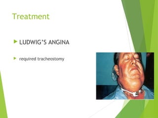
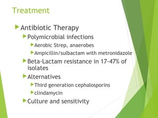
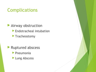
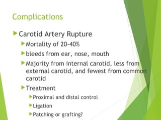
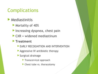
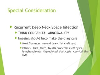
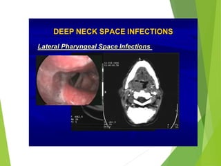
IN THE NAME OF
GOD`
Deep spaces of the neck
Arranged by :Dr.N.S.ValedSaravi
Shahid Beheshti Medical university
Department of Periodontics
Deep Neck Spaces
 Anatomy of the Cervical Fascia
 Anatomy of the Deep Neck Spaces
Cervical Fascia
 Superficial Layer
 Deep Layer
 Superficial
 Middle
 Deep
Cervical Fascia
 Superficial Layer
 Platysma
 Muscles of Facial
Expression
Cervical Fascia
 Superficial Layer of
the Deep Cervical
Fascia
 Muscles
 Sternocleidomastoid
 Trapezius
 Glands
 Submandibular
 Parotid
Cervical Fascia
 Middle Layer of the
Deep Cervical Fascia
 Muscular Division
 Infrahyoid Strap
Muscles
 Visceral Division
 Pharynx, Larynx,
Esophagus, Trachea,
Thyroid
 Buccopharyngeal
Fascia
Cervical Fascia
 Deep Layer of Deep
Cervical Fascia
 Alar Layer
 Posterior to visceral
layer of middle
fascia
 Anterior to
prevertebral layer
 Prevertebral Layer
 Vertebral bodies
 Deep muscles of the
neck
Cervical Fascia
 Carotid Sheath
 Formed by all
three layers of
deep fascia
 Contains carotid
artery, internal
jugular vein, and
vagus nerve
Deep Neck Spaces
 Described in
relation to the
hyoid
 Entire length of
the
neck
 Suprahyoid
 Infrahyoid
Deep Neck Spaces
 Entire Length of Neck:
Superficial Space
 Surrounds platysma
 Contains areolar tissue,
nodes, nerves and vessels
 Subplatysmal Flaps
 Involved with cellulitis
and superficial abscesses
 Treat with incision along
Langer’s lines, drainage
and antibiotics
Deep Neck Spaces
 Entire Length of
Neck:
Retropharyngeal
Space
 Posterior to pharynx
and esophagus
 Anterior to alar layer
of deep fascia
 Extends from skull
base to T1-T2
Deep Neck Spaces
 Entire Length of
Neck: Danger Space
 Anterior border is
alar layer of deep
fascia
 Posterior border is
prevertebral layer
 Extends from skull
base to diaphragm
Deep Neck Spaces
 Entire Length of
Neck: Prevertebral
Space
 Anterior border is
prevertebral fascia
 Posterior border is
vertebral bodies and
deep neck muscles
 Extends along entire
length of vertebral
column
Deep Neck Spaces
 Entire Length of
Neck: Visceral
Vascular Space
 Carotid Sheath
 “Lincoln’s Highway”
 Can become
secondarily involved
with any other deep
neck space infection
by direct spread
Deep Neck Spaces
 Suprahyoid:
Submandibular Space
 Anterior/Lateral—
mandible
 Superior—mucosa
 Inferior—superficial
layer of deep fascia
 Posterior/Inferior--
hyoid
Deep Neck Spaces
 Suprahyoid:
Submandibular Space
 Sublingual Space
 Areolar tissue
 Hypoglossal and lingual
nerves
 Sublingual gland
 Wharton’s duct
 Submylohyoid Space
 Anterior bellies of
digastrics
 Submandibular gland
Deep Neck Spaces
 Suprahyoid:
Parapharyngeal Space
 Superior—skull base
 Inferior—hyoid
 Anterior—
ptyergomandibular
raphe
 Posterior—prevertebral
fascia
 Medial—
buccopharyngeal fascia
 Lateral—superficial
layer of deep fascia
Deep Neck Spaces
 Suprahyoid:
Parapharyngeal Space
 Prestyloid
 Medial—tonsillar fossa
 Lateral—medial
pterygoid
 Contains fat,
connective tissue,
nodes
 Poststyloid
 Carotid sheath
 Cranial nerves IX, X,
XII
Deep Neck Spaces
 Suprahyoid:
Peritonsillar Space
 Medial—capsule of
palatine tonsil
 Lateral—superior
pharyngeal
constrictor
 Superior—anterior
tonsil pillar
 Inferior—posterior
tonsil pillar
Deep Neck Spaces
 Suprahyoid:
Masticator and
Temporal Spaces
 Formed by the
superficial layer of
deep cervical fascia
 Masseter and
pterygoids
 Temporalis
Deep Neck Spaces
 Suprahyoid: Parotid
Space
 Superficial layer of
deep fascia
 Dense septa from
capsule into gland
 Direct
communication to
parapharyngeal
space
Deep Neck Spaces
 Infrahyoid: Anterior
Visceral Space
 Middle layer of deep
fascia
 Contains thyroid,
trachea, esophagus
 Extends from thyroid
cartilage into
superior mediastinum
Deep Neck Space Infections
 Presentation/Origin of Infection
 Microbiology
 Imaging
 Treatment
 Complications
 Special Consideration
Presentation/Origin
 Retropharyngeal Abscess
 50% occur in patients 6-12 months of age
 96% occur before 6 years of age
 Children--fever, irritability, lymphadenopathy, torticollis, poor oral intake,
sore throat, drooling
 Adults--pain, dysphagia, anorexia, snoring, nasal obstruction, nasal
regurgitation
 Dyspnea and respiratory distress
 Lateral posterior oropharyngeal wall bulge
Presentation/Origin
 Pediatrics
 Cause—suppurative
process in lymph nodes
 Nose, adenoids,
nasopharynx, sinuses
 Adults
 Cause—trauma,
instrumentation,
extension from
adjoining deep neck
space
Presentation/Origin
 Danger Space
 Presentation and exam nearly identical to retropharyngeal
space infection
 Cause—extension from retropharyngeal, prevertebral or
parapharyngeal space
Presentation/Origin
 Prevertebral Space
 Back, shoulder, neck pain
made worse by deglutition
 Dysphagia or dyspnea
 Cause—Pott’s abscess, trauma,
osteomyelitis, extension from
retropharyngeal and danger spaces
Presentation/Origin
 Visceral Vascular Space
 Induration and tenderness over SCM
 Torticollis toward opposite side
 Spiking fevers, sepsis
 Cause—intravenous drug abuse, extension from other deep
neck spaces
Presentation/Origin
 Ludwig’s angina
 1. Cellulitis, not abscess
 2. Limited to SM space
 3. Foul serosanguinous fluid,
no frank purulence
 4. Fascia, muscle, connective
tissue involvement, sparing
glands
 5. Direct spread rather than
lymphatic spread
 Tender, firm anterior neck
edema without fluctuance
 “Hot potato” voice, drooling
 Tachypnea, dyspnea, stridor
Presentation/Origin
 Parapharyngeal Space
 Fever, chills, malaise
 Pain, dysphagia, trismus
 Medial bulge of lateral
pharyngeal wall
 Cause—infection of
pharynx, tonsil, adenoids,
dentition, parotid, mastoid,
suppurative lymphadenitis,
extension from other deep
neck spaces
Presentation/Origin
 Peritonsillar Space
 Fever, malaise
 Dysphagia, odynophagia
 “Hot-potato” voice,
trismus, bulging of
superior tonsil pole and
soft palate, deviation of
uvula
 Cause—extension from
tonsillitis
Presentation/Origin
 Masticator Temporal
Space
 Pain, trismus
 Posterior FOM edema
 Swelling along ramus
of mandible
 Cause—
odontogenic,
from third
molars
 Parotid Space
 Pain, trismus
 Medial bulge of
posterior lateral
pharyngeal wall
 Cause—parotitis,
sialolithiasis,
Sjogren’s
syndrome
Presentation/Origin
 Anterior Visceral Space
 Hoarseness, dyspnea, dysphagia,
odynophagia
 Erythema, edema of hypo pharynx,
may extend to include glottis and supra
glottis
 Anterior neck edema, pain, erythema,
crepitus
 Cause—foreign body, instrumentation,
extension of infection in thyroid
Microbiology
Pre antibiotic era—S.aureus
Currently—aerobic Strep species and non-
strep anaerobes
Gram-negatives uncommon
Almost always polymicrobial
Remember resistance
Imaging
 Lateral neck plain film
 Screening exam—mainly
for retropharyngeal and
pretracheal spaces
 Normal: 7mm at C-2,
14mm at C-6 for kids,
22mm at C-6 for adults
 Technique dependent
 Extension
 Inspiration
 Nagy, et al
 Sensitivity 83%,
compared to CT 100%
Imaging
High-resolution Ultrasound
Advantages
Avoids radiation
Portable
Disadvantages
Not widely accepted
Operator dependent
Inferior anatomic detail
Uses
Following infection during therapy
Image guided aspiration
Imaging
 Contrast enhanced CT
 Advantages
 Quick, easy
 Widely available
 Familiarity
 Superior anatomic
detail
 Differentiate abscess
and cellulitis
 Disadvantages
 Ionizing radiation
 Allergenic contrast
agent
 Soft tissue detail
 Artifact
Imaging
 MRI
 Advantages
 No radiation
 Safer contrast agent
 Better soft tissue detail
 Imaging in multiple planes
 No artifact by dental fillings
 Disadvantages
 Increased cost
 Increased exam time
 Dependent on patient
cooperation
 Availability
Treatment
 Airway protection
 Antibiotic therapy
 Surgical drainage
Treatment
 Airway protection
 Observation
 Intubation
 Direct laryngoscopy: possible risk of rupture and
aspiration
 Flexible fiberoptic
 Tracheostomy
 Ideally = planned, awake, local anesthesia
 Abscess may overlie trachea
 Distorted anatomy and tissue planes
Treatment
 LUDWIG’S ANGINA
 required tracheostomy
Treatment
Antibiotic Therapy
Cellulitis
Improvement in 24-48 hours
Abscess?
Mayor, et al: review of 31 patients, 19 with CT
evidence of abscess, 90% response
Nagy, et al: review of 47 pediatric patients,
51% response rate, only 7 of these had CT
evidence of abscess
Treatment
Antibiotic Therapy
Polymicrobial infections
Aerobic Strep, anaerobes
Ampicillin/sulbactam with metronidazole
Beta-Lactam resistance in 17-47% of
isolates
Alternatives
Third generation cephalosporins
clindamycin
Culture and sensitivity
 Surgical Drainage
 Transoral
 Preoperative CT—where are the great
vessels?
 Cruciate mucosal incision, blunt
spreading through superior pharyngeal
constrictor
 External
Treatment
Surgical Drainage
External
EXPOSURE
Levitt: anterior vs.
posterior
approach
Submandibular
incision
Submental incision
T-incision
Treatment
Image-guided Aspiration
Patient selection
Smaller abscesses, limited
extension, uniloculated
CT guided aspiration
Early specimen collection, reduced
expense, avoidance of neck scar
Ultrasound guided aspiration
Complications
 Airway obstruction
 Endotracheal intubation
 Tracheostomy
 Ruptured abscess
 Pneumonia
 Lung Abscess
Complications
Internal Jugular Vein Thrombosis
Lemierre’s syndrome
swelling and pain along SCM
Bacteremia, septic embolization, dural
sinus thrombosis
IV drug abusers
Treatment
IV antibiotic therapy
Anticoagulation?
Ligation and excision
Complications
Carotid Artery Rupture
Mortality of 20-40%
bleeds from ear, nose, mouth
Majority from internal carotid, less from
external carotid, and fewest from common
carotid
Treatment
Proximal and distal control
Ligation
Patching or grafting?
Complications
 Mediastinitis
 Mortality of 40%
 Increasing dyspnea, chest pain
 CXR = widened mediastinum
 Treatment
 EARLY RECOGNITION AND INTERVENTION
 Aggressive IV antibiotic therapy
 Surgical drainage
 Transcervical approach
 Chest tube vs. thoracotomy
Special Consideration
 Recurrent Deep Neck Space Infection
 THINK CONGENITAL ABNORMALITY
 Imaging should help make the diagnosis
 Most Common: second branchial cleft cyst
 Others: first, third, fourth branchial cleft cysts,
lymphangiomas, thyroglossal duct cysts, cervical thymic
cyst
Lateral pharyngeal
 start:Base skull .esphenoid bone
 end:hayoid bone
 Medial:medial trigoid muscle
 Lateral:superior constrictor
 Anterior:trigomandibular raphe
 Pesterior:retropharyngeal space
 Estyloid processs anterior= loose
connective tissue
 posterior=carotid
sleath,nerve 9,10,12

Anatomy

  • 1.
    IN THE NAMEOF GOD` Deep spaces of the neck Arranged by :Dr.N.S.ValedSaravi Shahid Beheshti Medical university Department of Periodontics
  • 2.
    Deep Neck Spaces Anatomy of the Cervical Fascia  Anatomy of the Deep Neck Spaces
  • 4.
    Cervical Fascia  SuperficialLayer  Deep Layer  Superficial  Middle  Deep
  • 5.
    Cervical Fascia  SuperficialLayer  Platysma  Muscles of Facial Expression
  • 6.
    Cervical Fascia  SuperficialLayer of the Deep Cervical Fascia  Muscles  Sternocleidomastoid  Trapezius  Glands  Submandibular  Parotid
  • 7.
    Cervical Fascia  MiddleLayer of the Deep Cervical Fascia  Muscular Division  Infrahyoid Strap Muscles  Visceral Division  Pharynx, Larynx, Esophagus, Trachea, Thyroid  Buccopharyngeal Fascia
  • 8.
    Cervical Fascia  DeepLayer of Deep Cervical Fascia  Alar Layer  Posterior to visceral layer of middle fascia  Anterior to prevertebral layer  Prevertebral Layer  Vertebral bodies  Deep muscles of the neck
  • 9.
    Cervical Fascia  CarotidSheath  Formed by all three layers of deep fascia  Contains carotid artery, internal jugular vein, and vagus nerve
  • 10.
    Deep Neck Spaces Described in relation to the hyoid  Entire length of the neck  Suprahyoid  Infrahyoid
  • 11.
    Deep Neck Spaces Entire Length of Neck: Superficial Space  Surrounds platysma  Contains areolar tissue, nodes, nerves and vessels  Subplatysmal Flaps  Involved with cellulitis and superficial abscesses  Treat with incision along Langer’s lines, drainage and antibiotics
  • 12.
    Deep Neck Spaces Entire Length of Neck: Retropharyngeal Space  Posterior to pharynx and esophagus  Anterior to alar layer of deep fascia  Extends from skull base to T1-T2
  • 13.
    Deep Neck Spaces Entire Length of Neck: Danger Space  Anterior border is alar layer of deep fascia  Posterior border is prevertebral layer  Extends from skull base to diaphragm
  • 14.
    Deep Neck Spaces Entire Length of Neck: Prevertebral Space  Anterior border is prevertebral fascia  Posterior border is vertebral bodies and deep neck muscles  Extends along entire length of vertebral column
  • 15.
    Deep Neck Spaces Entire Length of Neck: Visceral Vascular Space  Carotid Sheath  “Lincoln’s Highway”  Can become secondarily involved with any other deep neck space infection by direct spread
  • 16.
    Deep Neck Spaces Suprahyoid: Submandibular Space  Anterior/Lateral— mandible  Superior—mucosa  Inferior—superficial layer of deep fascia  Posterior/Inferior-- hyoid
  • 17.
    Deep Neck Spaces Suprahyoid: Submandibular Space  Sublingual Space  Areolar tissue  Hypoglossal and lingual nerves  Sublingual gland  Wharton’s duct  Submylohyoid Space  Anterior bellies of digastrics  Submandibular gland
  • 18.
    Deep Neck Spaces Suprahyoid: Parapharyngeal Space  Superior—skull base  Inferior—hyoid  Anterior— ptyergomandibular raphe  Posterior—prevertebral fascia  Medial— buccopharyngeal fascia  Lateral—superficial layer of deep fascia
  • 19.
    Deep Neck Spaces Suprahyoid: Parapharyngeal Space  Prestyloid  Medial—tonsillar fossa  Lateral—medial pterygoid  Contains fat, connective tissue, nodes  Poststyloid  Carotid sheath  Cranial nerves IX, X, XII
  • 20.
    Deep Neck Spaces Suprahyoid: Peritonsillar Space  Medial—capsule of palatine tonsil  Lateral—superior pharyngeal constrictor  Superior—anterior tonsil pillar  Inferior—posterior tonsil pillar
  • 21.
    Deep Neck Spaces Suprahyoid: Masticator and Temporal Spaces  Formed by the superficial layer of deep cervical fascia  Masseter and pterygoids  Temporalis
  • 22.
    Deep Neck Spaces Suprahyoid: Parotid Space  Superficial layer of deep fascia  Dense septa from capsule into gland  Direct communication to parapharyngeal space
  • 23.
    Deep Neck Spaces Infrahyoid: Anterior Visceral Space  Middle layer of deep fascia  Contains thyroid, trachea, esophagus  Extends from thyroid cartilage into superior mediastinum
  • 24.
    Deep Neck SpaceInfections  Presentation/Origin of Infection  Microbiology  Imaging  Treatment  Complications  Special Consideration
  • 25.
    Presentation/Origin  Retropharyngeal Abscess 50% occur in patients 6-12 months of age  96% occur before 6 years of age  Children--fever, irritability, lymphadenopathy, torticollis, poor oral intake, sore throat, drooling  Adults--pain, dysphagia, anorexia, snoring, nasal obstruction, nasal regurgitation  Dyspnea and respiratory distress  Lateral posterior oropharyngeal wall bulge
  • 26.
    Presentation/Origin  Pediatrics  Cause—suppurative processin lymph nodes  Nose, adenoids, nasopharynx, sinuses  Adults  Cause—trauma, instrumentation, extension from adjoining deep neck space
  • 27.
    Presentation/Origin  Danger Space Presentation and exam nearly identical to retropharyngeal space infection  Cause—extension from retropharyngeal, prevertebral or parapharyngeal space
  • 28.
    Presentation/Origin  Prevertebral Space Back, shoulder, neck pain made worse by deglutition  Dysphagia or dyspnea  Cause—Pott’s abscess, trauma, osteomyelitis, extension from retropharyngeal and danger spaces
  • 29.
    Presentation/Origin  Visceral VascularSpace  Induration and tenderness over SCM  Torticollis toward opposite side  Spiking fevers, sepsis  Cause—intravenous drug abuse, extension from other deep neck spaces
  • 30.
    Presentation/Origin  Ludwig’s angina 1. Cellulitis, not abscess  2. Limited to SM space  3. Foul serosanguinous fluid, no frank purulence  4. Fascia, muscle, connective tissue involvement, sparing glands  5. Direct spread rather than lymphatic spread  Tender, firm anterior neck edema without fluctuance  “Hot potato” voice, drooling  Tachypnea, dyspnea, stridor
  • 31.
    Presentation/Origin  Parapharyngeal Space Fever, chills, malaise  Pain, dysphagia, trismus  Medial bulge of lateral pharyngeal wall  Cause—infection of pharynx, tonsil, adenoids, dentition, parotid, mastoid, suppurative lymphadenitis, extension from other deep neck spaces
  • 32.
    Presentation/Origin  Peritonsillar Space Fever, malaise  Dysphagia, odynophagia  “Hot-potato” voice, trismus, bulging of superior tonsil pole and soft palate, deviation of uvula  Cause—extension from tonsillitis
  • 33.
    Presentation/Origin  Masticator Temporal Space Pain, trismus  Posterior FOM edema  Swelling along ramus of mandible  Cause— odontogenic, from third molars  Parotid Space  Pain, trismus  Medial bulge of posterior lateral pharyngeal wall  Cause—parotitis, sialolithiasis, Sjogren’s syndrome
  • 34.
    Presentation/Origin  Anterior VisceralSpace  Hoarseness, dyspnea, dysphagia, odynophagia  Erythema, edema of hypo pharynx, may extend to include glottis and supra glottis  Anterior neck edema, pain, erythema, crepitus  Cause—foreign body, instrumentation, extension of infection in thyroid
  • 35.
    Microbiology Pre antibiotic era—S.aureus Currently—aerobicStrep species and non- strep anaerobes Gram-negatives uncommon Almost always polymicrobial Remember resistance
  • 36.
    Imaging  Lateral neckplain film  Screening exam—mainly for retropharyngeal and pretracheal spaces  Normal: 7mm at C-2, 14mm at C-6 for kids, 22mm at C-6 for adults  Technique dependent  Extension  Inspiration  Nagy, et al  Sensitivity 83%, compared to CT 100%
  • 37.
    Imaging High-resolution Ultrasound Advantages Avoids radiation Portable Disadvantages Notwidely accepted Operator dependent Inferior anatomic detail Uses Following infection during therapy Image guided aspiration
  • 38.
    Imaging  Contrast enhancedCT  Advantages  Quick, easy  Widely available  Familiarity  Superior anatomic detail  Differentiate abscess and cellulitis  Disadvantages  Ionizing radiation  Allergenic contrast agent  Soft tissue detail  Artifact
  • 39.
    Imaging  MRI  Advantages No radiation  Safer contrast agent  Better soft tissue detail  Imaging in multiple planes  No artifact by dental fillings  Disadvantages  Increased cost  Increased exam time  Dependent on patient cooperation  Availability
  • 40.
    Treatment  Airway protection Antibiotic therapy  Surgical drainage
  • 41.
    Treatment  Airway protection Observation  Intubation  Direct laryngoscopy: possible risk of rupture and aspiration  Flexible fiberoptic  Tracheostomy  Ideally = planned, awake, local anesthesia  Abscess may overlie trachea  Distorted anatomy and tissue planes
  • 42.
  • 43.
    Treatment Antibiotic Therapy Cellulitis Improvement in24-48 hours Abscess? Mayor, et al: review of 31 patients, 19 with CT evidence of abscess, 90% response Nagy, et al: review of 47 pediatric patients, 51% response rate, only 7 of these had CT evidence of abscess
  • 44.
    Treatment Antibiotic Therapy Polymicrobial infections AerobicStrep, anaerobes Ampicillin/sulbactam with metronidazole Beta-Lactam resistance in 17-47% of isolates Alternatives Third generation cephalosporins clindamycin Culture and sensitivity
  • 46.
     Surgical Drainage Transoral  Preoperative CT—where are the great vessels?  Cruciate mucosal incision, blunt spreading through superior pharyngeal constrictor  External
  • 47.
    Treatment Surgical Drainage External EXPOSURE Levitt: anteriorvs. posterior approach Submandibular incision Submental incision T-incision
  • 48.
    Treatment Image-guided Aspiration Patient selection Smallerabscesses, limited extension, uniloculated CT guided aspiration Early specimen collection, reduced expense, avoidance of neck scar Ultrasound guided aspiration
  • 49.
    Complications  Airway obstruction Endotracheal intubation  Tracheostomy  Ruptured abscess  Pneumonia  Lung Abscess
  • 50.
    Complications Internal Jugular VeinThrombosis Lemierre’s syndrome swelling and pain along SCM Bacteremia, septic embolization, dural sinus thrombosis IV drug abusers Treatment IV antibiotic therapy Anticoagulation? Ligation and excision
  • 51.
    Complications Carotid Artery Rupture Mortalityof 20-40% bleeds from ear, nose, mouth Majority from internal carotid, less from external carotid, and fewest from common carotid Treatment Proximal and distal control Ligation Patching or grafting?
  • 52.
    Complications  Mediastinitis  Mortalityof 40%  Increasing dyspnea, chest pain  CXR = widened mediastinum  Treatment  EARLY RECOGNITION AND INTERVENTION  Aggressive IV antibiotic therapy  Surgical drainage  Transcervical approach  Chest tube vs. thoracotomy
  • 53.
    Special Consideration  RecurrentDeep Neck Space Infection  THINK CONGENITAL ABNORMALITY  Imaging should help make the diagnosis  Most Common: second branchial cleft cyst  Others: first, third, fourth branchial cleft cysts, lymphangiomas, thyroglossal duct cysts, cervical thymic cyst
  • 57.
    Lateral pharyngeal  start:Baseskull .esphenoid bone  end:hayoid bone  Medial:medial trigoid muscle  Lateral:superior constrictor  Anterior:trigomandibular raphe  Pesterior:retropharyngeal space  Estyloid processs anterior= loose connective tissue  posterior=carotid sleath,nerve 9,10,12