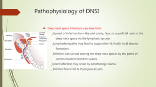
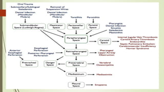
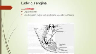
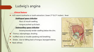
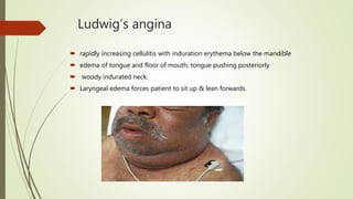
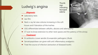
Deep neck space infections
Dr Ahlam alzuway
Objectives:
• Anatomy of Cervical fascia
• Anatomy of Deep neck spaces
• Deep neck space infections
Cervical fascia
*Deep fascia
.superficial layer(investing)
.middle layer(visceral &
muscular)
.deep layer (alar & prevertebral)
*Superficial fascia
Cervical fascia
SUPERFICIAL FASCIA
 Thin layer of connective tissue that lies between dermis
of the skin and investing layer of deep cervical fascia
Contains cutaneous nerves, blood and lymphatic
vessels, superficial lymph nodes and fat
extend from zygomatic process to thorax & axilla
 Anteriolaterally it contains the platysma
deep cervical fascia made up of 3 layers
1. Superficial investing layer
 superiorly it’s attached to the nuchal ridge ,mastoid ,zygoma ,mandible & hyoid
bone …inferiorly bounded by the sternum, clavicles, , acromion scapula
 Splits to enclose trapezius ,sternocleidomastoid ,
submandibular and parotid gland
2.middle layer of deep fascia
 It has 2 divisions
a. muscular division :surronds the strap muscles
b. visceral division :enclose thyroid ,larynx , trachea, pharynx
esophagus
 Its attached antero-superiorly to hyoid bone & thyroid cartilage ,postero-superiorly
to skull base , inferiorly to the sternum ,clavicle & scapula
3.deep layer of deep cervical fascia
 Splits into 2 layers at the transverse processes
a. Alar layer : lies between middle layer & prevertebral fascia
b. Prevertebral layer :envelopes vertebral bodies & deep muscle of the
neck
entire length space suprahyoid space infrahyoid space
Retropharyngeal space Peritonsillar space Pretracheal space
(anterior visceral space)
Danger space Parapharyngeal space
(lateral pharyngeal
,pharyngomaxillary)
Prevertebral space Submandibular space
visceral vascular space
(carotid sheath)
Submental space
Parotid space
Masticator space
Buccal space
deep neck spaces
Deep neck spaces
Entire Length of Neck:
1.Retropharyngeal Space
a. Posterior to pharynx and esophagus
b. Anterior to alar layer of deep fascia
c. Extends from skull base to T4
e. It has midline fibrous raphy dividing it into
2 spaces (spaces of Gillette)
2.Danger Space
a. Anterior border is alar layer of deep fascia
b. Posterior border is prevertebral layer
c. Extends from skull base to diaphragm and is so named
because it contains loose areolar tissue and offers little resistance
to the spread of infection.
 3.Prevertebral Space
a. Anterior border is prevertebral fascia
b. Posterior border is vertebral bodies
c. Extends along entire length of vertebral column.
 Infection in this space tends to stay localized due to the dense
fibrous attachments between the fascia and the deep muscles.
4.Visceral Vascular Space
 It is termed the “Lincolin’s highway” of the neck .
• It extends from the base of skull to the superior mediastinum
• because it receives contributions from all three layers of deep fascia it
can secondarily involved by infection in any other deep neck space
by direct spread.
 Carotid sheath and its contents
Suprahyoid spaces
1.Submandibular Space
 Anterior/Lateral: mandible
 Superior: floor of the mouth
 Inferior: superficial layer of deep fascia
 Postero-Inferior: hyoid bone
Submandibular Space divided into
Sublingual Space
• Areolar tissue
• Hypoglossal and lingual nerves
• Sublingual gland
• Wharton’s duct
• facial vessels
Submylohyoid Space (submaxillary space)
• Anterior & posterior bellies of digastrics
• Submandibular gland
• (These two subdivisions freely communicate
around the posterior border of the mylohyoid. )
Suprahyoid spaces
2. Parapharyngeal Space (pharyngomaxillary , lat. pharyngeal )
 Superior: skull base (petrous portion of temporal bone)
 Inferior: hyoid
 Anterior: ptyergomandibular raphe
 Posterior: prevertebral fascia
 Medial: buccopharyngeal fascia
 Lateral: superficial layer of deep fascia, medial pterygoid and sumandibulr G .
 The parapharyngeal space communicates with submandibular ,
retropharyngeal, parotid and masticator spaces with
important implications in spread of infection
the Styloid process divide the space into anterior and posterior
compartments
 Anterior compartment: medially related to the tonsillar fossa, and
laterally to the medial pterygoid muscle. Its contents:
_ Int. maxillary art
_ lingual ,inferior alveolar& auriculotemporal Ns
_ fat, connective tissues , LNs
 Posterior compartment: related medially to the posterior part of lateral
pharyngeal wall, and laterally to the parotid gland.
 The contents of posterior compartment
– Internal carotid artery
– Internal jugular vein
– Cranial nerves IX, X, XI and XII
– Sympathetic trunk
– Upper deep cervical nodes
Parotid space
Relations:
 Anteriorly: masticator space
 Medially: parapharyngeal space
 Laterally: superficial fascia & skin
 Superiorly: external auditory canal and mastoid tip
 Inferiorly: inferior mandibular margin& posterior end of sub-mandibular space.
Contents:
 parotid gland
 intraparotid lymph nodes
 intraparotid facial nerve
 external carotid artery
 retro-mandibular vein
Infrahyoid space
1.Anterior Visceral Space (pretracheal )
- Formed by middle layer of deep fascia
Contains thyroid, trachea, esophagus.
-This potential space runs from the thyroid
cartilage into the anterior superior
mediastinum to the arch of the aorta.
- Below the level of the thyroid gland
this space communicates laterally
with the retropharyngeal space .
Introduction to Deep neck space
infections
Pathophysiology of DNSI
 Deep neck space infections can arise from
_Spread of infection from the oral cavity, face, or superficial neck to the
deep neck space via the lymphatic system.
_Lymphadenopathy may lead to suppuration & finally focal abscess
formation.
_Infection can spread among the deep neck spaces by the paths of
communication between spaces.
_Direct infection may occur by penetrating trauma.
_ Infected branchial & thyroglossal cysts
 The signs and symptoms of a deep neck abscess develop because of the following:
-Mass effect of inflamed tissue or abscess cavity on surround structures
-Direct involvement of surrounding structures with the infectious process
 The microbiology of deep neck infections usually reveals mixed aerobic and anaerobic
organisms, often with a predominance of oral flora. Both gram +ve and gram -ve
organisms may be cultured.
 The spread of infection can vary based on:
- number & virulence of organisms
- host resistence
- anatomy of the involved area
Clinical Evaluation of patient with DNS infections
1. History
Inflammatory symptoms: pain, fever, swelling, redness.
Localizing symptoms: dysphagia/ odynophagia/drooling
(retropharyngeal abscess), hot potato voice/trismus (peritonsillar
abscess), hoarseness, dyspnea, neck swelling, otalgia.
Recent infection: dental, sinusitis, otitis
Recent trauma: IV drug use.
Recent surgery: dental, intubation, endoscopy.
Past medical history: antibiotic allergies.
Immuno-defciency (diabetes; HIV, autoimmune disorder,
hematologic malignancy; recent chemotherapy or steroid)
causes increased risk of atypical pathogens and rapidly
progressive symptoms.
2. Physical Examination
Palpation:
1. Localizing tenderness 2. Crepitus (gas- forming organism)
Otoscopy:
1. Otitis 2.Rhinosinusitis 3.Foreign body
Oral cavity and pharynx
1. Poor dentition (tooth infection; possible anaerobic organisms)
2. trismus (para-pharyngeal, pterygo-maxillary, masticator spaces)
3. Floor of mouth edema/ tongue swelling (sublingual and
submandibular spaces infections causing Ludwig angina and potential
airway emergency)
4. Purulent discharge from Wharton’s or Stenson’s duct (parotid and sublingual/
submandibular spaces; bimanual palpation for stones)
5. Unilateral tonsil swelling with deviation of uvula (peritonsillar abscess if
inflammation present; think tonsil or parapharyngeal space tumor if no
inflammation)
Cranial nerve examination
CN II, III, IV, V, VI can cause reduced vision and eye mobility via
retrograde flow of infection through valveless ophthalmic veins.
a wake flexible fiberoptic airway evaluation
1. Mandatory if hoarseness ,dyspnea ,stridor ,dysphagia,
odynophagia without obvious cause.
2. Identifes patients who may need intubation.
3 .Diagnostic testing
Blood tests
1. Leukocytosis common
2. Lack of leukocytosis (virus, immunodeciency, tumor,
congenital cyst)
3.blood sugar and electrolytes Basic electrolyte panel (glucose,
hydration level, renal function)
Plain film radiography
1. Jaw films (Panorex): thin bone at dental root (odontogenic abscess);
salivary stones
2.Lateral neck films: air- fluid level; prevertebral shadow > 5 mm
thickening (child) or > 7mm thickening (adult) at C2 (retropharyngeal
infection)
3. Chest films : widened mediastinum (mediastinitis); lower lobe infltrate
(aspiration pneumonia)
Computed tomography with IV contrast
1. Determines neck spaces requiring drainage.
2. Difficult to differentiate between abscess (pus) or phlegmon (edema).
3. Metastatic adenopathy ( from oropharyngeal primary) needs to be
rule out since this can mimic neck abscess on CT in adult patient.
 Magnetic resonance imaging
1. Helpful in select cases (intracranial complication; infection of
vertebral bodies)
2. MR angiography helpful if thrombi or pseudo-aneurysm of major vessel
suspected
 Ultrasonography
Advantages: noninvasive; no radiation; allows fine needle aspiration
Limitations: deeper abscesses; obese patients
. Treatment:
.Airway Management Loss of airway main source of mortality from deep neck
infection.
a. Fiberoptic exam evaluates at-risk airway
b. First-line therapy:
1. Oxygenated face tent with cool humidity
2. Intravenous steroids
3. Epinephrine nebulizers
4. ICU observation if stable and airway > 50 % normal caliber
c. Tracheotomy
1. Worsening stridor, dyspnea, or obstruction with < 50% of
normal caliber
2. Awake flexible intubation possible if glottis large enough to
pass adult flexible bronchoscope (6 mm)
3. Awake tracheotomy if patient not easily intubatable
4. Elective tracheotomy if prolonged (> 48 hr) airway edema
anticipated.
Fluid Resuscitation
a. Dehydration common due to dysphagia (peritonsillar, retropharyngeal).
b. Dehydration may be cause of sialadenitis.
c. Initial resuscitation: 1 to 2 L of isotonic IV fluids.
Intravenous Antibiotic Therapy
a. Broad-spectrum empiric therapy indicated at diagnosis (should not be
delayed or culture).
b. Clindamycin first-line therapy of choice for children < 2 years due to
increase rates of MRSA.
e. May delay need for surgical drainage in stable patient:
1. Contained abscess (intranodal) or phlegmon.
2. Most pediatric cases.
3. Abscess size < 2.5 cm contained within single neck space.
4. Patient kept NPO if surgery is considered.
5. Repeat imaging and/or surgical intervention indicated if no improvement
after 48 to 72 hours of therapy.
6. If improved, continue IV 24 hrs beyond symptom resolution followed
by 2 weeks of PO therapy
Surgical Management
a. Indications for surgical exploration:
1. Air-fluid level in neck or gas-forming organism
2. Abscess present in fascial spaces of neck
3. Failure to respond to 48 to 72 hours of IV antibiotics
b. Goals of surgical exploration:
1. Drainage of abscess
2. Fluid sampling for culture and sensitivity
3. Irrigation of involved neck space
4. Establishment of external drainage pathway to prevent recurrence.
5. Removal of infectious source (dental extraction, foreign body removal)
Surgical technique
1. Divide superficial cervical fascia and superficial layer of deep fascia.
2. Blunt dissection with hemostat and Kittner sponge into involved space.
3. Over dissection puts normal structures at risk and provides path or
infection to spread.
4. Culture inflammatory drainage or pus.
5. Copious irrigation of wound.
6. External drainage with Penrose or rubber band drain.
7. Loose closure of wound.
Deep neck space infections
Retropharyngeal space infection
Acute Retropharyngeal space infection
Acute Retropharyngeal abscess
 Common in children < 4yrs following suppuration of
retropharyngeal LNs secondary to adenoiditis , tonsillitis ,
Nasal sinuses or N. cavity infections
 In adults following foreign bodies impaction in the
cricopharynx or upper esophagus , penetrating injury of
posterior pharyngeal wall or cervical esophagus
 Extension from adjacent DNS
 Rarly as a complication of acute mastoiditis
Acute Retropharyngeal abscess
 Clinical features:
sore throat , child refuse feeding(acute odynophagia) ,fever,toxic look child
neck swelling (lymphadenopathy),slight neck rigidity& neck tilts to involved
side (torticollis) ,dribbling & pooling of saliva
Croupy cough may be present ,dyspnea, respiratory distress
bulge in posterior pharyngeal wall usually unilaterally ,gross inflamed
mucosa ,
larynx & trachea pushed forwards
, stridor +/- ,trismus is often absent
Acute Retropharyngeal abscess
Investigations
laboratory tests …CBC, CRP ,blood sugar, S electrolytes , TB test
Cultures: blood, needle aspirations
Panorex ( jaw film) …panoramic scanning dental x-ray
( for dental abscess).
X-ray soft tissue neck lateral view :
-widening of prevertebral soft tissue
shadow of 7 mm at C2, or 14mm in child
& 22mm in adults at C6
-displacement of larynx & trachea forwards
-straightening of C spine d/t prevertebral
muscle Spasm
-Air shadow in the space with or w/o fluid
level
Acute retropharyngeal abscess
X-ray chest: For assessing mediastinal silhouette for any widening.
USG :for seeing deep space neck abscesses & used to guide needle aspiration
CT and MRI:
- CT is usually the examination of choice though the MRI provides better for soft
tissues and involvement of great blood vessels ‘ internal jugular vein
thrombosis ’
- Tracheal compression
- Mediastinal spread
Treatment
 Hospitalization
 IV antibiotics, co-amoxyclav or a broad spectrum penicillin/cephalosporin and
metronidazole are the combination of choice.
 IV fluids
 Analgesics
 Anti inflammatory
 Tracheostomy in airway obstruction ,or stridor
 Endotracheal intubation is difficult to do ; because of inflamed & displaced larynx
& the risk of abscess rupture & aspiration of the pus into respiratory tract
Acute retropharyngeal abscess
Treatment: Tracheotomy ..in large abscess that cause airway obstruction &
laryngeal edema
 Incision & drainage :
 Done by transoral route ..with the patient in the head down Trendelenburg’s
position & intubated along the opposite side of the pharynx from the swelling if it
possible, needle aspiration for C&S ,and to minimize any risk of the patient
aspirating pus during the procedure
 Suction must be ready and handly to prevent aspiration of pus
Chronic retropharyngeal abscess
Aetiology:
 TB Spine(Pott’s Spine) where the pus collects in the prevertebral space (Cold abscess)
 TB of retropharyngeal LNs.
 Post traumatic vertebral fracture.
 Caries of cervical spine presents centrally behind the prevertebral fascia
 Spread from Parapharyngeal abscess
Symptoms :
 discomfort in throat
 Systemic features of TB +/-
 Painless lump in the throat
 Cervical pain may radiate to the upper limbs
Chronic retropharyngeal abscess
Signs:
 Median bulge on posterior pharyngeal wall
 No sings of acute inflammation
 Signs of cervical spine or LNs tuberculosis & neurological radiculopathies may be
present
Investigations:
 Plain X-ray ,CT & MRI scan show caries of the spine with vertebral body collapse,
prevertebral widening
 FNAC of the drained fluid show acid fast bacillus
& the cultures may be +ve for TB
Chronic retropharyngeal abscess
Treatment:
 Anti tubercular treatment
 Incison & drainage is done by a transcervical approach ,a vertical incision is made
along the anterior border (for low abscess) or posterior border (for high abscess) of
sternocleidomastoid muscle.
 Sequestrectomy performed by orthopedic surgeon or neurosurgeon
Parapharyngeal abscess
 Aetiology:
 Acute/Chronic infections of tonsils and adenoid, bursting of the peritonsillar abscess.
 Dental infection usually from the lower last molar.
 From otitis ,Bezold’s abscess or Petrositis ( rarly)
 Infections of parotid, retropharyngeal and submaxillary spaces.
 Penetrating injuries of neck
 Infection of the anterior compartment of the parapharyngeal space is more common
than the posterior compartment
Parapharyngeal abscess
 More common in adults
Clinical features: depend on the compartment involved
 Anterior compartment infection …triad of
 Trismus , prolapse of the tonsil & tonsillar fossa , external swelling behind the angle of the
mandible
 Posterior compartment infection…
 Bulge of the pharynx behind the tonsil, paralysis of CN 9, 10, 11 ,12 &sympathetic chain ,
Swelling of parotid region….minimal trismus or tonsillar prolapse
(Fever ,odynophagia , sore throat , torticollis & signs of toxaemia are common to
both compartment)
Diagnosis:
 Laboratory tests
 CT for location &extent of abscess , needle aspiration under CT or USG guidance
 Chest X-ray & CT for mediastinal spread
 Dental evaluation (odontogenic source)
Parapharyngeal space abscess
Treatment:
 IV antibiotics, iv fluids , analgesic &anti inflammatory
 Airway protection , Pre-op tracheostomy if trismus is marked
 Small loculated abscess.. conservatively
 Large collections .. external approach, medial to carotid sheath, insertion of drain
Incision and Drainage.
usually done under G.A, drained by :
 1. a horizontal incision made 2-3 cms below the angle of the mandible, Blunt dissection is
done along the inner surface of medial pterygoid towards styloid process and the abscess is
evacuated and a drain is inserted, (Transoral drainage should never be done due to the
danger of the great vessels which pass through this space)
 2. Mosher’s T-shape incision
Peritonsillar space infection
 Quinsy is a collection of pus in the peritonsillar space which lies
b/w the capsule of tonsil & the sup. constrictor muscle
Aetiology:
 follow acute Tonsillitis
Culture of pus show mixed growth of Strep. pyogenes, Staph.
aureus , aerobic and anaerobic organisms.
Clinical Features:
 fever ,chills and rigors, general malaise, headache, nausea
 Severe pain in throat;Usually unilateral ,trismus
 Tongue coated with White material ,halitosis
 odynophagia, hot potato voice, Ipsilateral otalgia
Peritonsillar abscess
clinical features:
 tonsil, pillars and soft palate on the involved
side are congested and swollen. Tonsil itself may not
appear enlarged as it gets buried in the oedematous
pillars
 Uvula is swollen and edematous and displaced to the opposite side.
 Bulging of the soft palate and anterior pillar above the tonsil.
 Mucopus may be seen covering the tonsillar region.
 Cervical lymphadnopathy is commonly seen ( jugulodigascric LNs)
 Torticollis.
Peritonsillar abscess
Investigations :
 Laboratory tests CBC, CRP ,S.electrolytes, c&s of pus ,BS
Treatment:
1. Hospitalisation.
2. IV fluids
3. IV Antibiotics . Broad spectrum to cover both aerobic and
anaerobic organisms
4. Analgesics
5. Oral hygiene
Peritonsillar abscess
Incision & drainage
 Peritonsillar abscess is opened at the point of maximum bulge above the
upper pole or just lateral to the point of junction of anterior pillar and a
horizontal line drawn through the base of the uvula
 Interval tonsillectomy.. Tonsils are removed 4 to 6 wks following an attack of
quinsy.
 Abscess or hot tonsillectomy.. Some surgeons do it instead of incision and
drainage, but it has the risk of abscess rupture during anaesthesia, and
excessive bleeding at the time of operation
Peritonsillar abscess
Complications
1. Parapharyngeal abscess .
2. edema of larynx. Tracheostomy may be required .
3. Septicemia.
4. IJV thrombosis.
5. Pneumonitis or lung abscess (aspiration of pus)
6. Spontaneous haemorrhage from CA or IJV.
Submandibular space abscess
Sublingual space abscess
 Source of infection premolar and less commonly from molar teeth when the
infection perforates the lingual cortex of the mandible above the attachment of the
Mylohyoid muscle.
 Indirectly the infection may spread from submental & submylohyoid spaces.
 Clinical features : erythematous swelling in floor of the mouth that may extend
through the midline since the barrier between the two sublingual spaces is weak,
usually there is elevation of the tongue.
 Treatment: IV Abs , treat the source of infection (infected tooth, S G stone)
 Incision& drainage: intraorally by incision through the mucosa only parallel to
Wharton's duct and lingual cortex in anteroposterior direction and away from the
sublingual fold.
 This space may be drained extraorally through submandibular and submental
incisions through the Mylohyoid muscle if the infection of these spaces is also
evident.
Submaxillary abscess(submylohyoid)
 Source of infection
 commonly 2nd & 3rd lower molars
 infected laceration of tongue, posterior part of the floor of the mouth, &
submandibular salivary gland infections.
 may spread from infected sublingual and submental spaces. Submandibular space
infections can spread posteriorly to the parapharyngeal space
 Clinical features: a firm or fluctuant erythematous swelling of the submandibular
region, tender swelling bulges over and obliterates the inferior border of the
mandible, there may be trismus, other signs and symptoms of infection may or
may not be present
 Investigations:
laboratory tests, dental x-ray
 Treatment:
IV Abs , treat the focus of infection
 Incision& drainage: extraoral horizontal incision about 2 cm below the inferior
border of the mandible to avoid injury to the marginal mandibular branch of the
facial N
Submental space infection
 Anatomical relations
 Aetiology: Odontogenic sources (ant. Teeth)
clinical features:
 Erythema, induration of skin in submental region, similar to Ludwig’s angina but
there is no swelling at floor of the mouth
 tenderness , Minimal respiratory distress &moderate dysphagia /odynophagia may
presents
Submental space abscess
Investigations:
 Laboratory tests
 jaw film
Treatment:
 IV Abs
 Horizontal incision at the site of maximum bulge after the infection localizes. It
may be drained intraorally through the Mentalis muscle via the labial vestibule, but
the dependent drainage can’t be established.
 Severe cellulitis involving the floor of the mouth (submandibular space)
 Initially infection in sublingual space then spreads to submaxillary &submental
spaces
 Gangrene with foul serosanguinous fluid on incision
 There is direct spread of infection rather than spread by
lymphatics
 The term angina is related to the sensation of suffocation
Aetiology:
 70% to 85% of these cases are odontogenic in origin,
 Sialadenitis
 Lymphadenitis
 Oral lacerations
 Mandibular fractures
Sub mandibular space infection
complication(Ludwig’s angina)
Ludwig’s angina
Aetiology:
 Lingual tonsillitis
 Mixed infection involve both aerobic and anaerobic pathogens
Ludwig’s angina
Clinical feature:
 H/O recent toothache or tooth extraction ( lower 2nd & 3rd molars) , fever
Sublingual space infection
-floor of mouth swelling.
-tongue pushed up & back .
Submaxillary space infection
-browny/woody tender swelling below the chin.
 Trismus, odynophagia, drooling
 muffled voice or trouble speaking and breathing
 Stridor.. due to falling back of tongue, laryngeal edema.
 Neck stifness
Ludwig’s angina
 rapidly increasing cellulitis with induration erythema below the mandible
 edema of tongue and floor of mouth; tongue pushing posteriorly
 woody indurated neck.
 Laryngeal edema forces patient to sit up & lean forwards
Ludwig’s angina
Diagnosis:
 Laboratory tests
 Jaw film
 Neck x-ray lat view volume increasing in the soft
tissues and if deviation of the trachea
 USG differentiate between cellulitis, abscess and adenopathy
 CT scan to know extension to other neck spaces and the patency of the airway
Treatment:
 IV antibiotics (cover aerobic & anaerobic pathogens ),fluids
 Serratiopeptidase can given with anti inflammatory analgesics
 Treat the source of infection (extraction of diseased tooth)
Thumb
sign
indicating
laryngeal
edema
Ludwig’s angina
 Surgical drainage is done by horizontal incision 2 fingers below mandible margin ,from one angel of
the mandible to the other ,over the area of induration .
 Pus is often not seen ,edematous fluid collection may be drained. debridement of necrotic tissue,
Irrigation with N/S
 Drainage tube or Abs soaked ribbon gauge is placed & the incision is not closed ,daily dressing ,
wound allowed to heal by 2ry intention
 Treatment of the underlying cause .
Ludwig’s angina
Complication:
 Airway obstruction.. requiring tracheotomy ,cricothyrotomy is sometimes
preformed instead of tracheotomy for lower risk of spreading infection to
mediastinum
 Aspiration pneumonia
 Lung abscess
 Tongue necrosis
 Spread to the other neck spaces
Parotid space infection
Aetiology:
 Parotitis ,Ascending of bacterial infection(Staphylococcus,
Streptococcus,Haemophilus) to a dehydrated parotid via Stenson’s duct from oral
cavity.
 Suppuration of intra-parotid LNs.
 Spread of infection from the auditory canal via the cartlaginous fissures of Santorini
or the bony foramen of Huschke.
Parotid space infection
Clinical features :
 Spontaneous onset of painful parotid enlargement followed by fever and cellulitis
which then turns into fluctuant parotid abscess.
 Pain and induration over the parotid gland & marked swelling over the jaw.
 Pitting edema over the parotid area differentiates parotid abscess from simple
parotitis, Parotid massage expresses pus into the oral cavity via the Stenson’s duct
,opposite the upper 2nd molar
 No trismus or pharyngeal swelling seen.
Parotid space infection (abscess)
Diagnosis:
 needle aspiration can be diagnostic.
 USG or CT/MRI can differentiate between parotidits & abscess.
Parotid space infection
Treatment:
 Maintinence of oral hygiene,
 IV antibiotics ,fluids
Incision and Drainage:-
-Modified Blair’s incision made..retromandibuar
incision extending from inf. aspect of ear lobule to angle of mandible
-Blunt dissection done to evacuate the pus, irrigation with N/S
-Drains are placed.
Parotid space infection (abscess)
Complications:
 Spread to parapharyngeal space through superomedial dehiscence
 Later it may involve other deep neck spaces & finally mediastinitis
Masticator space infections
 It’s formed as the deep cervical fascia covers the masseter muscle laterally & the
pterygoid muscle medially.
Contents:
 Ramus & body of mandible
 Muscles (masseter, med & lat pterygoids , tendon insertion of temporalis)
Aetiology :
 Infected tooth root of 2nd &3rd molar
 Infection following local anesthesia if asepsis is not maintained for inf.alveolar N
block
Masticator space infections
 Masticator space is subdivided into 3 spaces
Temporal space : divided into deep & superficial spaces by temporalis m
 Lies posterior & superior to masseteric & pterygomandibular space
 Deep temporal space contains interior max. art, mand. N & vessles
 Temporal space infection leads to trismus & induration over temporal area
posterior to orbital rim.
Masseteric space : lies on the lat aspect of mandible and masseter m
 Source of infection (3rd molar tooth or from buccal space)
 Induration of posteroinferior portion of face & mild trismus
Masticator space infections
Pterygomandibular space:
 lies between medial aspect of mandible &
medial pterygoid muscle
 Source of infection (3rd molar tooth ,spread from
submandibular/sublingual spaces)
 Marked trismus,without induration
Masticator space infections
Clinical features:
 Dysphagia ,odynophagia, marked trismus
 Posterior sublingual tissue induration
 Swelling over ramus of mandible
 If one space is involved rest of 3 spaces also may get involved eventually
Treatment:
 IV antibiotics
 Incision & drainage , incision externally below & behind angle of mandible
deepened to bone
 If abscess pointed lingually ,vertical intraoral incision along ant border of ramus of
mandible
 If temporal space , incision behind brow through skin & temporal fascia
Pretracheal space infection
 Synonyms ..anterior visceral space
 Extend from thyroid cartilage to anterior border of
superior mediastinum ( upper border of arch of aorta)
 Anterior boundary : strap muscles
 Posterior boundary: retropharyngeal space
 Lateral boundary: parapharyngeal space,
carotid sheath
 Continuous with retropharyngeal space
Aetiology
 tonsilits
 Thyroid G infections
 Trauma to larynx, hypopharynx, eosophagus
Pretracheal space infection
clinical features:
 Hoarseness &muffled voice
 Dyspnea & asphyxia
 Dysphagia
 Tenderness over larynx
 Pitting edema
 Subcutaneous crepitus indicating perforation of hollow viscous
Investigations:
 Laboratory tests
 X-ray neck lat view ..thickened retropharyngeal tissue & gas in soft tissue
 Gastrograffin defects to show site of perforation of esophagus
 Endoscopy to diagnose FB / laryngeal fracture
Pretracheal space infection
Treatment :
 IV Abs ,fluids , anti inflammatory
 Securing airway
 Incision & drainage
Complications of DNS infections
 Septicemia
 Meningitis
 osteomyelitis
 Metastatic abscess
 Rupture with aspiration pneumonia
 Lemierre syndrome
 Thrombophlebitis of the internal jugular vein.
 Most common organism: Fusobacterium necrophorum (anaerobic, gram-ve bacillus).
 Bacteria spreads to IJV from tonsillar vein where endotoxin causes platelet aggregation
 Associated with pharyngitis, fevers, lethargy, lateral neck tenderness, septic emboli
(nodular chest infiltrates and/or septic arthritis).
 Tobey-Ayer test: compression of the thrombosed IJV during spinal tap does not
increase CSF pressure as opposed to the contralateral side.
 CT with IV contrast can demonstrate filling defect in IJV.
 Intravenous beta-lactamase resistant antibiotics indicated for 2 to 3 weeks.
 Heparin anticoagulation can be considered.
 Vein ligation and excision indicated if clinical deterioration occurs.
 Cavernous sinus thrombosis
 Life-threatening condition with mortality rate of 30% to 40%
 Upper dentition common source of infection
 Retrograde spread via valveless ophthalmic veins to cavernous sinus
 Symptoms: fever, lethargy, orbital pain
 Signs: proptosis, reduced extraocular mobility, dilated pupil, reduced
pupillary light reflex.
 Carotid artery pseudoaneurysm or rupture
 Associated with infection of retropharyngeal or parapharyngeal space.
 Possible signs: pulsatile neck mass, Horner syndrome, palsies of CN IX-XII,
expanding hematoma, neck ecchymosis, sentinel bright red bleed from
nose or mouth, hemorrhagic shock
 Diagnosis: MRI or angiography.
 Treatment: Urgent ligation or stenting of carotid artery.
 Mediastinitis
 Infections of the retropharyngeal (most common; superior mediastinum)
and danger spaces (posterior mediastinum to diaphragm).
 Possible signs: diffuse neck edema, dyspnea, pleuritic chest pain,
tachycardia, hypoxia.
 CXR: mediastinal widening, pleural effusion.
 Improved survival with combined cervical and thoracic drainage (81%)
versus cervical drainage alone (53%).
 Necrotizing Fasciitis
 Mortality of 20% to 30% (highest with mediastinal extension).
 More common in older or immunocompromised patients.
 Dental infection most common cause; mixed aerobic and anaerobic flora.
 Signs: diffuse spreading erythematous pitting edema of neck with “orange
peel” appearance; subcutaneous crepitus.
 Neck CT shows tissue gas in 50% of cases.
 Treatment: critical care support; broad-spectrum antibiotics, surgical
exploration; hyperbaric oxygen.
 Surgery: debridement to bleeding tissue
The severity score of DNS infections
CONCLUSION
Its important to do dental examination as a routine in
any case with neck swelling , & its important to have
knowledge about face &neck spaces anatomy ,to
predict the pathway of infection spreads & to
perform correct incision & adequate drainage.
THANK YOU

Deep neck space infections

  • 1.
    Deep neck spaceinfections Dr Ahlam alzuway
  • 2.
    Objectives: • Anatomy ofCervical fascia • Anatomy of Deep neck spaces • Deep neck space infections
  • 3.
    Cervical fascia *Deep fascia .superficiallayer(investing) .middle layer(visceral & muscular) .deep layer (alar & prevertebral) *Superficial fascia
  • 4.
    Cervical fascia SUPERFICIAL FASCIA Thin layer of connective tissue that lies between dermis of the skin and investing layer of deep cervical fascia Contains cutaneous nerves, blood and lymphatic vessels, superficial lymph nodes and fat extend from zygomatic process to thorax & axilla  Anteriolaterally it contains the platysma
  • 5.
    deep cervical fasciamade up of 3 layers 1. Superficial investing layer  superiorly it’s attached to the nuchal ridge ,mastoid ,zygoma ,mandible & hyoid bone …inferiorly bounded by the sternum, clavicles, , acromion scapula  Splits to enclose trapezius ,sternocleidomastoid , submandibular and parotid gland
  • 6.
    2.middle layer ofdeep fascia  It has 2 divisions a. muscular division :surronds the strap muscles b. visceral division :enclose thyroid ,larynx , trachea, pharynx esophagus  Its attached antero-superiorly to hyoid bone & thyroid cartilage ,postero-superiorly to skull base , inferiorly to the sternum ,clavicle & scapula
  • 7.
    3.deep layer ofdeep cervical fascia  Splits into 2 layers at the transverse processes a. Alar layer : lies between middle layer & prevertebral fascia b. Prevertebral layer :envelopes vertebral bodies & deep muscle of the neck
  • 9.
    entire length spacesuprahyoid space infrahyoid space Retropharyngeal space Peritonsillar space Pretracheal space (anterior visceral space) Danger space Parapharyngeal space (lateral pharyngeal ,pharyngomaxillary) Prevertebral space Submandibular space visceral vascular space (carotid sheath) Submental space Parotid space Masticator space Buccal space deep neck spaces
  • 10.
    Deep neck spaces EntireLength of Neck: 1.Retropharyngeal Space a. Posterior to pharynx and esophagus b. Anterior to alar layer of deep fascia c. Extends from skull base to T4 e. It has midline fibrous raphy dividing it into 2 spaces (spaces of Gillette) 2.Danger Space a. Anterior border is alar layer of deep fascia b. Posterior border is prevertebral layer c. Extends from skull base to diaphragm and is so named because it contains loose areolar tissue and offers little resistance to the spread of infection.
  • 11.
     3.Prevertebral Space a.Anterior border is prevertebral fascia b. Posterior border is vertebral bodies c. Extends along entire length of vertebral column.  Infection in this space tends to stay localized due to the dense fibrous attachments between the fascia and the deep muscles.
  • 12.
    4.Visceral Vascular Space It is termed the “Lincolin’s highway” of the neck . • It extends from the base of skull to the superior mediastinum • because it receives contributions from all three layers of deep fascia it can secondarily involved by infection in any other deep neck space by direct spread.  Carotid sheath and its contents
  • 13.
    Suprahyoid spaces 1.Submandibular Space Anterior/Lateral: mandible  Superior: floor of the mouth  Inferior: superficial layer of deep fascia  Postero-Inferior: hyoid bone Submandibular Space divided into Sublingual Space • Areolar tissue • Hypoglossal and lingual nerves • Sublingual gland • Wharton’s duct • facial vessels
  • 14.
    Submylohyoid Space (submaxillaryspace) • Anterior & posterior bellies of digastrics • Submandibular gland • (These two subdivisions freely communicate around the posterior border of the mylohyoid. )
  • 15.
    Suprahyoid spaces 2. ParapharyngealSpace (pharyngomaxillary , lat. pharyngeal )  Superior: skull base (petrous portion of temporal bone)  Inferior: hyoid  Anterior: ptyergomandibular raphe  Posterior: prevertebral fascia  Medial: buccopharyngeal fascia  Lateral: superficial layer of deep fascia, medial pterygoid and sumandibulr G .  The parapharyngeal space communicates with submandibular , retropharyngeal, parotid and masticator spaces with important implications in spread of infection
  • 16.
    the Styloid processdivide the space into anterior and posterior compartments  Anterior compartment: medially related to the tonsillar fossa, and laterally to the medial pterygoid muscle. Its contents: _ Int. maxillary art _ lingual ,inferior alveolar& auriculotemporal Ns _ fat, connective tissues , LNs  Posterior compartment: related medially to the posterior part of lateral pharyngeal wall, and laterally to the parotid gland.  The contents of posterior compartment – Internal carotid artery – Internal jugular vein – Cranial nerves IX, X, XI and XII – Sympathetic trunk – Upper deep cervical nodes
  • 17.
    Parotid space Relations:  Anteriorly:masticator space  Medially: parapharyngeal space  Laterally: superficial fascia & skin  Superiorly: external auditory canal and mastoid tip  Inferiorly: inferior mandibular margin& posterior end of sub-mandibular space. Contents:  parotid gland  intraparotid lymph nodes  intraparotid facial nerve  external carotid artery  retro-mandibular vein
  • 18.
    Infrahyoid space 1.Anterior VisceralSpace (pretracheal ) - Formed by middle layer of deep fascia Contains thyroid, trachea, esophagus. -This potential space runs from the thyroid cartilage into the anterior superior mediastinum to the arch of the aorta. - Below the level of the thyroid gland this space communicates laterally with the retropharyngeal space .
  • 19.
    Introduction to Deepneck space infections
  • 20.
    Pathophysiology of DNSI Deep neck space infections can arise from _Spread of infection from the oral cavity, face, or superficial neck to the deep neck space via the lymphatic system. _Lymphadenopathy may lead to suppuration & finally focal abscess formation. _Infection can spread among the deep neck spaces by the paths of communication between spaces. _Direct infection may occur by penetrating trauma. _ Infected branchial & thyroglossal cysts
  • 21.
     The signsand symptoms of a deep neck abscess develop because of the following: -Mass effect of inflamed tissue or abscess cavity on surround structures -Direct involvement of surrounding structures with the infectious process  The microbiology of deep neck infections usually reveals mixed aerobic and anaerobic organisms, often with a predominance of oral flora. Both gram +ve and gram -ve organisms may be cultured.  The spread of infection can vary based on: - number & virulence of organisms - host resistence - anatomy of the involved area
  • 23.
    Clinical Evaluation ofpatient with DNS infections 1. History Inflammatory symptoms: pain, fever, swelling, redness. Localizing symptoms: dysphagia/ odynophagia/drooling (retropharyngeal abscess), hot potato voice/trismus (peritonsillar abscess), hoarseness, dyspnea, neck swelling, otalgia. Recent infection: dental, sinusitis, otitis Recent trauma: IV drug use. Recent surgery: dental, intubation, endoscopy. Past medical history: antibiotic allergies. Immuno-defciency (diabetes; HIV, autoimmune disorder, hematologic malignancy; recent chemotherapy or steroid) causes increased risk of atypical pathogens and rapidly progressive symptoms.
  • 24.
    2. Physical Examination Palpation: 1.Localizing tenderness 2. Crepitus (gas- forming organism) Otoscopy: 1. Otitis 2.Rhinosinusitis 3.Foreign body Oral cavity and pharynx 1. Poor dentition (tooth infection; possible anaerobic organisms) 2. trismus (para-pharyngeal, pterygo-maxillary, masticator spaces) 3. Floor of mouth edema/ tongue swelling (sublingual and submandibular spaces infections causing Ludwig angina and potential airway emergency) 4. Purulent discharge from Wharton’s or Stenson’s duct (parotid and sublingual/ submandibular spaces; bimanual palpation for stones) 5. Unilateral tonsil swelling with deviation of uvula (peritonsillar abscess if inflammation present; think tonsil or parapharyngeal space tumor if no inflammation)
  • 25.
    Cranial nerve examination CNII, III, IV, V, VI can cause reduced vision and eye mobility via retrograde flow of infection through valveless ophthalmic veins. a wake flexible fiberoptic airway evaluation 1. Mandatory if hoarseness ,dyspnea ,stridor ,dysphagia, odynophagia without obvious cause. 2. Identifes patients who may need intubation. 3 .Diagnostic testing Blood tests 1. Leukocytosis common 2. Lack of leukocytosis (virus, immunodeciency, tumor, congenital cyst) 3.blood sugar and electrolytes Basic electrolyte panel (glucose, hydration level, renal function)
  • 26.
    Plain film radiography 1.Jaw films (Panorex): thin bone at dental root (odontogenic abscess); salivary stones 2.Lateral neck films: air- fluid level; prevertebral shadow > 5 mm thickening (child) or > 7mm thickening (adult) at C2 (retropharyngeal infection) 3. Chest films : widened mediastinum (mediastinitis); lower lobe infltrate (aspiration pneumonia) Computed tomography with IV contrast 1. Determines neck spaces requiring drainage. 2. Difficult to differentiate between abscess (pus) or phlegmon (edema).
  • 27.
    3. Metastatic adenopathy( from oropharyngeal primary) needs to be rule out since this can mimic neck abscess on CT in adult patient.  Magnetic resonance imaging 1. Helpful in select cases (intracranial complication; infection of vertebral bodies) 2. MR angiography helpful if thrombi or pseudo-aneurysm of major vessel suspected  Ultrasonography Advantages: noninvasive; no radiation; allows fine needle aspiration Limitations: deeper abscesses; obese patients
  • 28.
    . Treatment: .Airway ManagementLoss of airway main source of mortality from deep neck infection. a. Fiberoptic exam evaluates at-risk airway b. First-line therapy: 1. Oxygenated face tent with cool humidity 2. Intravenous steroids 3. Epinephrine nebulizers 4. ICU observation if stable and airway > 50 % normal caliber c. Tracheotomy 1. Worsening stridor, dyspnea, or obstruction with < 50% of normal caliber 2. Awake flexible intubation possible if glottis large enough to pass adult flexible bronchoscope (6 mm) 3. Awake tracheotomy if patient not easily intubatable 4. Elective tracheotomy if prolonged (> 48 hr) airway edema anticipated.
  • 29.
    Fluid Resuscitation a. Dehydrationcommon due to dysphagia (peritonsillar, retropharyngeal). b. Dehydration may be cause of sialadenitis. c. Initial resuscitation: 1 to 2 L of isotonic IV fluids. Intravenous Antibiotic Therapy a. Broad-spectrum empiric therapy indicated at diagnosis (should not be delayed or culture). b. Clindamycin first-line therapy of choice for children < 2 years due to increase rates of MRSA. e. May delay need for surgical drainage in stable patient: 1. Contained abscess (intranodal) or phlegmon. 2. Most pediatric cases. 3. Abscess size < 2.5 cm contained within single neck space. 4. Patient kept NPO if surgery is considered. 5. Repeat imaging and/or surgical intervention indicated if no improvement after 48 to 72 hours of therapy. 6. If improved, continue IV 24 hrs beyond symptom resolution followed by 2 weeks of PO therapy
  • 30.
    Surgical Management a. Indicationsfor surgical exploration: 1. Air-fluid level in neck or gas-forming organism 2. Abscess present in fascial spaces of neck 3. Failure to respond to 48 to 72 hours of IV antibiotics b. Goals of surgical exploration: 1. Drainage of abscess 2. Fluid sampling for culture and sensitivity 3. Irrigation of involved neck space 4. Establishment of external drainage pathway to prevent recurrence. 5. Removal of infectious source (dental extraction, foreign body removal)
  • 31.
    Surgical technique 1. Dividesuperficial cervical fascia and superficial layer of deep fascia. 2. Blunt dissection with hemostat and Kittner sponge into involved space. 3. Over dissection puts normal structures at risk and provides path or infection to spread. 4. Culture inflammatory drainage or pus. 5. Copious irrigation of wound. 6. External drainage with Penrose or rubber band drain. 7. Loose closure of wound.
  • 33.
    Deep neck spaceinfections
  • 34.
    Retropharyngeal space infection AcuteRetropharyngeal space infection
  • 35.
    Acute Retropharyngeal abscess Common in children < 4yrs following suppuration of retropharyngeal LNs secondary to adenoiditis , tonsillitis , Nasal sinuses or N. cavity infections  In adults following foreign bodies impaction in the cricopharynx or upper esophagus , penetrating injury of posterior pharyngeal wall or cervical esophagus  Extension from adjacent DNS  Rarly as a complication of acute mastoiditis
  • 36.
    Acute Retropharyngeal abscess Clinical features: sore throat , child refuse feeding(acute odynophagia) ,fever,toxic look child neck swelling (lymphadenopathy),slight neck rigidity& neck tilts to involved side (torticollis) ,dribbling & pooling of saliva Croupy cough may be present ,dyspnea, respiratory distress bulge in posterior pharyngeal wall usually unilaterally ,gross inflamed mucosa , larynx & trachea pushed forwards , stridor +/- ,trismus is often absent
  • 37.
    Acute Retropharyngeal abscess Investigations laboratorytests …CBC, CRP ,blood sugar, S electrolytes , TB test Cultures: blood, needle aspirations Panorex ( jaw film) …panoramic scanning dental x-ray ( for dental abscess). X-ray soft tissue neck lateral view : -widening of prevertebral soft tissue shadow of 7 mm at C2, or 14mm in child & 22mm in adults at C6 -displacement of larynx & trachea forwards -straightening of C spine d/t prevertebral muscle Spasm -Air shadow in the space with or w/o fluid level
  • 38.
    Acute retropharyngeal abscess X-raychest: For assessing mediastinal silhouette for any widening. USG :for seeing deep space neck abscesses & used to guide needle aspiration CT and MRI: - CT is usually the examination of choice though the MRI provides better for soft tissues and involvement of great blood vessels ‘ internal jugular vein thrombosis ’ - Tracheal compression - Mediastinal spread
  • 39.
    Treatment  Hospitalization  IVantibiotics, co-amoxyclav or a broad spectrum penicillin/cephalosporin and metronidazole are the combination of choice.  IV fluids  Analgesics  Anti inflammatory  Tracheostomy in airway obstruction ,or stridor  Endotracheal intubation is difficult to do ; because of inflamed & displaced larynx & the risk of abscess rupture & aspiration of the pus into respiratory tract
  • 40.
    Acute retropharyngeal abscess Treatment:Tracheotomy ..in large abscess that cause airway obstruction & laryngeal edema  Incision & drainage :  Done by transoral route ..with the patient in the head down Trendelenburg’s position & intubated along the opposite side of the pharynx from the swelling if it possible, needle aspiration for C&S ,and to minimize any risk of the patient aspirating pus during the procedure  Suction must be ready and handly to prevent aspiration of pus
  • 42.
    Chronic retropharyngeal abscess Aetiology: TB Spine(Pott’s Spine) where the pus collects in the prevertebral space (Cold abscess)  TB of retropharyngeal LNs.  Post traumatic vertebral fracture.  Caries of cervical spine presents centrally behind the prevertebral fascia  Spread from Parapharyngeal abscess Symptoms :  discomfort in throat  Systemic features of TB +/-  Painless lump in the throat  Cervical pain may radiate to the upper limbs
  • 43.
    Chronic retropharyngeal abscess Signs: Median bulge on posterior pharyngeal wall  No sings of acute inflammation  Signs of cervical spine or LNs tuberculosis & neurological radiculopathies may be present Investigations:  Plain X-ray ,CT & MRI scan show caries of the spine with vertebral body collapse, prevertebral widening  FNAC of the drained fluid show acid fast bacillus & the cultures may be +ve for TB
  • 44.
    Chronic retropharyngeal abscess Treatment: Anti tubercular treatment  Incison & drainage is done by a transcervical approach ,a vertical incision is made along the anterior border (for low abscess) or posterior border (for high abscess) of sternocleidomastoid muscle.  Sequestrectomy performed by orthopedic surgeon or neurosurgeon
  • 45.
    Parapharyngeal abscess  Aetiology: Acute/Chronic infections of tonsils and adenoid, bursting of the peritonsillar abscess.  Dental infection usually from the lower last molar.  From otitis ,Bezold’s abscess or Petrositis ( rarly)  Infections of parotid, retropharyngeal and submaxillary spaces.  Penetrating injuries of neck  Infection of the anterior compartment of the parapharyngeal space is more common than the posterior compartment
  • 46.
    Parapharyngeal abscess  Morecommon in adults Clinical features: depend on the compartment involved  Anterior compartment infection …triad of  Trismus , prolapse of the tonsil & tonsillar fossa , external swelling behind the angle of the mandible  Posterior compartment infection…  Bulge of the pharynx behind the tonsil, paralysis of CN 9, 10, 11 ,12 &sympathetic chain , Swelling of parotid region….minimal trismus or tonsillar prolapse (Fever ,odynophagia , sore throat , torticollis & signs of toxaemia are common to both compartment) Diagnosis:  Laboratory tests  CT for location &extent of abscess , needle aspiration under CT or USG guidance  Chest X-ray & CT for mediastinal spread  Dental evaluation (odontogenic source)
  • 47.
    Parapharyngeal space abscess Treatment: IV antibiotics, iv fluids , analgesic &anti inflammatory  Airway protection , Pre-op tracheostomy if trismus is marked  Small loculated abscess.. conservatively  Large collections .. external approach, medial to carotid sheath, insertion of drain Incision and Drainage. usually done under G.A, drained by :  1. a horizontal incision made 2-3 cms below the angle of the mandible, Blunt dissection is done along the inner surface of medial pterygoid towards styloid process and the abscess is evacuated and a drain is inserted, (Transoral drainage should never be done due to the danger of the great vessels which pass through this space)  2. Mosher’s T-shape incision
  • 49.
    Peritonsillar space infection Quinsy is a collection of pus in the peritonsillar space which lies b/w the capsule of tonsil & the sup. constrictor muscle Aetiology:  follow acute Tonsillitis Culture of pus show mixed growth of Strep. pyogenes, Staph. aureus , aerobic and anaerobic organisms. Clinical Features:  fever ,chills and rigors, general malaise, headache, nausea  Severe pain in throat;Usually unilateral ,trismus  Tongue coated with White material ,halitosis  odynophagia, hot potato voice, Ipsilateral otalgia
  • 50.
    Peritonsillar abscess clinical features: tonsil, pillars and soft palate on the involved side are congested and swollen. Tonsil itself may not appear enlarged as it gets buried in the oedematous pillars  Uvula is swollen and edematous and displaced to the opposite side.  Bulging of the soft palate and anterior pillar above the tonsil.  Mucopus may be seen covering the tonsillar region.  Cervical lymphadnopathy is commonly seen ( jugulodigascric LNs)  Torticollis.
  • 51.
    Peritonsillar abscess Investigations : Laboratory tests CBC, CRP ,S.electrolytes, c&s of pus ,BS Treatment: 1. Hospitalisation. 2. IV fluids 3. IV Antibiotics . Broad spectrum to cover both aerobic and anaerobic organisms 4. Analgesics 5. Oral hygiene
  • 52.
    Peritonsillar abscess Incision &drainage  Peritonsillar abscess is opened at the point of maximum bulge above the upper pole or just lateral to the point of junction of anterior pillar and a horizontal line drawn through the base of the uvula  Interval tonsillectomy.. Tonsils are removed 4 to 6 wks following an attack of quinsy.  Abscess or hot tonsillectomy.. Some surgeons do it instead of incision and drainage, but it has the risk of abscess rupture during anaesthesia, and excessive bleeding at the time of operation
  • 53.
    Peritonsillar abscess Complications 1. Parapharyngealabscess . 2. edema of larynx. Tracheostomy may be required . 3. Septicemia. 4. IJV thrombosis. 5. Pneumonitis or lung abscess (aspiration of pus) 6. Spontaneous haemorrhage from CA or IJV.
  • 54.
    Submandibular space abscess Sublingualspace abscess  Source of infection premolar and less commonly from molar teeth when the infection perforates the lingual cortex of the mandible above the attachment of the Mylohyoid muscle.  Indirectly the infection may spread from submental & submylohyoid spaces.
  • 55.
     Clinical features: erythematous swelling in floor of the mouth that may extend through the midline since the barrier between the two sublingual spaces is weak, usually there is elevation of the tongue.  Treatment: IV Abs , treat the source of infection (infected tooth, S G stone)  Incision& drainage: intraorally by incision through the mucosa only parallel to Wharton's duct and lingual cortex in anteroposterior direction and away from the sublingual fold.  This space may be drained extraorally through submandibular and submental incisions through the Mylohyoid muscle if the infection of these spaces is also evident.
  • 56.
    Submaxillary abscess(submylohyoid)  Sourceof infection  commonly 2nd & 3rd lower molars  infected laceration of tongue, posterior part of the floor of the mouth, & submandibular salivary gland infections.  may spread from infected sublingual and submental spaces. Submandibular space infections can spread posteriorly to the parapharyngeal space  Clinical features: a firm or fluctuant erythematous swelling of the submandibular region, tender swelling bulges over and obliterates the inferior border of the mandible, there may be trismus, other signs and symptoms of infection may or may not be present
  • 57.
     Investigations: laboratory tests,dental x-ray  Treatment: IV Abs , treat the focus of infection  Incision& drainage: extraoral horizontal incision about 2 cm below the inferior border of the mandible to avoid injury to the marginal mandibular branch of the facial N
  • 58.
    Submental space infection Anatomical relations  Aetiology: Odontogenic sources (ant. Teeth) clinical features:  Erythema, induration of skin in submental region, similar to Ludwig’s angina but there is no swelling at floor of the mouth  tenderness , Minimal respiratory distress &moderate dysphagia /odynophagia may presents
  • 59.
    Submental space abscess Investigations: Laboratory tests  jaw film Treatment:  IV Abs  Horizontal incision at the site of maximum bulge after the infection localizes. It may be drained intraorally through the Mentalis muscle via the labial vestibule, but the dependent drainage can’t be established.
  • 60.
     Severe cellulitisinvolving the floor of the mouth (submandibular space)  Initially infection in sublingual space then spreads to submaxillary &submental spaces  Gangrene with foul serosanguinous fluid on incision  There is direct spread of infection rather than spread by lymphatics  The term angina is related to the sensation of suffocation Aetiology:  70% to 85% of these cases are odontogenic in origin,  Sialadenitis  Lymphadenitis  Oral lacerations  Mandibular fractures Sub mandibular space infection complication(Ludwig’s angina)
  • 61.
    Ludwig’s angina Aetiology:  Lingualtonsillitis  Mixed infection involve both aerobic and anaerobic pathogens
  • 62.
    Ludwig’s angina Clinical feature: H/O recent toothache or tooth extraction ( lower 2nd & 3rd molars) , fever Sublingual space infection -floor of mouth swelling. -tongue pushed up & back . Submaxillary space infection -browny/woody tender swelling below the chin.  Trismus, odynophagia, drooling  muffled voice or trouble speaking and breathing  Stridor.. due to falling back of tongue, laryngeal edema.  Neck stifness
  • 63.
    Ludwig’s angina  rapidlyincreasing cellulitis with induration erythema below the mandible  edema of tongue and floor of mouth; tongue pushing posteriorly  woody indurated neck.  Laryngeal edema forces patient to sit up & lean forwards
  • 64.
    Ludwig’s angina Diagnosis:  Laboratorytests  Jaw film  Neck x-ray lat view volume increasing in the soft tissues and if deviation of the trachea  USG differentiate between cellulitis, abscess and adenopathy  CT scan to know extension to other neck spaces and the patency of the airway Treatment:  IV antibiotics (cover aerobic & anaerobic pathogens ),fluids  Serratiopeptidase can given with anti inflammatory analgesics  Treat the source of infection (extraction of diseased tooth) Thumb sign indicating laryngeal edema
  • 65.
    Ludwig’s angina  Surgicaldrainage is done by horizontal incision 2 fingers below mandible margin ,from one angel of the mandible to the other ,over the area of induration .  Pus is often not seen ,edematous fluid collection may be drained. debridement of necrotic tissue, Irrigation with N/S  Drainage tube or Abs soaked ribbon gauge is placed & the incision is not closed ,daily dressing , wound allowed to heal by 2ry intention  Treatment of the underlying cause .
  • 66.
    Ludwig’s angina Complication:  Airwayobstruction.. requiring tracheotomy ,cricothyrotomy is sometimes preformed instead of tracheotomy for lower risk of spreading infection to mediastinum  Aspiration pneumonia  Lung abscess  Tongue necrosis  Spread to the other neck spaces
  • 67.
    Parotid space infection Aetiology: Parotitis ,Ascending of bacterial infection(Staphylococcus, Streptococcus,Haemophilus) to a dehydrated parotid via Stenson’s duct from oral cavity.  Suppuration of intra-parotid LNs.  Spread of infection from the auditory canal via the cartlaginous fissures of Santorini or the bony foramen of Huschke.
  • 68.
    Parotid space infection Clinicalfeatures :  Spontaneous onset of painful parotid enlargement followed by fever and cellulitis which then turns into fluctuant parotid abscess.  Pain and induration over the parotid gland & marked swelling over the jaw.  Pitting edema over the parotid area differentiates parotid abscess from simple parotitis, Parotid massage expresses pus into the oral cavity via the Stenson’s duct ,opposite the upper 2nd molar  No trismus or pharyngeal swelling seen.
  • 69.
    Parotid space infection(abscess) Diagnosis:  needle aspiration can be diagnostic.  USG or CT/MRI can differentiate between parotidits & abscess.
  • 70.
    Parotid space infection Treatment: Maintinence of oral hygiene,  IV antibiotics ,fluids Incision and Drainage:- -Modified Blair’s incision made..retromandibuar incision extending from inf. aspect of ear lobule to angle of mandible -Blunt dissection done to evacuate the pus, irrigation with N/S -Drains are placed.
  • 71.
    Parotid space infection(abscess) Complications:  Spread to parapharyngeal space through superomedial dehiscence  Later it may involve other deep neck spaces & finally mediastinitis
  • 72.
    Masticator space infections It’s formed as the deep cervical fascia covers the masseter muscle laterally & the pterygoid muscle medially. Contents:  Ramus & body of mandible  Muscles (masseter, med & lat pterygoids , tendon insertion of temporalis) Aetiology :  Infected tooth root of 2nd &3rd molar  Infection following local anesthesia if asepsis is not maintained for inf.alveolar N block
  • 73.
    Masticator space infections Masticator space is subdivided into 3 spaces Temporal space : divided into deep & superficial spaces by temporalis m  Lies posterior & superior to masseteric & pterygomandibular space  Deep temporal space contains interior max. art, mand. N & vessles  Temporal space infection leads to trismus & induration over temporal area posterior to orbital rim. Masseteric space : lies on the lat aspect of mandible and masseter m  Source of infection (3rd molar tooth or from buccal space)  Induration of posteroinferior portion of face & mild trismus
  • 74.
    Masticator space infections Pterygomandibularspace:  lies between medial aspect of mandible & medial pterygoid muscle  Source of infection (3rd molar tooth ,spread from submandibular/sublingual spaces)  Marked trismus,without induration
  • 75.
    Masticator space infections Clinicalfeatures:  Dysphagia ,odynophagia, marked trismus  Posterior sublingual tissue induration  Swelling over ramus of mandible  If one space is involved rest of 3 spaces also may get involved eventually Treatment:  IV antibiotics  Incision & drainage , incision externally below & behind angle of mandible deepened to bone  If abscess pointed lingually ,vertical intraoral incision along ant border of ramus of mandible  If temporal space , incision behind brow through skin & temporal fascia
  • 76.
    Pretracheal space infection Synonyms ..anterior visceral space  Extend from thyroid cartilage to anterior border of superior mediastinum ( upper border of arch of aorta)  Anterior boundary : strap muscles  Posterior boundary: retropharyngeal space  Lateral boundary: parapharyngeal space, carotid sheath  Continuous with retropharyngeal space Aetiology  tonsilits  Thyroid G infections  Trauma to larynx, hypopharynx, eosophagus
  • 77.
    Pretracheal space infection clinicalfeatures:  Hoarseness &muffled voice  Dyspnea & asphyxia  Dysphagia  Tenderness over larynx  Pitting edema  Subcutaneous crepitus indicating perforation of hollow viscous Investigations:  Laboratory tests  X-ray neck lat view ..thickened retropharyngeal tissue & gas in soft tissue  Gastrograffin defects to show site of perforation of esophagus  Endoscopy to diagnose FB / laryngeal fracture
  • 78.
    Pretracheal space infection Treatment:  IV Abs ,fluids , anti inflammatory  Securing airway  Incision & drainage
  • 79.
    Complications of DNSinfections  Septicemia  Meningitis  osteomyelitis  Metastatic abscess  Rupture with aspiration pneumonia
  • 80.
     Lemierre syndrome Thrombophlebitis of the internal jugular vein.  Most common organism: Fusobacterium necrophorum (anaerobic, gram-ve bacillus).  Bacteria spreads to IJV from tonsillar vein where endotoxin causes platelet aggregation  Associated with pharyngitis, fevers, lethargy, lateral neck tenderness, septic emboli (nodular chest infiltrates and/or septic arthritis).  Tobey-Ayer test: compression of the thrombosed IJV during spinal tap does not increase CSF pressure as opposed to the contralateral side.  CT with IV contrast can demonstrate filling defect in IJV.  Intravenous beta-lactamase resistant antibiotics indicated for 2 to 3 weeks.  Heparin anticoagulation can be considered.  Vein ligation and excision indicated if clinical deterioration occurs.
  • 81.
     Cavernous sinusthrombosis  Life-threatening condition with mortality rate of 30% to 40%  Upper dentition common source of infection  Retrograde spread via valveless ophthalmic veins to cavernous sinus  Symptoms: fever, lethargy, orbital pain  Signs: proptosis, reduced extraocular mobility, dilated pupil, reduced pupillary light reflex.
  • 82.
     Carotid arterypseudoaneurysm or rupture  Associated with infection of retropharyngeal or parapharyngeal space.  Possible signs: pulsatile neck mass, Horner syndrome, palsies of CN IX-XII, expanding hematoma, neck ecchymosis, sentinel bright red bleed from nose or mouth, hemorrhagic shock  Diagnosis: MRI or angiography.  Treatment: Urgent ligation or stenting of carotid artery.
  • 83.
     Mediastinitis  Infectionsof the retropharyngeal (most common; superior mediastinum) and danger spaces (posterior mediastinum to diaphragm).  Possible signs: diffuse neck edema, dyspnea, pleuritic chest pain, tachycardia, hypoxia.  CXR: mediastinal widening, pleural effusion.  Improved survival with combined cervical and thoracic drainage (81%) versus cervical drainage alone (53%).
  • 84.
     Necrotizing Fasciitis Mortality of 20% to 30% (highest with mediastinal extension).  More common in older or immunocompromised patients.  Dental infection most common cause; mixed aerobic and anaerobic flora.  Signs: diffuse spreading erythematous pitting edema of neck with “orange peel” appearance; subcutaneous crepitus.  Neck CT shows tissue gas in 50% of cases.  Treatment: critical care support; broad-spectrum antibiotics, surgical exploration; hyperbaric oxygen.  Surgery: debridement to bleeding tissue
  • 85.
    The severity scoreof DNS infections
  • 86.
    CONCLUSION Its important todo dental examination as a routine in any case with neck swelling , & its important to have knowledge about face &neck spaces anatomy ,to predict the pathway of infection spreads & to perform correct incision & adequate drainage.
  • 87.