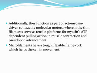
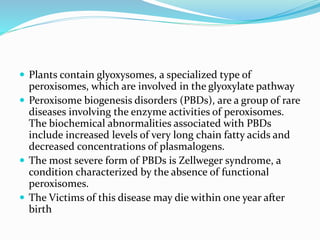
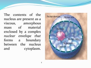
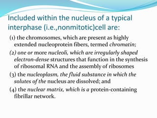
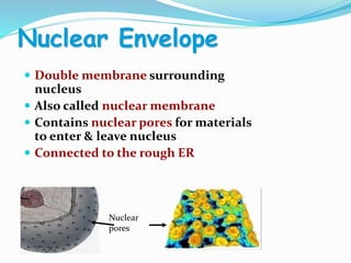
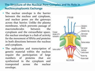
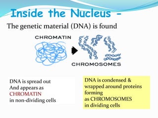
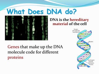
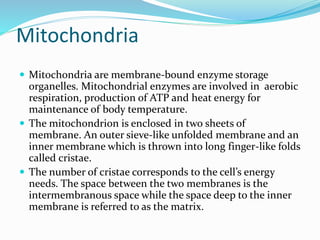
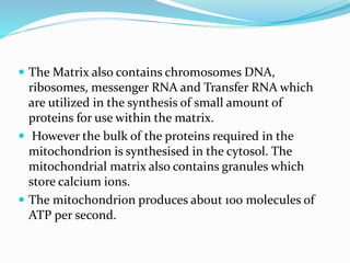
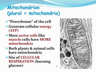
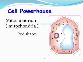
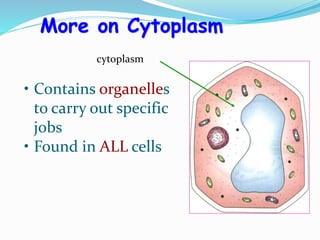
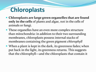
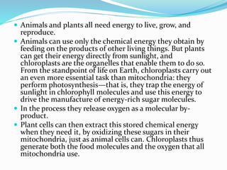
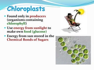
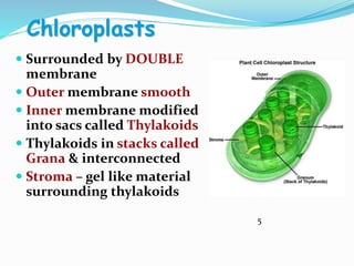
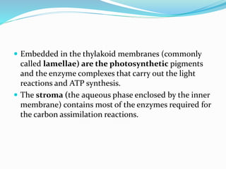
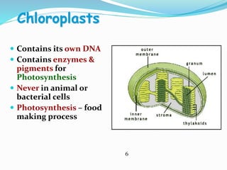
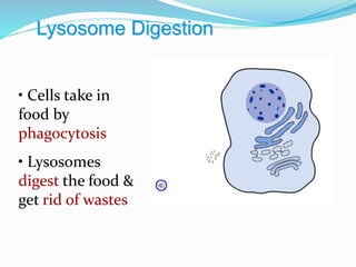
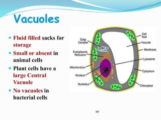
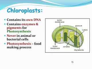
Cellular Biochemistry
UNIT- 2
B.Sc Biochemistry II
Ultra structure of cell
 Ultrastructure (or ultra-structure) is the
nanostructure of a biological specimen, such as a cell,
tissue, or organ, at scales smaller than can be viewed
with light microscopy. It is viewed with ultra
microscopy or electron microscopy.
 Such cellular structures as organelles, which allow the
cell to function properly within its specified
environment, can be examined at the ultrastructural
level.
CELL:
 The cell is the living functional unit of all organisms.
An organism may be composed of one cell only
(Unicellular) e.g. Bacteria and Algae or of several cells
(Multicellular) e.g. Man. The cell exists in two forms:
1.Eukaryotic cell, which has a nucleus that is enclosed in
a nuclear envelope and several membrane- limited
compartments e.g. the human cell.
2.Prokaryotic cell which has no nucleus and is devoid of
membrane-limited compartments e.g. the bacterial
cell.
NUCLEUS
 This is the largest organelle of the cell often located in
the central part of the cytoplasm and enclosed in a
double-layered nuclear membrane. Its shape usually
corresponds to the shape of the cell in which it is
found.
 It contains a nucleolus/nucleoli (which produces
ribosomal subunits) and chromatin (DNA).
 The latter is the genetic material implicated in cell
division and in the synthesis of several molecules
particularly proteins.
The contents of the
nucleus are present as a
viscous, amorphous
mass of material
enclosed by a complex
nuclear envelope that
forms a boundary
between the nucleus
and cytoplasm.
1
Included within the nucleus of a typical
interphase (i.e.,nonmitotic)cell are:
(1) the chromosomes, which are present as highly
extended nucleoprotein fibers, termed chromatin;
(2) one or more nucleoli, which are irregularly shaped
electron-dense structures that function in the synthesis
of ribosomal RNA and the assembly of ribosomes
(3) the nucleoplasm, the fluid substance in which the
solutes of the nucleus are dissolved; and
(4) the nuclear matrix, which is a protein-containing
fibrillar network.
Nuclear Envelope
 Double membrane surrounding
nucleus
 Also called nuclear membrane
 Contains nuclear pores for materials
to enter & leave nucleus
 Connected to the rough ER
Nuclear
pores
The Nuclear Envelope
 The separation of a cell’s genetic material from the
surrounding cytoplasm may be the single most important
feature that distinguishes eukaryotes from prokaryotes,
which makes the appearance of the nuclear envelope a
landmark in biological evolution.
 The nuclear envelope consists of two cellular
membranes arranged parallel to one another and separated
by 10 to 50 nm
 The membranes of the nuclear envelope serve as a barrier
that keeps ions, solutes, and macromolecules from passing
freely between the nucleus and cytoplasm.
 The two membranes are fused at sites forming circular
pores that contain complex assemblies of proteins. The
average mammalian cell contains several thousand nuclear
pores.
The Structure of the Nuclear Pore Complex and Its Role in
Nucleocytoplasmic Exchange
 The nuclear envelope is the barrier
between the nucleus and cytoplasm,
and nuclear pores are the gateways
across that barrier. Unlike the plasma
membrane, which prevents passage of
macromolecules between the
cytoplasm and the extracellular space,
the nuclear envelope is a hub of activity
for the movement of RNAs and proteins
in both directions between the nucleus
and cytoplasm.
 The replication and transcription of
genetic material within the nucleus
require the participation of large
numbers of proteins that are
synthesized in the cytoplasm and
transported across the nuclear
envelope.
Inside the Nucleus -
The genetic material (DNA) is found
DNA is spread out
And appears as
CHROMATIN
in non-dividing cells
DNA is condensed &
wrapped around proteins
forming
as CHROMOSOMES
in dividing cells
What Does DNA do?
DNA is the hereditary
material of the cell
Genes that make up the DNA
molecule code for different
proteins
Mitochondria
 Mitochondria are membrane-bound enzyme storage
organelles. Mitochondrial enzymes are involved in aerobic
respiration, production of ATP and heat energy for
maintenance of body temperature.
 The mitochondrion is enclosed in two sheets of
membrane. An outer sieve-like unfolded membrane and an
inner membrane which is thrown into long finger-like folds
called cristae.
 The number of cristae corresponds to the cell’s energy
needs. The space between the two membranes is the
intermembranous space while the space deep to the inner
membrane is referred to as the matrix.
 The Matrix also contains chromosomes DNA,
ribosomes, messenger RNA and Transfer RNA which
are utilized in the synthesis of small amount of
proteins for use within the matrix.
 However the bulk of the proteins required in the
mitochondrion is synthesised in the cytosol. The
mitochondrial matrix also contains granules which
store calcium ions.
 The mitochondrion produces about 100 molecules of
ATP per second.
Mitochondrion
(plural = mitochondria)
 “Powerhouse” of the cell
 Generate cellular energy
(ATP)
 More active cells like
muscle cells have MORE
mitochondria
 Both plants & animal cells
have mitochondria
 Site of CELLULAR
RESPIRATION (burning
glucose) 2
MITOCHONDRIA
Surrounded by a DOUBLE
membrane
Folded inner membrane called CRISTAE
(increases surface area
for more chemical
Reactions)
Has its own DNA
Interior called MATRIX
3
Interesting Fact ---
 Mitochondria Come
from cytoplasm in the
EGG cell during
fertilization
Therefore …
 You inherit your
mitochondria from
your mother!
 Muscle tissues are most commonly affected by
mitochondrial deficiency diseases because of their
high-energy metabolism.
 Most mitochondrial diseases often result from
chromosomal defect in the nucleus or in the
mitochondrion.
 Hereditary mitochondrial diseases are usually
maternal in origin because only very few paternal
mitochondria are left in the zygote following
fertilization.
Rod shape
Cell Powerhouse
Mitochondrion
( mitochondria )
4
THE ENDOPLASMIC RETICULUM (ER)
 This organelle is made up of anastomosing network of
intercommunicating channels/cisternae/sacs enclosed in a continuous
membrane.
 ER occurs in two forms, namely Rough and Smooth which are also
interconnected. While the cisternae of smooth ER are tubular in shape,
those of Rough ER are flattened. The roughness on the surface of rough
ER is due to the adsorption of polyribosome on their outer surface.
 Polyribosome also impacts the basophilic staining characteristic on RER.
Furthermore, its membrane is continuous with that of the nuclear
envelope.
Distribution and Functions of RER
 RER is prominent in protein synthesising cells such as; Pancreatic acinar
cells, cells of the endocrine glands, plasma cells, fibroblast etc.
 Proteins synthesised in RER are stored in Lysosomes or granules; stored
temporarily before exocytosis or used as integral membrane proteins.
Smooth Endoplasmic Reticulum (SER)
 This is ER not bund to polyribosomes but continuous with RER
and are less abundant is cell containing RER.
Distribution and Functions of SER
 SER is found in all cells where they are involved in: ‘The
synthesis phospholipids and cholesterol used in all cellular
membranes including membranes of organelles.
 They occur in abundance in other cells where they are involved
in: Sequestration and release of Calcium ions a vital process in
muscular contraction Biosynthesis of Lipids required for
synthesis of steroid hormones
 Detoxification of potentially harmful compounds such as alcohol
and barbiturates
RIBOSOMES
 Ribosomes are small,
electron-dense particles
not enclosed in
membrane and are
located in the cytosol.
 Measuring about 20-30
nanometer they are
basophilic and stained
by all basic dyes.
 Ribosome is composed of rRNA and about 80 different
proteins. It usually occur in two subunits, large and
small subunits.
 The rRNA of the ribosome is synthesised in the nucleus
while its protein is synthesised in the cytosol.
 Ribosomes are involved in protein synthesis. While
cytosolic proteins (free proteins) are synthesised by
polyribosomes, secretory and endoplasmic reticulum
proteins are synthesised on the membrane of rough
endoplasmic reticulum.
THE CYTOPLASM
 The fluid component of the cytoplasm is the Cytosol
(pH 7.2) while the metabolically active contents of the
cytoplasm are the Organelles.
 Apart from being metabolically active, organelles are
permanent residents of the cell which would survive
cell division i.e. they reappear in the daughter cells
following cell division.
 Organelles occur in two forms, freely within the
cytosol or enclosed in membrane. The cytoplasm also
contains substances which are not metabolically
active.
• Jelly-like substance
enclosed by cell
membrane
• Provides a medium
for chemical reactions
to take place
Cytoplasm of a Cell
cytoplasm
• Contains organelles
to carry out specific
jobs
• Found in ALL cells
More on Cytoplasm
cytoplasm
Chloroplasts
 Chloroplasts are large green organelles that are found
only in the cells of plants and algae, not in the cells of
animals or fungi.
 These organelles have an even more complex structure
than mitochondria: in addition to their two surrounding
membranes, chloroplasts possess internal stacks of
membranes containing the green pigment chlorophyll
 When a plant is kept in the dark, its greenness fades; when
put back in the light, its greenness returns. This suggests
that the chlorophyll—and the chloroplasts that contain it
 Animals and plants all need energy to live, grow, and
reproduce.
 Animals can use only the chemical energy they obtain by
feeding on the products of other living things. But plants
can get their energy directly from sunlight, and
chloroplasts are the organelles that enable them to do so.
From the standpoint of life on Earth, chloroplasts carry out
an even more essential task than mitochondria: they
perform photosynthesis—that is, they trap the energy of
sunlight in chlorophyll molecules and use this energy to
drive the manufacture of energy-rich sugar molecules.
 In the process they release oxygen as a molecular by-
product.
 Plant cells can then extract this stored chemical energy
when they need it, by oxidizing these sugars in their
mitochondria, just as animal cells can. Chloroplasts thus
generate both the food molecules and the oxygen that all
mitochondria use.
Chloroplasts
 Found only in producers
(organisms containing
chlorophyll)
 Use energy from sunlight to
make own food (glucose)
 Energy from sun stored in the
Chemical Bonds of Sugars
Chloroplasts
 Surrounded by DOUBLE
membrane
 Outer membrane smooth
 Inner membrane modified
into sacs called Thylakoids
 Thylakoids in stacks called
Grana & interconnected
 Stroma – gel like material
surrounding thylakoids
5
 Like mitochondria, they are surrounded by two
membranes, an outer membrane that is permeable to
small molecules and ions, and an inner membrane
that encloses the internal compartment.
 This compartment contains many flattened,
membrane-surrounded vesicles or sacs, the
thylakoids, usually arranged in stacks called
grana.
 Embedded in the thylakoid membranes (commonly
called lamellae) are the photosynthetic pigments
and the enzyme complexes that carry out the light
reactions and ATP synthesis.
 The stroma (the aqueous phase enclosed by the inner
membrane) contains most of the enzymes required for
the carbon assimilation reactions.
Chloroplasts
 Contains its own DNA
 Contains enzymes &
pigments for
Photosynthesis
 Never in animal or
bacterial cells
 Photosynthesis – food
making process
6
Cytoskeleton
 Helps cell maintain cell
shape
 Also help move organelles
around
 Made of proteins
 Microfilaments are
threadlike & made of
ACTIN
 Microtubules are tubelike
& made of TUBULIN
Centrioles
 Found only in animal
cells
 Paired structures near
nucleus
 Made of bundle of
microtubules
 Appear during cell
division forming mitotic
spindle
 Help to pull chromosome
pairs apart to opposite
ends of the cell
7
Golgi Bodies
Look like a stack of pancakes
Modify, sort, & package
molecules from ER
for storage OR
transport out of cell
Golgi Animation
Materials are transported from Rough ER to Golgi to the cell membrane by
VESICLES
Lysosomes
• Contain digestive
enzymes
• Break down food,
bacteria, and worn out
cell parts for cells
• Programmed for cell
death (AUTOLYSIS)
• Lyse (break open) &
release enzymes to break
down & recycle cell parts)
Lysosome Digestion
• Cells take in
food by
phagocytosis
• Lysosomes
digest the food &
get rid of wastes
Cilia & Flagella:
 Made of protein tubes called
microtubules
 Microtubules arranged (9 + 2
arrangement)
 Function in moving cells, in
moving fluids, or in small
particles across the cell
surface
8
Cilia & Flagella
 Cilia are shorter
and more
numerous on cells
 Flagella are longer
and fewer (usually
1-3) on cells 9
Vacuoles
 Fluid filled sacks for
storage
 Small or absent in
animal cells
 Plant cells have a
large Central
Vacuole
 No vacuoles in
bacterial cells
10
Vacuoles
 In plants, they store Cell Sap
 Includes storage of sugars,
proteins, minerals, lipids,
wastes, salts, water, and
enzymes
Contractile Vacuole
 Found in unicellular protists
like paramecia
 Regulate water intake by
pumping out excess
(homeostasis)
 Keeps the cell from lysing
(bursting)
43
Contractile vacuole animation
11
Chloroplasts
 Found only in producers
(organisms containing
chlorophyll)
 Use energy from sunlight to
make own food (glucose)
 Energy from sun stored in the
Chemical Bonds of Sugars
Chloroplasts
 Surrounded by DOUBLE
membrane
 Outer membrane smooth
 Inner membrane modified
into sacs called Thylakoids
 Thylakoids in stacks called
Grana & interconnected
 Stroma – gel like material
surrounding thylakoids
12
 Like mitochondria, they are surrounded by two
membranes, an outer membrane that is permeable to
small molecules and ions, and an inner membrane
that encloses the internal compartment.
 This compartment contains many flattened,
membrane-surrounded vesicles or sacs, the
thylakoids, usually arranged in stacks called
grana.
 Embedded in the thylakoid membranes (commonly
called lamellae) are the photosynthetic pigments
and the enzyme complexes that carry out the light
reactions and ATP synthesis.
 The stroma (the aqueous phase enclosed by the inner
membrane) contains most of the enzymes required for
the carbon assimilation reactions.
Chloroplasts:
 Contains its own DNA
 Contains enzymes &
pigments for
Photosynthesis
 Never in animal or
bacterial cells
 Photosynthesis – food
making process
13
Vacuoles:
 Fluid filled sacks
for storage
 Small or absent in
animal cells
 Plant cells have a
large Central
Vacuole
 No vacuoles in
bacterial cells
Vacuoles
 In plants, they store Cell Sap
 Includes storage of sugars,
proteins, minerals, lipids,
wastes, salts, water, and
enzymes
Contractile Vacuole
 Found in unicellular protists
like paramecia
 Regulate water intake by
pumping out excess
(homeostasis)
 Keeps the cell from lysing
(bursting)
51
Contractile vacuole animation
Microtubules:
 Microtubules are a component of the cytoskeleton,
found throughout the cytoplasm.
 These tubular polymers of tubulin can grow as long as
50 micrometres, with an average length of 25 µm, and
are highly dynamic.
 The outer diameter of a microtubule is about 24 nm
while the inner diameter is about 12 nm. They are
found in eukaryotic cells and are formed by the
polymerization of a dimer of two globular proteins,
alpha and beta tubulin.
 Microtubules are very important in a number of
cellular processes. They are involved in maintaining
the structure of the cell and, together with
microfilaments and intermediate filaments, they form
the cytoskeleton. They also make up the internal
structure of cilia and flagella.
 They provide platforms for intracellular transport and
are involved in a variety of cellular processes, including
the movement of secretory vesicles, organelles, and
intracellular substances (see entries for dynein and
kinesin).
 They are also involved in cell division (mitosis and
meiosis), including the formation of mitotic spindles,
which are used to pull apart eukaryotic chromosomes.
 Microtubules are nucleated and organized in
microtubule organizing centres (MTOCs), such as the
centrosome or the basal bodies found in cilia and
flagella. These MTOCs may or may not possess
centrioles.
 There are many proteins that bind to microtubules,
including motor proteins such as kinesin and dynein,
severing proteins like katanin, and other proteins
important for regulating microtubule dynamics
Microfilaments:
 Microfilaments or actin filaments are the thinnest
filaments of the cytoskeleton, a structure found in the
cytoplasm of eukaryotic cells.
 These linear polymers of actin subunits are flexible and
relatively strong, resisting buckling by multi-piconewton
compressive forces and filament fracture by nanonewton
tensile forces.
 Microfilaments are highly versatile, functioning in
cytokinesis, amoeboid movement, and changes in cell
shape. In inducing this cell motility, one end of the actin
filament elongates while the other end contracts,
presumably by myosin II molecular motors.
 Additionally, they function as part of actomyosin-
driven contractile molecular motors, wherein the thin
filaments serve as tensile platforms for myosin's ATP-
dependent pulling action in muscle contraction and
pseudopod advancement.
 Microfilaments have a tough, flexible framework
which helps the cell in movement.
Peroxisomes and glyoxysomes:
 Peroxisomes, also known as microbodies, are single
membrane cellular organelles. They are spherical or
oval in shape and contain the enzyme catalase.
 Catalase protects the cell from the toxic effects of
H2O2, by converting it to H2O and O2.
 Peroxisomes are also involved in the oxidation of long
chain fatty acids (> C18), and synthesis of plasmalogens
and glycolipids.
 Plants contain glyoxysomes, a specialized type of
peroxisomes, which are involved in the glyoxylate pathway
 Peroxisome biogenesis disorders (PBDs), are a group of rare
diseases involving the enzyme activities of peroxisomes.
The biochemical abnormalities associated with PBDs
include increased levels of very long chain fatty acids and
decreased concentrations of plasmalogens.
 The most severe form of PBDs is Zellweger syndrome, a
condition characterized by the absence of functional
peroxisomes.
 The Victims of this disease may die within one year after
birth
References Images references:
1. http://www.uic.edu/classes/bios/bios100/summer2002/nucleus.jpg
2. http://pixgood.com/electron-micrograph-mitochondria.html
3. https://www.pinterest.com/tserex/mitochondrial-disease/
4. http://www.slideshare.net/tas11244/cell-structure-lecture
5. http://en.wikipedia.org/wiki/Chloroplast
6. http://creationrevolution.com/chloroplasts-%E2%80%93-simple-cell-part-15/
7. http://www.bioanim.com/CellTissueHumanBody6/O3mitosisMeiosisChrom/celCentriol1lgws.html
8. http://biology4isc.weebly.com/cell-organelles.html
9. http://engineering.curiouscatblog.net/2006/12/12/micro-robots-to-swim-through-veins/
10. http://torresbioclan.pbworks.com/w/page/22377199/Prokaryotes%20and%20Eukaryotes
11. http://www.sparknotes.com/biology/microorganisms/protista/section2.rhtml
12. http://en.wikipedia.org/wiki/Chloroplast
 Reading references:
 Cell and Molecular Biology, 6th Ed By Karp
 Molecular Cell Biology by Lodish 5th Edition
B.Sc. Biochemistry II Cellular Biochemistry Unit 2 Cellular components

B.Sc. Biochemistry II Cellular Biochemistry Unit 2 Cellular components

  • 1.
  • 2.
    Ultra structure ofcell  Ultrastructure (or ultra-structure) is the nanostructure of a biological specimen, such as a cell, tissue, or organ, at scales smaller than can be viewed with light microscopy. It is viewed with ultra microscopy or electron microscopy.  Such cellular structures as organelles, which allow the cell to function properly within its specified environment, can be examined at the ultrastructural level.
  • 3.
    CELL:  The cellis the living functional unit of all organisms. An organism may be composed of one cell only (Unicellular) e.g. Bacteria and Algae or of several cells (Multicellular) e.g. Man. The cell exists in two forms: 1.Eukaryotic cell, which has a nucleus that is enclosed in a nuclear envelope and several membrane- limited compartments e.g. the human cell. 2.Prokaryotic cell which has no nucleus and is devoid of membrane-limited compartments e.g. the bacterial cell.
  • 4.
    NUCLEUS  This isthe largest organelle of the cell often located in the central part of the cytoplasm and enclosed in a double-layered nuclear membrane. Its shape usually corresponds to the shape of the cell in which it is found.  It contains a nucleolus/nucleoli (which produces ribosomal subunits) and chromatin (DNA).  The latter is the genetic material implicated in cell division and in the synthesis of several molecules particularly proteins.
  • 5.
    The contents ofthe nucleus are present as a viscous, amorphous mass of material enclosed by a complex nuclear envelope that forms a boundary between the nucleus and cytoplasm. 1
  • 6.
    Included within thenucleus of a typical interphase (i.e.,nonmitotic)cell are: (1) the chromosomes, which are present as highly extended nucleoprotein fibers, termed chromatin; (2) one or more nucleoli, which are irregularly shaped electron-dense structures that function in the synthesis of ribosomal RNA and the assembly of ribosomes (3) the nucleoplasm, the fluid substance in which the solutes of the nucleus are dissolved; and (4) the nuclear matrix, which is a protein-containing fibrillar network.
  • 7.
    Nuclear Envelope  Doublemembrane surrounding nucleus  Also called nuclear membrane  Contains nuclear pores for materials to enter & leave nucleus  Connected to the rough ER Nuclear pores
  • 8.
    The Nuclear Envelope The separation of a cell’s genetic material from the surrounding cytoplasm may be the single most important feature that distinguishes eukaryotes from prokaryotes, which makes the appearance of the nuclear envelope a landmark in biological evolution.  The nuclear envelope consists of two cellular membranes arranged parallel to one another and separated by 10 to 50 nm  The membranes of the nuclear envelope serve as a barrier that keeps ions, solutes, and macromolecules from passing freely between the nucleus and cytoplasm.  The two membranes are fused at sites forming circular pores that contain complex assemblies of proteins. The average mammalian cell contains several thousand nuclear pores.
  • 9.
    The Structure ofthe Nuclear Pore Complex and Its Role in Nucleocytoplasmic Exchange  The nuclear envelope is the barrier between the nucleus and cytoplasm, and nuclear pores are the gateways across that barrier. Unlike the plasma membrane, which prevents passage of macromolecules between the cytoplasm and the extracellular space, the nuclear envelope is a hub of activity for the movement of RNAs and proteins in both directions between the nucleus and cytoplasm.  The replication and transcription of genetic material within the nucleus require the participation of large numbers of proteins that are synthesized in the cytoplasm and transported across the nuclear envelope.
  • 10.
    Inside the Nucleus- The genetic material (DNA) is found DNA is spread out And appears as CHROMATIN in non-dividing cells DNA is condensed & wrapped around proteins forming as CHROMOSOMES in dividing cells
  • 11.
    What Does DNAdo? DNA is the hereditary material of the cell Genes that make up the DNA molecule code for different proteins
  • 12.
    Mitochondria  Mitochondria aremembrane-bound enzyme storage organelles. Mitochondrial enzymes are involved in aerobic respiration, production of ATP and heat energy for maintenance of body temperature.  The mitochondrion is enclosed in two sheets of membrane. An outer sieve-like unfolded membrane and an inner membrane which is thrown into long finger-like folds called cristae.  The number of cristae corresponds to the cell’s energy needs. The space between the two membranes is the intermembranous space while the space deep to the inner membrane is referred to as the matrix.
  • 13.
     The Matrixalso contains chromosomes DNA, ribosomes, messenger RNA and Transfer RNA which are utilized in the synthesis of small amount of proteins for use within the matrix.  However the bulk of the proteins required in the mitochondrion is synthesised in the cytosol. The mitochondrial matrix also contains granules which store calcium ions.  The mitochondrion produces about 100 molecules of ATP per second.
  • 14.
    Mitochondrion (plural = mitochondria) “Powerhouse” of the cell  Generate cellular energy (ATP)  More active cells like muscle cells have MORE mitochondria  Both plants & animal cells have mitochondria  Site of CELLULAR RESPIRATION (burning glucose) 2
  • 15.
    MITOCHONDRIA Surrounded by aDOUBLE membrane Folded inner membrane called CRISTAE (increases surface area for more chemical Reactions) Has its own DNA Interior called MATRIX 3
  • 16.
    Interesting Fact --- Mitochondria Come from cytoplasm in the EGG cell during fertilization Therefore …  You inherit your mitochondria from your mother!
  • 17.
     Muscle tissuesare most commonly affected by mitochondrial deficiency diseases because of their high-energy metabolism.  Most mitochondrial diseases often result from chromosomal defect in the nucleus or in the mitochondrion.  Hereditary mitochondrial diseases are usually maternal in origin because only very few paternal mitochondria are left in the zygote following fertilization.
  • 18.
  • 19.
    THE ENDOPLASMIC RETICULUM(ER)  This organelle is made up of anastomosing network of intercommunicating channels/cisternae/sacs enclosed in a continuous membrane.  ER occurs in two forms, namely Rough and Smooth which are also interconnected. While the cisternae of smooth ER are tubular in shape, those of Rough ER are flattened. The roughness on the surface of rough ER is due to the adsorption of polyribosome on their outer surface.  Polyribosome also impacts the basophilic staining characteristic on RER. Furthermore, its membrane is continuous with that of the nuclear envelope. Distribution and Functions of RER  RER is prominent in protein synthesising cells such as; Pancreatic acinar cells, cells of the endocrine glands, plasma cells, fibroblast etc.  Proteins synthesised in RER are stored in Lysosomes or granules; stored temporarily before exocytosis or used as integral membrane proteins.
  • 20.
    Smooth Endoplasmic Reticulum(SER)  This is ER not bund to polyribosomes but continuous with RER and are less abundant is cell containing RER. Distribution and Functions of SER  SER is found in all cells where they are involved in: ‘The synthesis phospholipids and cholesterol used in all cellular membranes including membranes of organelles.  They occur in abundance in other cells where they are involved in: Sequestration and release of Calcium ions a vital process in muscular contraction Biosynthesis of Lipids required for synthesis of steroid hormones  Detoxification of potentially harmful compounds such as alcohol and barbiturates
  • 21.
    RIBOSOMES  Ribosomes aresmall, electron-dense particles not enclosed in membrane and are located in the cytosol.  Measuring about 20-30 nanometer they are basophilic and stained by all basic dyes.
  • 22.
     Ribosome iscomposed of rRNA and about 80 different proteins. It usually occur in two subunits, large and small subunits.  The rRNA of the ribosome is synthesised in the nucleus while its protein is synthesised in the cytosol.  Ribosomes are involved in protein synthesis. While cytosolic proteins (free proteins) are synthesised by polyribosomes, secretory and endoplasmic reticulum proteins are synthesised on the membrane of rough endoplasmic reticulum.
  • 23.
    THE CYTOPLASM  Thefluid component of the cytoplasm is the Cytosol (pH 7.2) while the metabolically active contents of the cytoplasm are the Organelles.  Apart from being metabolically active, organelles are permanent residents of the cell which would survive cell division i.e. they reappear in the daughter cells following cell division.  Organelles occur in two forms, freely within the cytosol or enclosed in membrane. The cytoplasm also contains substances which are not metabolically active.
  • 24.
    • Jelly-like substance enclosedby cell membrane • Provides a medium for chemical reactions to take place Cytoplasm of a Cell cytoplasm
  • 25.
    • Contains organelles tocarry out specific jobs • Found in ALL cells More on Cytoplasm cytoplasm
  • 26.
    Chloroplasts  Chloroplasts arelarge green organelles that are found only in the cells of plants and algae, not in the cells of animals or fungi.  These organelles have an even more complex structure than mitochondria: in addition to their two surrounding membranes, chloroplasts possess internal stacks of membranes containing the green pigment chlorophyll  When a plant is kept in the dark, its greenness fades; when put back in the light, its greenness returns. This suggests that the chlorophyll—and the chloroplasts that contain it
  • 27.
     Animals andplants all need energy to live, grow, and reproduce.  Animals can use only the chemical energy they obtain by feeding on the products of other living things. But plants can get their energy directly from sunlight, and chloroplasts are the organelles that enable them to do so. From the standpoint of life on Earth, chloroplasts carry out an even more essential task than mitochondria: they perform photosynthesis—that is, they trap the energy of sunlight in chlorophyll molecules and use this energy to drive the manufacture of energy-rich sugar molecules.  In the process they release oxygen as a molecular by- product.  Plant cells can then extract this stored chemical energy when they need it, by oxidizing these sugars in their mitochondria, just as animal cells can. Chloroplasts thus generate both the food molecules and the oxygen that all mitochondria use.
  • 28.
    Chloroplasts  Found onlyin producers (organisms containing chlorophyll)  Use energy from sunlight to make own food (glucose)  Energy from sun stored in the Chemical Bonds of Sugars
  • 29.
    Chloroplasts  Surrounded byDOUBLE membrane  Outer membrane smooth  Inner membrane modified into sacs called Thylakoids  Thylakoids in stacks called Grana & interconnected  Stroma – gel like material surrounding thylakoids 5
  • 30.
     Like mitochondria,they are surrounded by two membranes, an outer membrane that is permeable to small molecules and ions, and an inner membrane that encloses the internal compartment.  This compartment contains many flattened, membrane-surrounded vesicles or sacs, the thylakoids, usually arranged in stacks called grana.
  • 31.
     Embedded inthe thylakoid membranes (commonly called lamellae) are the photosynthetic pigments and the enzyme complexes that carry out the light reactions and ATP synthesis.  The stroma (the aqueous phase enclosed by the inner membrane) contains most of the enzymes required for the carbon assimilation reactions.
  • 32.
    Chloroplasts  Contains itsown DNA  Contains enzymes & pigments for Photosynthesis  Never in animal or bacterial cells  Photosynthesis – food making process 6
  • 33.
    Cytoskeleton  Helps cellmaintain cell shape  Also help move organelles around  Made of proteins  Microfilaments are threadlike & made of ACTIN  Microtubules are tubelike & made of TUBULIN
  • 34.
    Centrioles  Found onlyin animal cells  Paired structures near nucleus  Made of bundle of microtubules  Appear during cell division forming mitotic spindle  Help to pull chromosome pairs apart to opposite ends of the cell 7
  • 35.
    Golgi Bodies Look likea stack of pancakes Modify, sort, & package molecules from ER for storage OR transport out of cell
  • 36.
    Golgi Animation Materials aretransported from Rough ER to Golgi to the cell membrane by VESICLES
  • 37.
    Lysosomes • Contain digestive enzymes •Break down food, bacteria, and worn out cell parts for cells • Programmed for cell death (AUTOLYSIS) • Lyse (break open) & release enzymes to break down & recycle cell parts)
  • 38.
    Lysosome Digestion • Cellstake in food by phagocytosis • Lysosomes digest the food & get rid of wastes
  • 39.
    Cilia & Flagella: Made of protein tubes called microtubules  Microtubules arranged (9 + 2 arrangement)  Function in moving cells, in moving fluids, or in small particles across the cell surface 8
  • 40.
    Cilia & Flagella Cilia are shorter and more numerous on cells  Flagella are longer and fewer (usually 1-3) on cells 9
  • 41.
    Vacuoles  Fluid filledsacks for storage  Small or absent in animal cells  Plant cells have a large Central Vacuole  No vacuoles in bacterial cells 10
  • 42.
    Vacuoles  In plants,they store Cell Sap  Includes storage of sugars, proteins, minerals, lipids, wastes, salts, water, and enzymes
  • 43.
    Contractile Vacuole  Foundin unicellular protists like paramecia  Regulate water intake by pumping out excess (homeostasis)  Keeps the cell from lysing (bursting) 43 Contractile vacuole animation 11
  • 44.
    Chloroplasts  Found onlyin producers (organisms containing chlorophyll)  Use energy from sunlight to make own food (glucose)  Energy from sun stored in the Chemical Bonds of Sugars
  • 45.
    Chloroplasts  Surrounded byDOUBLE membrane  Outer membrane smooth  Inner membrane modified into sacs called Thylakoids  Thylakoids in stacks called Grana & interconnected  Stroma – gel like material surrounding thylakoids 12
  • 46.
     Like mitochondria,they are surrounded by two membranes, an outer membrane that is permeable to small molecules and ions, and an inner membrane that encloses the internal compartment.  This compartment contains many flattened, membrane-surrounded vesicles or sacs, the thylakoids, usually arranged in stacks called grana.
  • 47.
     Embedded inthe thylakoid membranes (commonly called lamellae) are the photosynthetic pigments and the enzyme complexes that carry out the light reactions and ATP synthesis.  The stroma (the aqueous phase enclosed by the inner membrane) contains most of the enzymes required for the carbon assimilation reactions.
  • 48.
    Chloroplasts:  Contains itsown DNA  Contains enzymes & pigments for Photosynthesis  Never in animal or bacterial cells  Photosynthesis – food making process 13
  • 49.
    Vacuoles:  Fluid filledsacks for storage  Small or absent in animal cells  Plant cells have a large Central Vacuole  No vacuoles in bacterial cells
  • 50.
    Vacuoles  In plants,they store Cell Sap  Includes storage of sugars, proteins, minerals, lipids, wastes, salts, water, and enzymes
  • 51.
    Contractile Vacuole  Foundin unicellular protists like paramecia  Regulate water intake by pumping out excess (homeostasis)  Keeps the cell from lysing (bursting) 51 Contractile vacuole animation
  • 52.
    Microtubules:  Microtubules area component of the cytoskeleton, found throughout the cytoplasm.  These tubular polymers of tubulin can grow as long as 50 micrometres, with an average length of 25 µm, and are highly dynamic.  The outer diameter of a microtubule is about 24 nm while the inner diameter is about 12 nm. They are found in eukaryotic cells and are formed by the polymerization of a dimer of two globular proteins, alpha and beta tubulin.
  • 53.
     Microtubules arevery important in a number of cellular processes. They are involved in maintaining the structure of the cell and, together with microfilaments and intermediate filaments, they form the cytoskeleton. They also make up the internal structure of cilia and flagella.  They provide platforms for intracellular transport and are involved in a variety of cellular processes, including the movement of secretory vesicles, organelles, and intracellular substances (see entries for dynein and kinesin).  They are also involved in cell division (mitosis and meiosis), including the formation of mitotic spindles, which are used to pull apart eukaryotic chromosomes.
  • 54.
     Microtubules arenucleated and organized in microtubule organizing centres (MTOCs), such as the centrosome or the basal bodies found in cilia and flagella. These MTOCs may or may not possess centrioles.  There are many proteins that bind to microtubules, including motor proteins such as kinesin and dynein, severing proteins like katanin, and other proteins important for regulating microtubule dynamics
  • 55.
    Microfilaments:  Microfilaments oractin filaments are the thinnest filaments of the cytoskeleton, a structure found in the cytoplasm of eukaryotic cells.  These linear polymers of actin subunits are flexible and relatively strong, resisting buckling by multi-piconewton compressive forces and filament fracture by nanonewton tensile forces.  Microfilaments are highly versatile, functioning in cytokinesis, amoeboid movement, and changes in cell shape. In inducing this cell motility, one end of the actin filament elongates while the other end contracts, presumably by myosin II molecular motors.
  • 56.
     Additionally, theyfunction as part of actomyosin- driven contractile molecular motors, wherein the thin filaments serve as tensile platforms for myosin's ATP- dependent pulling action in muscle contraction and pseudopod advancement.  Microfilaments have a tough, flexible framework which helps the cell in movement.
  • 57.
    Peroxisomes and glyoxysomes: Peroxisomes, also known as microbodies, are single membrane cellular organelles. They are spherical or oval in shape and contain the enzyme catalase.  Catalase protects the cell from the toxic effects of H2O2, by converting it to H2O and O2.  Peroxisomes are also involved in the oxidation of long chain fatty acids (> C18), and synthesis of plasmalogens and glycolipids.
  • 58.
     Plants containglyoxysomes, a specialized type of peroxisomes, which are involved in the glyoxylate pathway  Peroxisome biogenesis disorders (PBDs), are a group of rare diseases involving the enzyme activities of peroxisomes. The biochemical abnormalities associated with PBDs include increased levels of very long chain fatty acids and decreased concentrations of plasmalogens.  The most severe form of PBDs is Zellweger syndrome, a condition characterized by the absence of functional peroxisomes.  The Victims of this disease may die within one year after birth
  • 59.
    References Images references: 1.http://www.uic.edu/classes/bios/bios100/summer2002/nucleus.jpg 2. http://pixgood.com/electron-micrograph-mitochondria.html 3. https://www.pinterest.com/tserex/mitochondrial-disease/ 4. http://www.slideshare.net/tas11244/cell-structure-lecture 5. http://en.wikipedia.org/wiki/Chloroplast 6. http://creationrevolution.com/chloroplasts-%E2%80%93-simple-cell-part-15/ 7. http://www.bioanim.com/CellTissueHumanBody6/O3mitosisMeiosisChrom/celCentriol1lgws.html 8. http://biology4isc.weebly.com/cell-organelles.html 9. http://engineering.curiouscatblog.net/2006/12/12/micro-robots-to-swim-through-veins/ 10. http://torresbioclan.pbworks.com/w/page/22377199/Prokaryotes%20and%20Eukaryotes 11. http://www.sparknotes.com/biology/microorganisms/protista/section2.rhtml 12. http://en.wikipedia.org/wiki/Chloroplast  Reading references:  Cell and Molecular Biology, 6th Ed By Karp  Molecular Cell Biology by Lodish 5th Edition