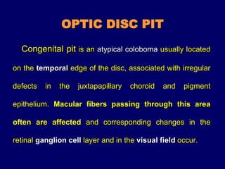
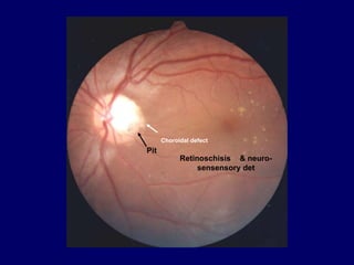
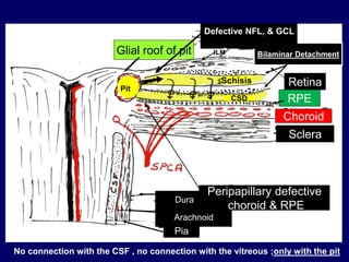
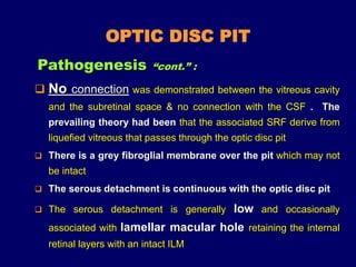
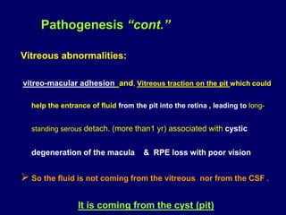
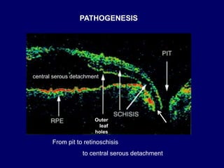
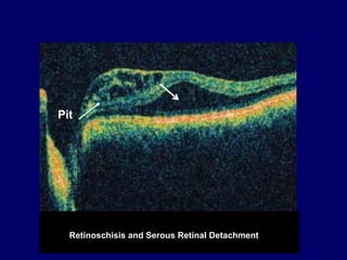
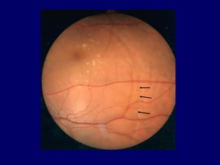
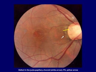
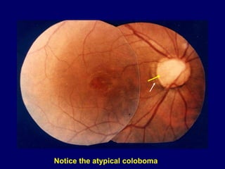
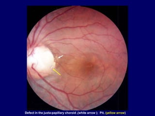
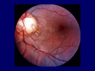
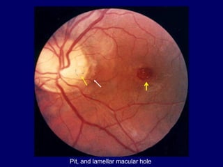
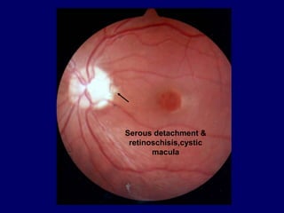
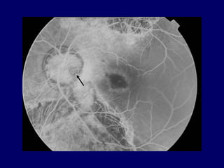
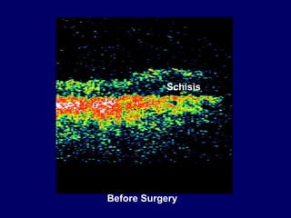
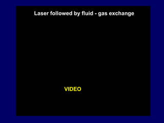
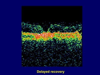
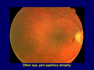
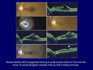
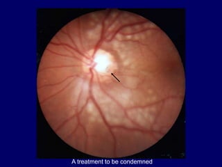
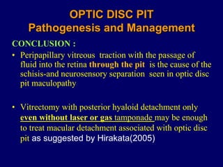
Abdel-Latif Siam
Ain Shams University
Cairo Egypt
OPTIC DISC PIT
Pathogenesis and Management
OPTIC DISC PIT
Congenital pit is an atypical coloboma usually located
on the temporal edge of the disc, associated with irregular
defects in the juxtapapillary choroid and pigment
epithelium. Macular fibers passing through this area
often are affected and corresponding changes in the
retinal ganglion cell layer and in the visual field occur.
Pit
Choroidal defect
Retinoschisis & neuro-
sensensory det
OPTIC DISC PIT
• First described 1882
• 1 in 10,000 eyes
• 85 % unilateral
• 70 % on the temporal side
• 20 % centrally
• Few reports demonstrate autosomal dominant
OPTIC DISC PIT
 When the pit is located temporally on the edge of the disc it
may become associated with abnormalities in the macula
 In the vast majority of cases there is only one pit and is
unilateral , varying in size between 150 to 500 m in diam.
 Principal threat is sensory macular retinal detachment. The
detachment always communicating with the disc unlike
central serous choroidopathy
Pit
Choroidal defect
Retinoschisis & neuro-
sensensory det
The macular elevation occurs later in life in 25 – 75 % of cases ,
and represents splitting of the retina (retinoschisis), and
sub-retinal fluid accumulation
OCT confirmed the two-layer structure of the serous
detachment and the connection of the pit with the
retinoschisis which can be multi-layered
OPTIC DISC PIT
OPTIC DISC PIT
 Recent OCT studies seem to confirm that the pit is a cyst from
which fluid passes into the sensory retina leading to
retinoschisis. Fluid passing through outer leaf holes causes
detachment of the neurosensory retina from the RPE
 All OCT studies confirm the bilaminar structure of the neurosensory
detachment
 Vitrectomy procedures demonstrated the importance of vitreoretinal
traction in the pathogenesis of the retinoschisis and neurosensory
detachment associated with optic disc pit
Pathogenesis :
Bilaminar Detachment
Retina
Pia
Dura
Arachnoid
Choroid
Sclera
Glial roof of pit
Peripapillary defective
choroid & RPE
Defective NFL, & GCL
RPE
Schisis
CSD
No connection with the CSF , no connection with the vitreous ;only with the pit
Pit
ILM
OPTIC DISC PIT
 No connection was demonstrated between the vitreous cavity
and the subretinal space & no connection with the CSF . The
prevailing theory had been that the associated SRF derive from
liquefied vitreous that passes through the optic disc pit
 There is a grey fibrogIial membrane over the pit which may not
be intact
 The serous detachment is continuous with the optic disc pit
 The serous detachment is generally low and occasionally
associated with lamellar macular hole retaining the internal
retinal layers with an intact ILM
Pathogenesis “cont.” :
Vitreous abnormalities:
vitreo-macular adhesion and, Vitreous traction on the pit which could
help the entrance of fluid from the pit into the retina , leading to long-
standing serous detach. (more than1 yr) associated with cystic
degeneration of the macula & RPE loss with poor vision
 So the fluid is not coming from the vitreous nor from the CSF .
It is coming from the cyst (pit)
Pathogenesis “cont.”
From pit to retinoschisis
to central serous detachment
Outer
leaf
holes
central serous detachment
PATHOGENESIS
Retinoschisis and Serous Retinal Detachment
Pit
Pit
Choroidal defect
Retinoschisis & neuro-
sensensory det
Defect in the juxta-papillary choroid (white arrow); Pit, yellow arrow
Notice the atypical coloboma
Defect in the juxta-papillary choroid ,(white arrow ); Pit, (yellow arrow)
Pit, and lamellar macular hole
Serous detachment &
retinoschisis,cystic
macula
FLUOROGRAPHIC FEATURES
 Early hypofluorescence
 Late hyperfluorescence due to leakage at the
margins of, or from the depth of the pit.
 RPE window defects are observed with RPE atrophy
DIFFERENTIAL DIAGNOSIS
 Central serous retinopathy
 Macular hole with surrounding detachment
 Coloboma of the optic disc
Central serous choroidopathy
A misdiagnosed case of optic disc pit with serous macular detachment as being
a case of central ret. det due to a macular hole & treated as such !!
TREATMENT
The most recent treatment is by pars plana
vitrectomy, posterior hyaloid detachment & preferably
with ILM peeling , laser on the temporal edge of the
optic disc followed by fluid-gas exchange.
This provides the best structural and functional
results by relieving vitreoretinal interface traction
& closing the connection with the pit.
TREATMENT (cont.)
 Macular buckling was reported to give good anatomical and
functional results
 Older techniques include photocoagulation on the temporal edge of
the disc & scleral buckling procedures.
Suggested prophylaxis for pits without detachment.
is by laser treatment temporal to the edge of the
optic disc ?
• Can recover vision with spontaneous resolution of the serous
detach. within a relatively short period of time.
• Following surgical intervention resolution of the
detachment may take 10-12 months.
• Hirakata(2005) suggests that PVD without laser or gas injection
may be sufficient since recovery may be long delayed
Makes life easy !!
TREATMENT (Continued.)
A misdiagnosed case of optic disc pit with serous macular detachment as being
a case of central ret. det due to a macular hole & treated as such !!
Macular laser scar !1
Schisis
Before Surgery
VIDEO
Laser followed by fluid - gas exchange
Delayed recovery
Other eye, peri papillary atrophy
Shukla (Retina 2012 )suggested leaving a small central island of ILM over the
fovea “to avoid iatrogenic macular hole as ILM is being removed
A treatment to be condemned
OPTIC DISC PIT
Pathogenesis and Management
CONCLUSION :
• Peripapillary vitreous traction with the passage of
fluid into the retina through the pit is the cause of the
schisis-and neurosensory separation seen in optic disc
pit maculopathy
• Vitrectomy with posterior hyaloid detachment only
even without laser or gas tamponade may be enough
to treat macular detachment associated with optic disc
pit as suggested by Hirakata(2005)
TREATMENT
The most recent treatment is by pars plana
vitrectomy, posterior hyaloid detachment & preferably
with ILM peeling , laser on the temporal edge of the
optic disc followed by fluid-gas exchange.
This provides the best structural and functional
results by relieving vitreoretinal interface traction
& closing the connection with the pit.

OPTIC DISC PIT Pathogenesis and Management

  • 1.
    Abdel-Latif Siam Ain ShamsUniversity Cairo Egypt OPTIC DISC PIT Pathogenesis and Management
  • 2.
    OPTIC DISC PIT Congenitalpit is an atypical coloboma usually located on the temporal edge of the disc, associated with irregular defects in the juxtapapillary choroid and pigment epithelium. Macular fibers passing through this area often are affected and corresponding changes in the retinal ganglion cell layer and in the visual field occur.
  • 3.
  • 4.
    OPTIC DISC PIT •First described 1882 • 1 in 10,000 eyes • 85 % unilateral • 70 % on the temporal side • 20 % centrally • Few reports demonstrate autosomal dominant
  • 5.
    OPTIC DISC PIT When the pit is located temporally on the edge of the disc it may become associated with abnormalities in the macula  In the vast majority of cases there is only one pit and is unilateral , varying in size between 150 to 500 m in diam.  Principal threat is sensory macular retinal detachment. The detachment always communicating with the disc unlike central serous choroidopathy
  • 6.
  • 7.
    The macular elevationoccurs later in life in 25 – 75 % of cases , and represents splitting of the retina (retinoschisis), and sub-retinal fluid accumulation OCT confirmed the two-layer structure of the serous detachment and the connection of the pit with the retinoschisis which can be multi-layered OPTIC DISC PIT
  • 8.
    OPTIC DISC PIT Recent OCT studies seem to confirm that the pit is a cyst from which fluid passes into the sensory retina leading to retinoschisis. Fluid passing through outer leaf holes causes detachment of the neurosensory retina from the RPE  All OCT studies confirm the bilaminar structure of the neurosensory detachment  Vitrectomy procedures demonstrated the importance of vitreoretinal traction in the pathogenesis of the retinoschisis and neurosensory detachment associated with optic disc pit Pathogenesis :
  • 9.
    Bilaminar Detachment Retina Pia Dura Arachnoid Choroid Sclera Glial roofof pit Peripapillary defective choroid & RPE Defective NFL, & GCL RPE Schisis CSD No connection with the CSF , no connection with the vitreous ;only with the pit Pit ILM
  • 10.
    OPTIC DISC PIT No connection was demonstrated between the vitreous cavity and the subretinal space & no connection with the CSF . The prevailing theory had been that the associated SRF derive from liquefied vitreous that passes through the optic disc pit  There is a grey fibrogIial membrane over the pit which may not be intact  The serous detachment is continuous with the optic disc pit  The serous detachment is generally low and occasionally associated with lamellar macular hole retaining the internal retinal layers with an intact ILM Pathogenesis “cont.” :
  • 11.
    Vitreous abnormalities: vitreo-macular adhesionand, Vitreous traction on the pit which could help the entrance of fluid from the pit into the retina , leading to long- standing serous detach. (more than1 yr) associated with cystic degeneration of the macula & RPE loss with poor vision  So the fluid is not coming from the vitreous nor from the CSF . It is coming from the cyst (pit) Pathogenesis “cont.”
  • 12.
    From pit toretinoschisis to central serous detachment Outer leaf holes central serous detachment PATHOGENESIS
  • 13.
    Retinoschisis and SerousRetinal Detachment Pit
  • 14.
  • 16.
    Defect in thejuxta-papillary choroid (white arrow); Pit, yellow arrow
  • 17.
  • 18.
    Defect in thejuxta-papillary choroid ,(white arrow ); Pit, (yellow arrow)
  • 20.
    Pit, and lamellarmacular hole
  • 21.
  • 22.
    FLUOROGRAPHIC FEATURES  Earlyhypofluorescence  Late hyperfluorescence due to leakage at the margins of, or from the depth of the pit.  RPE window defects are observed with RPE atrophy
  • 23.
    DIFFERENTIAL DIAGNOSIS  Centralserous retinopathy  Macular hole with surrounding detachment  Coloboma of the optic disc
  • 24.
  • 25.
    A misdiagnosed caseof optic disc pit with serous macular detachment as being a case of central ret. det due to a macular hole & treated as such !!
  • 26.
    TREATMENT The most recenttreatment is by pars plana vitrectomy, posterior hyaloid detachment & preferably with ILM peeling , laser on the temporal edge of the optic disc followed by fluid-gas exchange. This provides the best structural and functional results by relieving vitreoretinal interface traction & closing the connection with the pit.
  • 27.
    TREATMENT (cont.)  Macularbuckling was reported to give good anatomical and functional results  Older techniques include photocoagulation on the temporal edge of the disc & scleral buckling procedures. Suggested prophylaxis for pits without detachment. is by laser treatment temporal to the edge of the optic disc ?
  • 28.
    • Can recovervision with spontaneous resolution of the serous detach. within a relatively short period of time. • Following surgical intervention resolution of the detachment may take 10-12 months. • Hirakata(2005) suggests that PVD without laser or gas injection may be sufficient since recovery may be long delayed Makes life easy !! TREATMENT (Continued.)
  • 29.
    A misdiagnosed caseof optic disc pit with serous macular detachment as being a case of central ret. det due to a macular hole & treated as such !! Macular laser scar !1
  • 31.
  • 32.
    VIDEO Laser followed byfluid - gas exchange
  • 33.
  • 34.
    Other eye, peripapillary atrophy
  • 35.
    Shukla (Retina 2012)suggested leaving a small central island of ILM over the fovea “to avoid iatrogenic macular hole as ILM is being removed
  • 36.
    A treatment tobe condemned
  • 38.
    OPTIC DISC PIT Pathogenesisand Management CONCLUSION : • Peripapillary vitreous traction with the passage of fluid into the retina through the pit is the cause of the schisis-and neurosensory separation seen in optic disc pit maculopathy • Vitrectomy with posterior hyaloid detachment only even without laser or gas tamponade may be enough to treat macular detachment associated with optic disc pit as suggested by Hirakata(2005)
  • 39.
    TREATMENT The most recenttreatment is by pars plana vitrectomy, posterior hyaloid detachment & preferably with ILM peeling , laser on the temporal edge of the optic disc followed by fluid-gas exchange. This provides the best structural and functional results by relieving vitreoretinal interface traction & closing the connection with the pit.