Downloaded 123 times
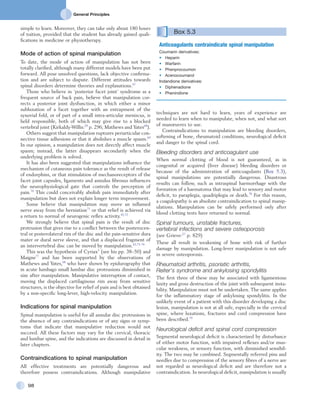


This document outlines five principles of treatment for orthopedic problems: techniques, passive movements, active movements, injection and infiltration, and deep transverse friction massage. It describes the indications, contraindications, and techniques for deep transverse friction massage. This type of connective tissue massage was developed by Cyriax to treat soft tissue injuries from trauma or overuse. While the exact mechanism is unknown, it is believed to provide pain relief and better alignment of connective tissue fibers. When applied correctly, deep transverse friction massage is usually not painful and can help resolve soft tissue issues without steroid injections.


