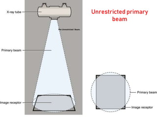
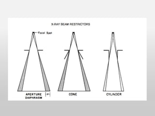
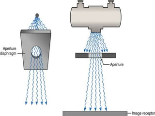
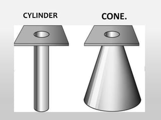
BEAM LIMITING
TECHNIQUE AND DEVICES
Precentor: Vani pushpa
M.Sc 1 st year RIT
Contents
• Introduction
• Beam-Restricting Devices
• Advantages and disadvantages
• Technique
• Filters
Beam restriction:
• The beam restriction and collimation are refer to
decrease in the size and shape of the projected
radiation field.
• Collimation decreases the risk of radiation and
minimizes scattered radiation with better contrast.
The Unrestricted Beam.
Unrestricted primary
beam
Beam-Restricting Devices
• An x-ray beam restrictor is a device that is
attached to the opening in the x-ray tube housing
to regulate the size and shape of an x-ray beam.
Importance of beam limiting
devices :
Beam restriction serves for three purposes:
• Limiting patient exposure
• Reducing the amount of scatter radiation
• Focus on the correct anatomy .
Limitation:
Reduce intensity by blocking incoming radiation.
The relationship between collimation
(field size) and quantity of scatter
radiation
• Increasing collimation means
decreasing field size,
and decreasing
collimation means
increasing field size.
• Collimation and
scatter radiation are
Inversely proportional.
Material used as collimator?
• Composed of high density (atomic number)
materials which is required to "collimate" the
incoming photons
• Lead is the most common element and the least
expensive
• However, collimators can also be made of tungsten,
gold, depleted uranium, all of which are better
attenuator when compared to lead.
• Cost is another significant factor
Types :
• All beam-restricting devices are made of metal or
a combination of metals that readily absorb
x-rays.
Classified into 3 categories:
• Aperture Diaphragms
• Cones and Cylinders
• Collimators
Aperture Diaphragms
• An aperture diaphragm is a thin sheet of lead
(diaphragm) with a hole (aperture) in center.
• Aperture diaphragms are easy to use.
• They are placed directly below the x-ray tube window.
• An aperture diaphragm can be made by cutting
rubberized lead to the size needed.
Application:
😊 Used in dental radiography with rectangular
collimation.
😊 In addition, it is used in trauma and chest radiography.
😊 angio
😊
Aperture Diaphragms contd..
Advantages
• Lead is soft, so the aperture can be easily altered to
any desired size or shape.
Disadvantages:
• Large penumbra—can be reduced by keeping
Aperture Diaphragm far away from target.
Cones and Cylinders
• Cones and cylinders are shaped differently.
• Both have extended metal structures (10-20inch) that
restrict the useful circular beam to require size.
• Positioning and size of the distal end determines the field
size.
• Cone is a ideal beam restrictor, but the flare of cone is
greater than the flare of x ray beam.
• If the x ray source, film and cone are not aligned properly,
then one side of the film may not be exposed, which is
called cone cutting.
• Advantage: Reduces the penumbra (as they placed far)
• Disadvantage: These systems provide only limited number of
field sizes.
CYLINDER CONE.
Application:
• Designed to operate most
effectively at a designed SID
• Sinuses
• Dental x ray and treatment
Collimators
• The most useful, and accepted beam-restricting
device is the collimator.
• The terms collimation and beam restriction are
used interchangeably.
It has two advantages over the other types:
 it provides an infinite variety of rectangular x-ray
fields
 a light beam shows the center and exact
configuration of the x-ray field.
Structure of the collimator:
• 2 sets of shutters control the dimensions.
• Each shutter contains 4 or more lead plates of 3mm
thickness, which move in independent pairs.
• When the shutters are closed, they meet at the
center.
• Collimator also has a light and mirror arrangement
to create radiation and optical coincidence.
• The light beam is deflected by a mirror mounted in
the path of x ray beam at an angle of 45 degree.
• The target of x ray tube and the light bulb should
be the exactly same distance from the center of the
mirror.
• The light field and radiation field should match
exactly with each other.
Quality assurance :
• Do collimator –beam alignment check at least every
six months
• Do check of alignment of center of x ray beam at
least every six months.
X RAY FILTERS
• When the X ray beam is produced many energies of
photons exit (polychromatic). Many are of such low
energies that they were offered nothing to
production of that radiograph. So, removal of low
energy x rays from beam spectrum is called
filtration.
• Metals such as aluminum will absorb the soft low
energy rays.
• This reduces the patient exposure
why we use filters in the
diagnostic radiology?
In diagnostic radiology, if the filters are not used,
• The patient acts as a filter,
• Which will result in absorption of X rays by the
tissues.
• Unnecessary dosage of radiation to the patient.
• To Increase the tube age
Types of rays:
• The radiation emitted by the X ray tube consists of
heterogeneous beam of various wavelengths that
are (longer and shorter wavelengths both soft and
hard rays respectively).
• As the soft rays (long wave lengths) are harmful to
skin and other tissues these rays shall be filtered off
to get better penetrated X ray and to avoid damage
to skin.
• If not filtered the patient may get skin burns during
fluoroscopic examination.
Preferred metal:
• Copper (Z= 29)
• Aluminum (Z=13)
most commonly preferred in diagnostic radiology
low atomic number therefore excellent material for
absorbing soft rays.
low energy X ray photons low in weight therefore
make the extra tube lighter in weight and easily
handling
Types
Inherent filtration
• Absorption of X rays as they pass through
The window in the Glass envelope (thin beryllium 1mm)
The insulating oil with in the shield
thickness 0.5mm aluminum equivalent
About three together added up to a filter which is able to
remove the longest wavelength in the this is called inherent
filtration of x ray tube.
Added filtration
Filters are used to remove unwanted bremsstrahlung
x rays outside the X ray tube housing is called added
filters
 Copper
Aluminum
Combination of cu+Al
Rhodium
Molybdenum
Silver
Applications:
 Copper- 0.1-0.9mm- general radiography x ray
tubes- in pediatric applications
 Aluminum-0.1mm-general radiography x ray tubes
Rhodium - 0.025mm- mammo with Rh anode
Molybdenum - 0.03mm- Mammography
Silver –used in mammo with tungsten anode
• Filter X ray beam = decrease photon intensity +
increase X ray beam energy.
• Total filtration :Combination of inherent and added
Minimum total filtration of 2.5mm of Al eq is required
for x ray tube operating above 70 kVp
Disadvantages
• Reduction in the intensity of X ray photons but not
no. of photons.
• Lengthen the time required to make an exposure
• May absorb primary X ray beam during excessive
filtration
• Increase tube loading (due to more mAs)
Reference :
• The Physics of Radiology and imaging by K.Thayalan
• radiologykey.com
• Christensen’s Physics of Diagnostic Radiology
• Handbook of medical radiography C Ramamohan
Queries
• Difference between collimation
and filtration?
• Why can’t we use lead as a filter material ?
Thank you for your attention

Beam restriction technique and devices.pptx

  • 1.
    BEAM LIMITING TECHNIQUE ANDDEVICES Precentor: Vani pushpa M.Sc 1 st year RIT
  • 2.
    Contents • Introduction • Beam-RestrictingDevices • Advantages and disadvantages • Technique • Filters
  • 3.
    Beam restriction: • Thebeam restriction and collimation are refer to decrease in the size and shape of the projected radiation field. • Collimation decreases the risk of radiation and minimizes scattered radiation with better contrast.
  • 4.
  • 5.
    Beam-Restricting Devices • Anx-ray beam restrictor is a device that is attached to the opening in the x-ray tube housing to regulate the size and shape of an x-ray beam.
  • 6.
    Importance of beamlimiting devices : Beam restriction serves for three purposes: • Limiting patient exposure • Reducing the amount of scatter radiation • Focus on the correct anatomy . Limitation: Reduce intensity by blocking incoming radiation.
  • 7.
    The relationship betweencollimation (field size) and quantity of scatter radiation • Increasing collimation means decreasing field size, and decreasing collimation means increasing field size. • Collimation and scatter radiation are Inversely proportional.
  • 8.
    Material used ascollimator? • Composed of high density (atomic number) materials which is required to "collimate" the incoming photons • Lead is the most common element and the least expensive • However, collimators can also be made of tungsten, gold, depleted uranium, all of which are better attenuator when compared to lead. • Cost is another significant factor
  • 9.
    Types : • Allbeam-restricting devices are made of metal or a combination of metals that readily absorb x-rays. Classified into 3 categories: • Aperture Diaphragms • Cones and Cylinders • Collimators
  • 11.
    Aperture Diaphragms • Anaperture diaphragm is a thin sheet of lead (diaphragm) with a hole (aperture) in center. • Aperture diaphragms are easy to use. • They are placed directly below the x-ray tube window. • An aperture diaphragm can be made by cutting rubberized lead to the size needed. Application: 😊 Used in dental radiography with rectangular collimation. 😊 In addition, it is used in trauma and chest radiography. 😊 angio 😊
  • 13.
    Aperture Diaphragms contd.. Advantages •Lead is soft, so the aperture can be easily altered to any desired size or shape. Disadvantages: • Large penumbra—can be reduced by keeping Aperture Diaphragm far away from target.
  • 14.
    Cones and Cylinders •Cones and cylinders are shaped differently. • Both have extended metal structures (10-20inch) that restrict the useful circular beam to require size. • Positioning and size of the distal end determines the field size. • Cone is a ideal beam restrictor, but the flare of cone is greater than the flare of x ray beam. • If the x ray source, film and cone are not aligned properly, then one side of the film may not be exposed, which is called cone cutting. • Advantage: Reduces the penumbra (as they placed far) • Disadvantage: These systems provide only limited number of field sizes.
  • 15.
  • 16.
    Application: • Designed tooperate most effectively at a designed SID • Sinuses • Dental x ray and treatment
  • 17.
    Collimators • The mostuseful, and accepted beam-restricting device is the collimator. • The terms collimation and beam restriction are used interchangeably. It has two advantages over the other types:  it provides an infinite variety of rectangular x-ray fields  a light beam shows the center and exact configuration of the x-ray field.
  • 18.
    Structure of thecollimator: • 2 sets of shutters control the dimensions. • Each shutter contains 4 or more lead plates of 3mm thickness, which move in independent pairs. • When the shutters are closed, they meet at the center. • Collimator also has a light and mirror arrangement to create radiation and optical coincidence.
  • 19.
    • The lightbeam is deflected by a mirror mounted in the path of x ray beam at an angle of 45 degree. • The target of x ray tube and the light bulb should be the exactly same distance from the center of the mirror. • The light field and radiation field should match exactly with each other.
  • 22.
    Quality assurance : •Do collimator –beam alignment check at least every six months • Do check of alignment of center of x ray beam at least every six months.
  • 23.
    X RAY FILTERS •When the X ray beam is produced many energies of photons exit (polychromatic). Many are of such low energies that they were offered nothing to production of that radiograph. So, removal of low energy x rays from beam spectrum is called filtration. • Metals such as aluminum will absorb the soft low energy rays. • This reduces the patient exposure
  • 24.
    why we usefilters in the diagnostic radiology? In diagnostic radiology, if the filters are not used, • The patient acts as a filter, • Which will result in absorption of X rays by the tissues. • Unnecessary dosage of radiation to the patient. • To Increase the tube age
  • 25.
    Types of rays: •The radiation emitted by the X ray tube consists of heterogeneous beam of various wavelengths that are (longer and shorter wavelengths both soft and hard rays respectively). • As the soft rays (long wave lengths) are harmful to skin and other tissues these rays shall be filtered off to get better penetrated X ray and to avoid damage to skin. • If not filtered the patient may get skin burns during fluoroscopic examination.
  • 26.
    Preferred metal: • Copper(Z= 29) • Aluminum (Z=13) most commonly preferred in diagnostic radiology low atomic number therefore excellent material for absorbing soft rays. low energy X ray photons low in weight therefore make the extra tube lighter in weight and easily handling
  • 27.
  • 28.
    Inherent filtration • Absorptionof X rays as they pass through The window in the Glass envelope (thin beryllium 1mm) The insulating oil with in the shield thickness 0.5mm aluminum equivalent About three together added up to a filter which is able to remove the longest wavelength in the this is called inherent filtration of x ray tube.
  • 29.
    Added filtration Filters areused to remove unwanted bremsstrahlung x rays outside the X ray tube housing is called added filters  Copper Aluminum Combination of cu+Al Rhodium Molybdenum Silver
  • 30.
    Applications:  Copper- 0.1-0.9mm-general radiography x ray tubes- in pediatric applications  Aluminum-0.1mm-general radiography x ray tubes Rhodium - 0.025mm- mammo with Rh anode Molybdenum - 0.03mm- Mammography Silver –used in mammo with tungsten anode
  • 31.
    • Filter Xray beam = decrease photon intensity + increase X ray beam energy. • Total filtration :Combination of inherent and added Minimum total filtration of 2.5mm of Al eq is required for x ray tube operating above 70 kVp
  • 32.
    Disadvantages • Reduction inthe intensity of X ray photons but not no. of photons. • Lengthen the time required to make an exposure • May absorb primary X ray beam during excessive filtration • Increase tube loading (due to more mAs)
  • 33.
    Reference : • ThePhysics of Radiology and imaging by K.Thayalan • radiologykey.com • Christensen’s Physics of Diagnostic Radiology • Handbook of medical radiography C Ramamohan
  • 34.
    Queries • Difference betweencollimation and filtration? • Why can’t we use lead as a filter material ?
  • 35.
    Thank you foryour attention