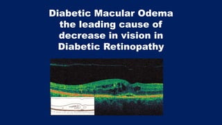
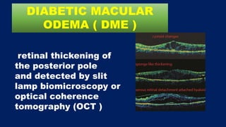
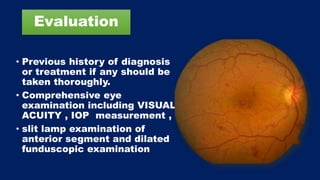
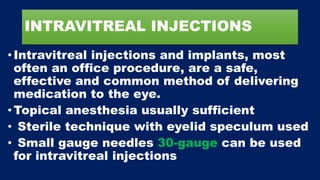
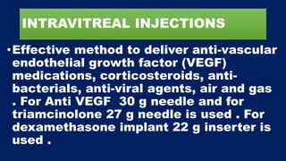
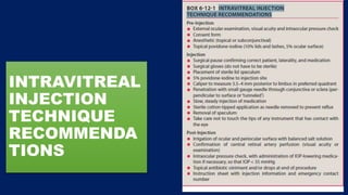
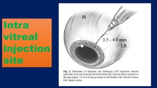
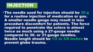
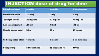
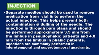
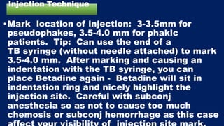
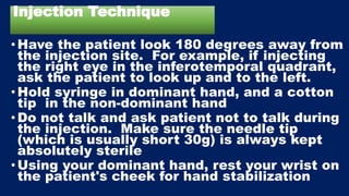
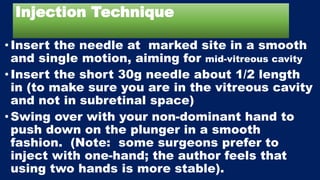
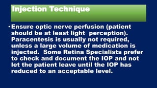
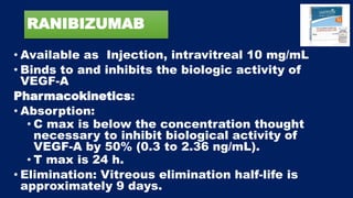
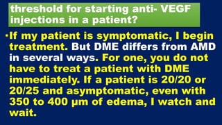
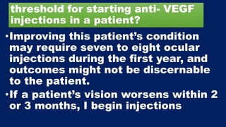
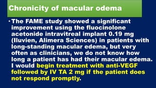
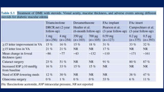
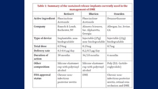
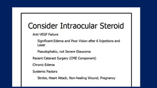
This document discusses diabetic macular edema (DME), including its prevalence, evaluation, treatment options, and intravitreal injection technique. DME is a leading cause of vision loss in diabetic retinopathy. Evaluation involves visual acuity testing, slit lamp examination, and imaging like OCT and FA. Treatment includes tight blood sugar and blood pressure control, as well as therapies like anti-VEGF agents, corticosteroids, and laser photocoagulation. Intravitreal injections are a common method to deliver these medications, with 30-gauge needles typically used for anti-VEGF drugs and 27-gauge for corticosteroids like triamcinolone.
4. •In western countries, the expected
prevalence of diabetic retinopathy is
close to 35 % in the diabetic
population, while DME may be present
in 7–10 % of patients . After 2 years,
over half of patients with DME will
lose two or more lines of visual acuity
(VA) . Worldwide, it has been
estimated that 21 million people have
DME
5. Treatment of DME
• Treatment for DME includes treatment of
the systemic disease by maintaining tight
blood sugar and blood pressure control,
weight loss, and the lowering of circulating
triglycerides and cholesterol.
•However, once there is progression to
DME,therapy is indicated to slow the rate
of vision loss and to attempt to improve
the long- term prognosis.
6. retinal thickening of
the posterior pole
and detected by slit
lamp biomicroscopy or
optical coherence
tomography (OCT )
DIABETIC MACULAR
ODEMA ( DME )
7. Evaluation
• Previous history of diagnosis
or treatment if any should be
taken thoroughly.
• Comprehensive eye
examination including VISUAL
ACUITY , IOP measurement ,
• slit lamp examination of
anterior segment and dilated
funduscopic examination
8. Evaluation
• ophthalmoscopy in a dilated pupil
is helpful.
• posterior pole examination is
best done with slit lamp
biomicroscopy with accessory
lenses .
• imaging modalities commonly
used in the management are
fundus photography , FA & OCT
9. OCT and FA
are the most useful investigations in DME
• Confirm presence macular edema
• Know type of macular edema
• Assess macular thickness
• Know response to intravitreal
pharmacotherapy
• For follow up and documentation
OCT role in DME
10. FA role
• Type of leak focal
or diffuse
• Rule out macular
ischaemia
Increasingly OCT is being used
for evaluation of macular edema .
Spectral domain ( SD OCT ) has replaced
time domain OCT ( TD OCT ) .
11. Diabetic macular odema
• Slit lamp biomicroscopy , color fundus
photography or ophthalmoscopy may not be
able to detect mild DME . Here OCT is very
helpful in measuring central foveal thikness.
• OCT has become gold standard in
monitoring the progression and treatment
response in DME
• give micrometer sensitive measurements in
central retinal thickness.
12. DME
• One in every 25 diabetic patients of age 40 or
older experiences diabetic macular edema
(DME). Although there has been a paradigm
shift in their management with the advent of
anti-VEGF agents, a sizeable number of
patients with DME are resistant to these
agents despite frequent (monthly) intravitreal
injections. Several recent studies have shown
incomplete response to such agents despite
rigorous study protocols, illustrating the problem
of persistent DME. The management of these
patients is challenging and frustrating
17. OCT can also produce a
retinal thickness map.
The OCT software
automatically de-
termines the inner and
outer retinal boundaries
and produces a false-
color topographic map
showing areas of
increased thickening in
brighter colors and areas
of lesser thickening in
darker colors
18. An assessment of macular volume can
also be obtained from the retinal
thickness map. By evaluating
differences in retinal volume over time,
the clinician can judge the efficacy of
therapy
RETINAL THICKNESS MAP
19.
20.
21. OCT gold standard in monitoring the
progression and treatment response in DME
patients .
Retinal thickness is the most commonly used
quantitative parameter.
CIRRHUS measures the retinal thickness
between ILM & anterior edge of RPE layer .
Normal subjects central retinal thickness is
265 µm with CIRRHUS OCT .
22. Why macula only & not other parts
of retina become odematous
•Anatomically, the macula ( area centralis ) is
defined as the central retinal region
containing more than one layer of ganglion
cells and presenting xanthophyll pigment,
which is located mainly in the layer of the
fibers of Henle and in the inner nuclear layer
. At its center lies the fovea, a 1.5 mm
depression that is devoid of ganglion cells
and thinnest at its central area, the umbo.
23. Why macula only & not other parts
of retina become odematous
. The macula has several anatomical features
that diverge from remaining retina; these
include a high cell density with almost no
extracellular space , loose arrangement of
thick fibers in Henle’s layer, & avascularity of
its central zone, which is a watershed zone
between choroidal and retinal circulations
primarily supplied by choriocapillaris.
24. Why macula only & not other parts
of retina become odematous
. These unique anatomical
characteristics of macula,
combined with its high
metabolic demand, make it
a preferential site for fluid
accumulation and edema
25. Definition of Macular Edema
• Macular edema is defined as accumulation
of fluid in macula. Histologically, fluid
accumulation may occur in intercellular
space or may be intracellular, causing
cellular swelling. Clinically, ME can be
classified as diffuse, with generalized
leakage throughout the posterior pole, or
focal, if discrete areas of retinal thickening
are present (usually caused by leakage from
microaneurysms or dilated capillaries).
26. Definition of Macular Edema
•In cystoid ME, clear fluid-filled
cystoid spaces are present in the
retina, most commonly in the outer
plexiform and inner nuclear layers,
and they are visible by
ophthalmoscopy, OCT, and/or FA ,
which shows a typical petaloid
pattern on FA
27. Pathophysiology of
Macular Edema
•Macular edema (ME) is final common pathway
of numerous retinal diseases, and a major
cause of blindness . In a physiological setting,
the blood-retinal barrier (BRB), is largely
formed by retinal pigment epithelium and
retinal capillary endothelial cells. When this
barrier is broken, water and proteins can
enter the retinal extra- and intracellular
space, with fluid accumulation leading to
edema and vision loss.
31. Mechanisms in DR
Leading to DME
•For decades DR has been mainly considered
simply a microvascular disorder, caused by
endothelial cell damage, pericyte loss, and
secondary breakdown of the inner blood-
retinal barrier (BRB), leading to DME
formation. Other factors such as hypoxia,
altered blood flow, retinal ischemia, and
inflammation are also associated with the
progression of DR and DME
32. Mediators in DME (VEGF)
• Vascular Endothelial Growth Factor (VEGF) is increased in
human ocular fluids and plays a crucial role in ischemic
retinal diseases, such as DR and retinal vein occlusions . The
VEGF family includes different isoforms: VEGF-A has been
shown to be upregulated in hypoxic tissues. In DR, the loss of
retinal capillaries is believed to lead to hypoxia, which is the
main stimulus for increased retinal expression of VEGF-A,
mediated through hypoxia-inducible factors. VEGF-A has an
angiogenic role that is responsible for the progression of DR
to the proliferative stage. Apart from its angiogenic role,
VEGF-A increases vascular permeability . Thus, the role of
VEGF-A may be central to DME pathogenesis. Moreover,
several studies have demonstrated the efficacy of anti-VEGF
treatment of DME, thus supporting the role of VEGF.
33. Mediators in DME
The Inflammatory Cascade
• Efficacy of anti-VEGF treatment of DME
support role of VEGF. Funk demonstrated
that while anti- VEGF treatment decreases
aqueous VEGF to sub-physiologic levels, this
effect does not induce a complete resolution
of DME. This study also demonstrated
correlation between high intraocular cytokine
levels and presence of DME. These data
suggest that VEGF alone cannot be unique
driving factor for development of DME
34. •DME is a result of endothelial dysfunction,
increased vascular permeability, and release
of several cytokines. Currently, anti-VEGF
agents are the first-line therapy—and the
gold standard—for managing DME. Although
these agents are effective in several cases,
some patients can be resistant to treatment.
It is thought that inhibition of VEGF alone is
insufficient, and that other proinflammatory
cytokines and pathways may play more
important roles in such resistant cases.
35. •Corticosteroids have multiple mechanisms of
action, including inhibition of VEGF synthesis
and leukocyte migration, down-regulation of
intercellular adhesion molecule-1
expression, antagonism of prostaglandins
and other cytokines, and an antipermeability
effect, thereby enhancing the barrier
function of vascular tight junctions.
•The superiority of the dexamethasone
implant over bevacizumab in macular edema
control in some cases is thought to be
related to its myriad mechanisms of action,
as opposed to the sole action of VEGF
36. •Ranibizumab in 2012 & Aflibercept in 2014
were approved for treatment of DME by the
FDA .chronic use of the anti-VEGF agents in
this patient population warrants close
monitoring by FA and OCT .
•In the phase 3 RISE and RIDE studies that
compared ranibizumab 0.3 and 0.5 mg, the
higher dose was associated with more
deaths without providing any efficacy
advantage compared to the lower dose. As a
result of these studies, Ranibizumab 0.3 mg
is the approved dose for treating DME.
37. •when an eye fails to respond to anti-
VEGF agents as expected (within the
first 3-6 months), then we have a low
threshold to switch to corticosteroids,
with the goal of achieving macular
deturgence at the earliest point in time.
This threshold is even lower for
pseudophakic eyes. An additional
advantage is the need for fewer
injections, and therefore fewer patient
visits.
38. •As we know, corticosteroid agents are
not without attendant risks. IOP
elevation and cataract formation are
most notable side effects, for which we
regularly monitor our patients started on
steroids . In our experience, these side
effects, although common, are
effectively managed with topical
glaucoma medications and cataract
surgery, respectively .
39. DEXAMETHASONE AND
FLUCINOLONE IMPLANT
• The dexamethasone intravitreal implant 0.7 mg was
approved in 2014 for the treatment of DME, but was
initially granted approval only in select patient groups
(pseudophakes and phakic patients scheduled to
undergo cataract surgery). Approval had been limited
because of the increased number of adverse events—
namely, cataract formation and iop spikes. The FDA
removed restrictions on lens status in October 2014.
• Fluocinolone acetonide 0.19 mg has been approved for
the treatment of DME. As with the other steroids,
however, this implant is associated with increased
cataract formation, increased IOP, and the necessity for
surgical treatment of elevated IOP. As a result, its
approval has been limited to patients who have
previously shown no significant rise in IOP.
40. Do use fluorescein angiography
(FA) at baseline in your patients?
•Yes For most patients, particularly
those with macular edema, I will
use FA look for areas of retinal
neovascularization that cannot be
detected clinically and areas of
nonperfusion.
41. Do you do laser
•Although laser usage has decreased, it has
not been totally eliminated. I still use the
laser for high-risk, extra-foveal areas with
circinate exudates. Since the introduction of
anti-VEGF, my laser use has significantly
decreased. LASER is also used after anti
vegf and steroid therapy to supplement there
effect and to reduce the number of injections
. If DME is not invoving centre and pt is
asymptomatic then also we may apply laser .
43. INTRAVITREAL INJECTIONS
•Intravitreal injections and implants, most
often an office procedure, are a safe,
effective and common method of delivering
medication to the eye.
•Topical anesthesia usually sufficient
• Sterile technique with eyelid speculum used
• Small gauge needles 30-gauge can be used
for intravitreal injections
44. INTRAVITREAL INJECTIONS
•Effective method to deliver anti-vascular
endothelial growth factor (VEGF)
medications, corticosteroids, anti-
bacterials, anti-viral agents, air and gas
. For Anti VEGF 30 g needle and for
triamcinolone 27 g needle is used . For
dexamethasone implant 22 g inserter is
used .
45. Intravitreal Injections Introduction
•The office delivery of medication directly
into the vitreous cavity via injection or
implant has become commonplace in
ophthalmology. This method allows for higher
concentrations in the eye with less systemic
absorption compared to other methods of
medication administration.
•In 1911, the injection of air into the vitreous
cavity for retinal detachment (RD) repair was
initially reported.
48. PRE-INJECTION PREPARATION
•Treatment of overt, active blepharitis
should be done prior to injection to attempt
to decrease bacterial load, which may
increase risk of infection. Patients with
bacterial or viral conjunctivitis should be
treated to manage infection and have their
injection rescheduled. As with any surgical
procedure all patients should sign an
informed consent after being explained the
risks, benefits, and alternatives to injection
49. INJECTION
•The needle used for injection should be 30 g
for a routine injection of medication or gas.
A smaller needle gauge may result in less
procedure discomfort for patients. The force
required to penetrate the sclera is almost
twice as much using a 27-gauge needle
compared to 30- or 31-gauge needles.
Needle length should be 1/2 to 5/8 inches to
prevent globe trauma.
50. INJECTION which gauge of needle
•The majority of retinal specialists
surveyed in 2010 used a 30-gauge
needle for BEVACIZUMAB and
RANIBIZUMAB, and a 27-gauge
needle for TRIAMCINOLONE.
Triamcinolone can precipitate
within the syringe and clog a needle
smaller than 27-gauge.
51. INJECTION dose of drug for dme
Intravitreal drug avastin lucentis triamcinolone
Intravitreal dose 1.25 mg .3 mg 2 mg
strength in vial 25 mg / ml 10 mg / ml 40 mg / ml
Amt in cc injected .05 ml .03 ml .05 ml
Needle gauge used 30 g 30 g 27 gauge
To be repeated after 1 month 1 month 3 to 4 months
Cost per inj 5 thousand rs 25 thousand rs 100 rs
52. INJECTION
• Separate needles should be used to remove
medication from vial & to perform the
actual injection. This helps prevent both
contamination & dulling of the needle. The
injection is through pars plana and should
be performed approximately 3.5 mm from
the limbus in pseudophakic patients and 4.0
mm from the limbus in phakic patients.
Injections are commonly performed in
inferotemporal and superotemporal quadrants.
53. INJECTION
The quadrant of injection may be influenced
by indication for injection or other factors.
For example, injecting an opaque steroid
superiorly may obscure the visual axis more
initially than injecting inferiorly.
Similarly, a patient with a large
inferotemporal retinoschisis cavity may
benefit from having their injection in another
quadrant
54. INJECTION
•The traditional injection method involves
inserting needle perpendicular to the ocular
scleral surface in a single plane to penetrate
globe. Another tunneling method has been
described which involves needle insertion at
a 30 degree angle initially with repositioning
of the needle to perpendicular and towards
the center of the globe. Final needle
direction should always be towards the
center of the eye to prevent damage to other
intraocular contents.
55. INJECTION
•Inserting needle perpendicular to eye is
preferred over other techniques .
•The injection should be done in a slow,
steady maneuver to prevent a sudden
flux through the vitreous cavity, as this
may disrupt vitreoretinal adhesions.
•A sterile cotton tip applicator may be
used to prevent reflux for injections
56. Injection Technique
• Confirm informed consent obtained
• Surgical time-out to confirm correct
medication and correct eye
•Place patient in near supine position; make
sure the headrest of the chair is stable
• Topical proparacaine (wait 10-15 seconds
before placing Betadine)
• 10% Betadine swab to inferior cul-de-sac, to
allow Betadine to start working; ask patient
to blink multiple times to spread Betadine .
57. Injection Technique
•Place 2% lidocaine jelly on the eye, focusing
on inferotemporal quadrant.
• (Note: Betadine placed prior to lidocaine
jelly to sterilize the globe prior to placing the
jelly so as not to have bacteria trapped in the
jelly and possibly increase risk of
endophthalmitis).
• Ask patient to close their eyes, and return in
2-5 minutes.
58. Injection Technique
•Apply Betadine swab again to inferotemporal
quadrant and inferior cul-de-sac, as well as to
eyelashes (do not manipulate much so as not
to liberate bacteria from the lashes)
• Place sterile closed-blade eyelid speculum
(careful not to cause corneal abrasion and
save yourself a phone call from the patient
with a painful corneal abrasion).
• Clean again with Betadine
59. Injection Technique
• Mark location of injection: 3-3.5mm for
pseudophakes, 3.5-4.0 mm for phakic
patients. Tip: Can use the end of a
TB syringe (without needle attached) to mark
3.5-4.0 mm. After marking and causing an
indentation with the TB syringe, you can
place Betadine again - Betadine will sit in
indentation ring and nicely highlight the
injection site. Careful with subconj
anesthesia so as not to cause too much
chemosis or subconj hemorrhage as this case
60. Injection Technique
•Have the patient look 180 degrees away from
the injection site. For example, if injecting
the right eye in the inferotemporal quadrant,
ask the patient to look up and to the left.
•Hold syringe in dominant hand, and a cotton
tip in the non-dominant hand
•Do not talk and ask patient not to talk during
the injection. Make sure the needle tip
(which is usually short 30g) is always kept
absolutely sterile
•Using your dominant hand, rest your wrist on
the patient's cheek for hand stabilization
61. Injection Technique
•Insert the needle at marked site in a smooth
and single motion, aiming for mid-vitreous cavity
•Insert the short 30g needle about 1/2 length
in (to make sure you are in the vitreous cavity
and not in subretinal space)
•Swing over with your non-dominant hand to
push down on the plunger in a smooth
fashion. (Note: some surgeons prefer to
inject with one-hand; the author feels that
using two hands is more stable).
62. Injection Technique
• Do not move the needle while inside
the eye so as to not cause traction on
the vitreous and potentially cause a
retinal tear / detachment.
•As you remove the needle out, cover
the injection site with a cotton tip
that is in your non-dominant hand
•Rinse the Betadine off of the patients
eye
63. Injection Technique
•Ensure optic nerve perfusion (patient
should be at least light perception).
Paracentesis is usually not required,
unless a large volume of medication is
injected. Some Retina Specialists prefer
to check and document the IOP and not
let the patient leave until the IOP has
reduced to an acceptable level.
64. BEVACIZUMAB
• Recombinant humanized monoclonal antibody that
blocks angiogenesis by inhibiting VEGF-A
• It received its first approval in 2004, for
combination use with standard chemotherapy for
metastatic colon cancer
• It has since been approved for use in
• Certain lung cancers,
• Renal cancers,
• Ovarian cancers
• Glioblastoma multiforme of the brain
65. ADMINISTRATION AND DOSING
•In ophthalmology, Bevacizumab is typically
given by transconjunctival intravitreal
injections into the posterior segment
• Intravitreal injections for retinal pathologies
are typically administered at 4-6 week
intervals, although this varies widely based
on disease and response.
•DOSE: The typical dose is 1.25mg in 0.05ml
in adults,and half that dose in babies.
67. RANIBIZUMAB
• Available as Injection, intravitreal 10 mg/mL
• Binds to and inhibits the biologic activity of
VEGF-A
Pharmacokinetics:
• Absorption:
• C max is below the concentration thought
necessary to inhibit biological activity of
VEGF-A by 50% (0.3 to 2.36 ng/mL).
• T max is 24 h.
• Elimination: Vitreous elimination half-life is
approximately 9 days.
68.
69. AFLIBERCEPT
• Recombinant fusion protein consisting
of VEGF-binding portions from the
extracellular domains of human
VEGF receptors 1 and 2, that are fused
to the Fc portion of the human
IgG1 immunoglobulin
70. Based on recent clinical trial data,
which anti-VEGF do you use first?
• I start almost every patient on
bevacizumab because of affordability .
Patients with relatively good vision and
not a great deal of edema would probably
do well on any of the anti-VEGF drugs.
(DRCR protocol T data) . Overall,
aflibercept (Eylea, Regeneron) has proved
best for patients with diabetic
retinopathy and vision less than 20/40 or
with >400 μm of edema.
71. Based on recent clinical trial data,
which anti-VEGF do you use first?
• I would, however, switch any
nonresponders with severe edema to
another anti-VEGF because they have
different mechanism of action . I might
also switch a patient with poor vision
(20/200) to aflibercept if they had more
than 400 μm of edema, based on Protocol
T findings ( )
72. threshold for starting anti- VEGF
injections in a patient?
•If my patient is symptomatic, I begin
treatment. But DME differs from AMD
in several ways. For one, you do not
have to treat a patient with DME
immediately. If a patient is 20/20 or
20/25 and asymptomatic, even with
350 to 400 μm of edema, I watch and
wait.
73. threshold for starting anti- VEGF
injections in a patient?
•Improving this patient’s condition
may require seven to eight ocular
injections during the first year, and
outcomes might not be discernable
to the patient.
•If a patient’s vision worsens within 2
or 3 months, I begin injections
74. NON RESPONDER
•For me, a nonresponder is someone who has
had less than a 10% or 20% improvement on
their OCT within the first month of
treatment. Even though anti-VEGF might
have had a minimal effect on the macular
edema in 4 or 5 months, you might see a
profound effect on the patient’s level of
retinopathy, making the case for continuing
the anti-VEGF or combining it with a steroid
where required for edema.
75. Switching or combine steroid
• Switching is not the issue. I think it is more a
question of continuing the anti-VEGF and
then adding a drug. Some patients are pure
anti-VEGF responders, while others do
extremely well on steroids alone. Some do
well on both. In some cases after steroid
injections, patients re-accumulate fluid
quickly (within 8 weeks) and require anti-
VEGF therapy again. Instead of switching, it
might be more a question of continuing the
anti-VEGF and then periodically giving
patients a bolus dose of a steroid.
76. If you choose to include steroids in
your treatment regimens, which one
do you use first
•I use triamcinolone suspension, 1 mg
to 2 mg as recommended by the
Protocol B and the SCORE studies. I
used to use a 4-mg dose, but now we
know that the same biologic effect
occurs with 1 mg, and that greatly
reduces the complication rate.
77. Chronicity of macular odema
• Does length of time a patient has had macular
edema & their history of treatment make you move
to steroids faster than you would for a patient with
recent-onset macular edema?
• I am certainly more likely to require
something additional for long-standing
edema, as borne out in the FAME study. Any
patient with chronic edema has a problem,
whether it is denial or lack of follow-up. Thus,
it is important to consider using something
that will be less of a burden on these patients .
78. Chronicity of macular odema
•The FAME study showed a significant
improvement using the fluocinolone
acetonide intravitreal implant 0.19 mg
(Iluvien, Alimera Sciences) in patients with
long-standing macular edema, but very
often as clinicians, we do not know how
long a patient has had their macular edema.
I would begin treatment with anti-VEGF
followed by IV TA 2 mg if the patient does
not respond promptly.
79. Use of flucinolone
•It is important to provide a steroid challenge
before resorting to fluocinolone. You have to
know if a patient will respond to a steroid.
Bear in mind that patients would need
multiple injections with an intravitreal
before we should start fluocinolone, since a
significant number of patients will not get
any steroid rise, even after their first,
second, or sometimes after their third
injection.
80. STEROID IN DME
•For younger patients who do not have
cataracts, clinicians must explain that using
a steroid will hasten the formation of a
cataract. As we dry out their retina, their
vision may worsen, necessitating cataract
surgery. However, the 20-year insulin-
dependent diabetic might not respond to
anti-VEGF until you add the steroid. At the
same time, you will be putting them on a
road you do not want them to travel.
81. STEROID IN DME
• True, but I assure my patients that
cataracts will not make them go blind, and
they can be fixed; however, if their
macular edema persists, it will cause
substantial vision loss .
• In Protocol I, pseudophakic patients do as
well with steroids as they do with the anti-
VEGF and require only three injections in
the first year. So, I think there is a place
for steroids
82. IST YEAR ANTI DME TREATMENT
• The great outcomes in RISE/RIDE, VIVID/VISTA and
Protocol I require a very heavy burden, especially in that
first year. That is one of the real key points, as there is a
fair amount of undertreatment in the community in the first
year. In AMD, the initiation phase/loading dose has usually
involved three injections; and people have been applying
this concept to DME. However, in DME, there is a much
heavier injection burden in the first year. The visual acuity
curves in RISE/RIDE show an improvement after the first
three injections, but the curve continues to gradually go up
over the first 18 months. It is a very steady, gradual
improvement in both visual acuity and OCT outcomes
83. DRCR.net Protocol I
• DRCR.net Protocol I, patients were
started off with a loading dose of four
injections, but if patients did not have
20/20 vision and no edema, they were
given an additional two injections. So in
reality, it was a six-dose loading phase.
Comparatively speaking, in AMD it is
traditionally a three-dose loading
phase.
84. DRCR.net Protocol I
•Most of the AMD studies that utilized a p.r.n.
dosing strategy required six to seven
injections in the first year in comparison to
Protocol I and T, where the average was
higher with nine to 10 injections . Protocol I
was instrumental in showing us that monthly
treatment is not necessary to achieve vision
stabilization and letter gain comparable with
the more continuous treatment regimens in
RISE/RIDE .
85. DRCR .net protocol I
intravitreal anti vegf
agents
with early or
deferred laser
Superior
Over
laser alone
laser with steroids
steroids alone
or
or
86. DRCR .net protocol T
• All Three Anti Vegf Agents RANIBIZUMAB ,
BEVACIMIZUMAB & AFLIBERCEPT Are
Effective In Management Of DME .
•RANIBIZUMAB And AFLIBERCEPT Are Slightly
Better Than BEVACIMIZUMAB if vision is less
But Latter Is More Economical .
87. TREATMENT OF DME VS ARMD
• It is important to distinguish wet AMD from
DME—these are, indeed, two very different
diseases. For patients with wet AMD, we are
treating choroidal neovascularization with
exudation. For patients with DME, we are
only treating edema, the source of which is
retinovascular incompetence. While we are
trying to get that macula to a satisfactory
dry status, the burden over time tends to fall
off in DME.
88. TREATMENT OF DME VS ARMD
•In wet AMD, there is more of a
continuous burden year after year for
an indefinite time frame in most
patients. In DME, studies with
discontinuous or P.R.N. style therapy
such as Protocol I and Protocol T,
many patients were eventually able to
cease therapy .
89. TREATMENT OF DME VS ARMD
•With wet AMD, whether it is P.R.N. or TREAT-
AND-EXTEND (TAE), the mean number of
treatments will typically reach a plateau after
the first year. It does not tend to decrease
much thereafter. But with DME, P.R.N.
studies that we have seen, Protocol I in
particular, started with a mean of eight to
nine injections in year 1, then three to four in
year 2, two to three in year 3 and by years 4
and 5, many had no further treatment .
90. How to reduce numer of injections
•Steroids can be used for reduced burden of
treatment as a combination therapy, but
probably not as a low-burden monotherapy
for most diabetic patients. There may be
special cases where steroids are best
choice as a monotherapy, such as in a newly
pseudophakic patient who develops macular
edema post-operatively, but results are simply
not as robust as with anti VEGF therapy.
91. DME BUT STILL NO TREATMENT
• Protocol I allowed for patients to have DME if
it was stable without a decrease in vision. It
was possible to have patients with persistent
DME, as long as vision did not drop and there
was not an increase in the OCT thickness. In
DME, we now know that persistent DME does
not translate into lost vision. With results
reported out to 5 years, Protocol I showed those
great visual improvements at year 1 could be
maintained with an injection burden that
dramatically decreased in years 2 through 5.
92. Type of response to anti VEGF
•My patients typically fall into one of three
categories. The first is what we all want—a
patient who is exquisitely sensitive to anti-
VEGF therapy and needs only one injection to
have a very dramatic improvement. The
second group is what we dread—no
measurable response with anti-VEGFs after
three or four injections. The third group,
however, is what most of us see—people who
have some response to the anti-VEGF therapy.
93. COMBINATION THERAPY IN DME
• Monotherapy with anti-VEGF agents or sequential
monotherapy, anti-VEGF followed by steroids, gives
us a good result. There is a role for laser at some
point in the process in selected case such as when
there is persistent or refractory edema and you
see an obvious source of leakage on fluorescein
angiography (FA) with large, leaking microaneurysms.
Targeted focal laser can provide adequate drying
and stabilization, so I do consider it. I almost never
use it for center-involving DME, but there is a role
for laser as second-line treatment .
94. final
• After all the studies, I think it is safe to say anti-VEGF is
our first-line defense. We have long-term outcomes from
the studies, and those results are extremely efficacious.
But it is a complex disease. Even when you look at the
results from Protocol T, the agent that worked the best
(aflibercept) still left one-third of the patients with more
than 250 microns of edema at 1 year. And that range was
as high as 60% with bevacizumab. Most of us are using
bevacizumab, and 60% of our patients are going to have
persistent fluid after 10 treatments in the first year. That
means there’s a role for other treatments. We don’t have
large studies to dictate whether or not the intravitreal
triamcinolone should be our preferred second-line agent.
There are some new studies being undertaken by the
DRCR.net on the combination of anti-VEGF and steroid
95. ROLE OF LASER
• Laser may be appropriate in patients with focal
extrafoveal macular edema secondary to focal
microaneurysms. If one laser treatment could
spare them the burden of a series of multiple
injections, it may be worthwhile to consider.
• The gold standard based on evidence accumulated
over the past few years is that for center-involving,
symptomatic DME, pharmacotherapy is the gold
standard. Laser has a role for the center-involving
patient as a deferred therapy, and is still a primary
therapy for non-center-involving.
96. How to tell pt requirement of so
many injections .
• I discuss that anti-VEGF treatment is an
ongoing process and in some ways
analagous to chemotherapy. I emphasize
that regular injections often keep the
disease process under control, may be
able to improve the vision. While the
initial injections occur monthly, it may be
possible to reduce the number of visits or
injections over time
97. How to tell pt requirement of so
many injections
• There is a different conversation for the AMD patient and
the DME patient. I tell the wet AMD patient that these
injections serve as disease control and there is a good
chance for good outcomes, but it is going to be an
indefinite, ongoing treatment process. I do let them know
that, while rare, some patients do improve to the point
where we can stop treatment altogether.
• With DME, I tell patients there will be a lot of treatment up
front, a lot of visits initially, as the disease is usually slow
to respond. But I also tell them that compared with AMD,
there is a better chance of coming off therapy
98. How to tell pt requirement of so
many injections
• For DME, I explain to patients that we will require monthly
injections for a prolonged period of time, and that as the
disease stabilizes, there may be an opportunity for us to
increase the interval between injections. I do alert them
that there will be multiple injections during that first year
but that too will vary from one patient to the next. Once I
have a patient’s visual acuity and edema within a normal
range, I begin a TAE regimen. I inform him or her that we
may need to cut back interval between injections if there
is a recurrence. If they have stable vision but thick
edema, I will discuss need to introduce combination therapy
99. COMBINATION THERAPY IN DME
• We begin with an anti-VEGF and are
guided by Protocol T. If the patient cannot
demonstrate improvement after three
intravitreal injections, we consider adding
a steroid. We are constantly walking the
line between pushing the VEGF or
inflammatory component. So we continue
with the anti-VEGF in those that do
respond until we hit the maximum effect
& Once the patient has stable vision .
100. COMBINATION THERAPY IN DME
• If there is continued macular thickening,
we consider introducing either a steroid
or laser therapy. FAME did show one
subgroup with chronic DME for more than
3 years did a lot better on steroids, so
that may be a key differentiating point. It
is a nice option to have steroids for
people who don’t respond to our previous
treatments.
101. SWITCHING
•I switch between anti-VEGF agents
first because ranibizumab and
aflibercept have different
mechanisms of effect. If that does
not work, I might switch to a steroid,
combine medications, or consider a
laser, either extrafoveal focal or
micropulse
102. STEROIDS
•Steroids also can be considered for patients
who have an increased risk of
thromboembolic events and want to avoid
the theoretical risk with Anti-VEGF agents,
for treating pregnant women with DME
rather than potentially exposing the fetus to
an anti-VEGF agent, and finally for people
who are not willing to tolerate the high
injection burden (nine to 10 treatments in
the first year) with anti-angiogenic therapy
103. STEROIDS
•I like adding steroids in pseudophakic
patients. I am more apt to try another anti-
angiogenic agent in a phakic patient before
going straight to a steroid. I like steroids
for DME, but it is not without a cost. I will
probably do at least one antiangiogenic
switch between agents after Protocol T
before going to steroid in phakic patients.
But it will not be a rapid switch
105. Intravitreal Steroids
•Corticosteroids are well known for their
antiangiogenic, antiedematous, and anti-infl
ammatory properties. Consequently, these
have been widely used in treatment of many
retinal diseases since the early 1950s. Later,
intravitreal steroids emerged as an essential
tool, allowing for optimal drug efficacy when
given locally. The topical or local route offers
the great advantage that there is no
significant systemic side effect
106. Intravitreal Steroids
• Randomized studies have properly shown that
steroids are very effective in the treatment of
posterior segment diseases such as retinal
vascular occlusion, diabetic macular edema,
and uveitis. This is true for both, disease
activity and visual outcome. Today because
of the anti-infl ammatory, antiangiogenic, and
antipermeability properties, steroids are an
attractive therapeutic option for vascular and
inflammatory retinal diseases
107. Intravitreal Steroids
•Recent advances in ocular drug delivery
methods led to the development of
intraocular implants, which help to provide
prolonged treatment with a continuous drug
release. An increasing number of ophthalmologists
use intravitreal steroids for the treatment of
various posterior segment disorders, not only
when traditional therapeutic methods have
failed, but more and more as a first-line
therapy .
109. Treatment of DME with
Triamcinolone
•Triamcinolone has been shown to
reduce VEGF production, decrease
prostaglandin production, and stabilize
the blood–retinal barrier . This action of
decreasing vascular permeability has
led investigators to try triamcinolone in
the treatment of DME .
110. Treatment of DME with
Triamcinolone
•studies have shown the efficacy of IVTA to
temporarily reduce DME and increase visual
acuity . The mean reduction in macular
thickening reaches 85 % 3 months after
injection with a mean two-line improvement
of visual acuity . However, recurrence of
DME occurs between 3 and 6 months after
injection.
111. Treatment of DME with
Triamcinolone
•Recurrence of DME occurs between 3
and 6 months after injection. Due to the
high rate of complications and the
absence of evidence of its superiority
over laser photocoagulation, IVTA is
generally reserved for patients
refractory to focal/grid laser therapy and
anti-VEGF agents .
112. Treatment of DME with
Dexamethasone
• The dexamethasone 700 μg posterior segment
drug delivery system (Ozurdex®) is a bioerodible
Implant delivered intravitreally using a 22-gauge
injector .
• Considering the higher cost but similar efficacy ,
side effect profile and frequency of
administration we generally prefer 2mg dose of
Triamcinolone over Dexamethasone implant
(Ozurdex) .
113. Treatment of DME with Dexamethasone
• treatment was well tolerated and had an
acceptable safety profile. Similar to IVTA,
DEX-DDS has the advantage of repeatability,
as long as the IOP and cataract effects are
mitigated. The advantages of DEXDDS over
intravitreal triamcinolone include a possible
longer duration of action, absence of
postinjection “visual clouding” or floater-like
symptoms, and a lower rate of IOP increase.
month 12.
114. Treatment of DME with Fluocinolone
Acetonide FAc Insert
• Two fluocinolone acetonide (FAc) intravitreal
devices have been studied for the treatment
of DME: a FAc implant (Retisert) and a FAc
insert (ILUVIEN®).
• ILUVIEN is a non-bioerodible insert implanted
in the eye via injection through the pars plana
using a 25-gauge needle. ILUVIEN contains
0.19 mg of Flucinolone acetonide .
115. Treatment of DME with Fluocinolone
Acetonide FAc Insert FAME
•The Fluocinolone Acetonide for Macular
Edema (FAME) studies were two large
prospective, randomized, controlled studies
that followed 956 eyes randomized to receive
0.2-μg/day (lowdose) or 0.50-μg/day (high-
dose) inserts or sham. The most common
adverse event was cataract, and almost all
phakic patients in the FAc insert groups
developed cataract
116. Treatment of DME with Fluocinolone
Acetonide FAc Insert FAME
•The incidence of increased IOP (37.1
and 45.5 %, in the low-dose and high-
dose insert groups, respectively), need
for IOP-lowering medication (38.4 and
47.3 %, respectively), incisional
glaucoma surgery (4.8 and 8.1 %,
respectively), and trabeculoplasty (1.3
and 2.5 %, respectively)
117. Treatment of DME with steroids.
adverse events different steroids
118.
119.
120. Conclusions
•Interestingly, reductions in OCT central
subfield thickness in ptotocol I were similar
between ranibizumab and triamcinolone
groups, suggesting the visual benefits of
intravitreal corticosteroids may be tempered by
their side-effects of cataract and elevated IOP .
• Many randomized clinical studies have shown the
clinical efficacy of steroid injections to treat DME,
but their use is limited by their adverse events,
including cataract progression and intraocular
pressure (IOP) increase .
121. Role For Surgery In Patients With DME
• Vitreomacular traction (VMT) is something we
watch very carefully; however, we have been
surprised to see some patients with epiretinal
membrane (ERM) dry out that we suspected would
need vitrectomy surgery. In the patients with ERMs,
edema tends to recur quickly, but we do much less
vitrectomy surgery in these patients now because
the anti-VEGFs actually improve the anatomic
configuration of the retina, even in presence of an
ERM. Some patients have taught hyaloids or ERM
that refuse to dry out despite intravitreal anti-VEGF
and/or steroid injections you may consider surgery.
128. Treatment details avastin
•The 1.25 mg dosage is administered
monthly . "Another consideration with
bevacizumab is the preparation of doses
for intravitreal injection. A common
approach used in the clinical studies is for
a pharmacy department to aseptically
prepare a batch of syringes for use in a
later clinic with one study reporting
storage of such syringes for up to 14 days
in a refrigerator.
129. Treatment details avastin
• Each vial of Avastin® contains 100 mg
of bevacizumab, sufficient for 80 doses
however it would be difficult to extract
this exact quantity.
• One method employed is to fill syringes
with between 2.0 and 2.5 mg of
bevacizumab (0.08 to 0.10 millilitre)
resulting in up to 50 doses per vial of
Avastin®."
130. Treatment details avastin
• "An alternative is for the clinical team to
prepare doses in the treatment room
immediately prior to administration using
aseptic technique but this option may
present greater risk of cross contamination.
The number of doses extracted per vial could
be reduced to a minimum of one, which will
reduce the risk of cross contamination but
will also eliminate some of the cost benefits
presented by use of bevacizumab."
131. Treatment details avastin
• "Pre-packaged syringes of bevacizumab for
intravitreal use are available to purchase from
special manufacturing units. Moorfields
Pharmaceuticals, of Moorfields NHS hospital, can
supply syringes of 1.25 mg in 0.05 ml at £85 per
syringe, excluding VAT and delivery charges. The
syringes must be stored in a refrigerator and have
an expiry date of six weeks from the date of
manufacture, which would mean an effective
expiry of about two to four weeks from receipt of
delivery."
132. Treatment details avastin
•"Any compounding of a single vial of drug
into multiple dose units will carry some risk
of microbial and particulate cross
contamination beyond that associated with
preparation of a single dose. This risk can be
minimised by performing the procedure in an
aseptic clean room using trained staff and
storing the finished product in a refrigerator."
133. Safety Of Bevacizumab For The
Treatment Of AMD
• The systematic review concluded that adverse events
were rare amongst 1,400 patients who had received a
total of several thousand intravitreal injections of
bevacizumab. In the former report, with a mean follow-up
of 3.5 months, the maximum incidence of any single event
was 0.21% . In the latter report, that included a majority
proportion of non-AMD patients, the total incidence of
systemic adverse events was 1.5% (18 patients) and
included hypertension, stroke, myocardial infarction,
aneurysm, digit amputation, and fatalities, although none
were conclusively and causally linked to bevacizumab.
• The review authors state that the type and incidence of
effects ‘does not seem to be very different from … two
large RCTs of ranibizumab
136. summary
•For patients in whom macular edema has been
confirmed clinically, we usually order OCT to
get a baseline retinal thickness & volume.
• If there are moderate to severe diabetic changes in
the periphery, we do a FA to establish degree of
nonperfusion and to rule out neovascularization
elsewhere. If we were considering any form of focal
laser therapy for extra foveal leakage, we would
also do an angiogram. we also check HbA1c
hemoglobin levels to determine if the patient’s
diabetes is under control .
137. summary
• For symptomatic patients or patients with some
vision loss and central retinal thickening on OCT,
our first-line treatment regimen is
therapy. For asymptomatic patients who have good
vision, we , as outer-retinal disease
affords you the luxury of time. If a patient’s
condition worsens over a period of time,
particularly if they have 350 μm or more of fluid,
we start treatment—usually with bevacizumab
(Avastin)—even if they have good vision. we also
try to get all patients to improve their control of
their blood pressure, blood sugar, and lipids.
138. DME
• Our strategy in management of DME is as
follows – for cases of centre involving DME
with diffuse leak or leak close to the fovea,
intravitreal anti-VEGF
followed by laser is used.
.
.
In cases of DME where the leak is away from
macular centre without significant thickening
of fovea laser alone is preferred .
139. •The major problems with anti-VEGF therapy
are cost and frequency of administration. At
present, ranibizumab at a dose of 0.3 mg has
undergone the most rigorous testing, but due
to cost issues, worldwide, BEVACIZUMAB
1.25 mg is the drug of choice for DME . .
• Triamcinolone 2 mg is used to enhance the
effect or to decrease number of required
injections of anti vegf drugs keeping in mind
its complications of cataract and glaucoma .
• Focal and grid laser should be used where
indicated .