CRANIAL NERVES
PRESENTED BY : HINA KHALID
INTRODUCTION
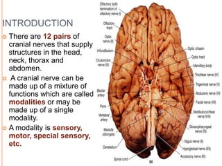
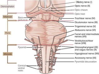
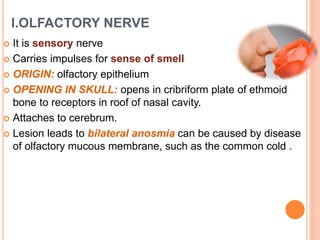
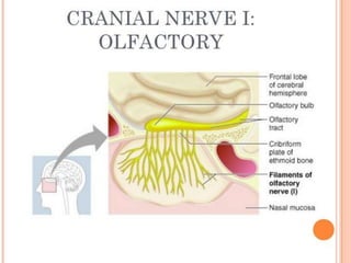
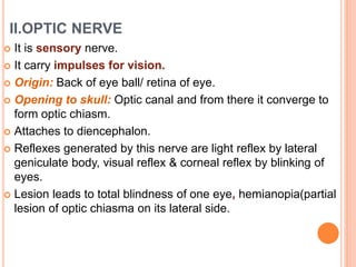
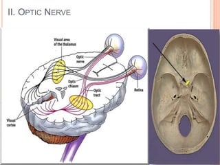
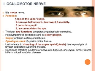
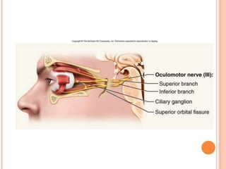
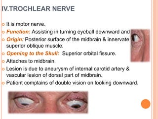
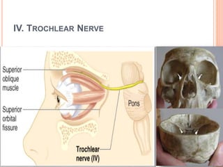
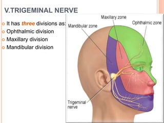
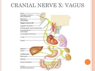
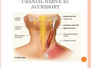
 There are 12 pairs of
cranial nerves that supply
structures in the head,
neck, thorax and
abdomen.
 A cranial nerve can be
made up of a mixture of
functions which are called
modalities or may be
made up of a single
modality.
 A modality is sensory,
motor, special sensory,
etc.
I.OLFACTORY NERVE
 It is sensory nerve
 Carries impulses for sense of smell
 ORIGIN: olfactory epithelium
 OPENING IN SKULL: opens in cribriform plate of ethmoid
bone to receptors in roof of nasal cavity.
 Attaches to cerebrum.
 Lesion leads to bilateral anosmia can be caused by disease
of olfactory mucous membrane, such as the common cold .
II.OPTIC NERVE
 It is sensory nerve.
 It carry impulses for vision.
 Origin: Back of eye ball/ retina of eye.
 Opening to skull: Optic canal and from there it converge to
form optic chiasm.
 Attaches to diencephalon.
 Reflexes generated by this nerve are light reflex by lateral
geniculate body, visual reflex & corneal reflex by blinking of
eyes.
 Lesion leads to total blindness of one eye, hemianopia(partial
lesion of optic chiasma on its lateral side.
II. OPTIC NERVE
III.OCULOMOTOR NERVE
 It is motor nerve.
 Function:
1.raises the upper eyelid.
2.turn eye ball upward, downward & medially.
3.constricts pupil.
4. accommodates the eye
o The later two functions are parasympathetically controlled.
o Parasympathetic cell bodies are in ciliary ganglia.
o Origin: anterior surface of midbrain.
o Opening in skull: Superior orbital fissure.
o Lesion leads to drooping of the upper eyelid(ptosis) due to paralysis of
levator palpebrae superioris muscle.
o Conditions effecting oculomotor nerve are diabetes, aneurysm, tumor, trauma,
inflammation& vascular disease
IV.TROCHLEAR NERVE
 It is motor nerve.
 Function: Assisting in turning eyeball downward and laterally
 Origin: Posterior surface of the midbrain & innervate the
superior oblique muscle.
 Opening to the Skull: Superior orbital fissure.
 Attaches to midbrain.
 Lesion is due to aneurysm of internal carotid artery &
vascular lesion of dorsal part of midbrain.
 Patient complains of double vision on looking downward.
IV. TROCHLEAR NERVE
V.TRIGEMINAL NERVE
 It has three divisions as:
 Ophthalmic division
 Maxillary division
 Mandibular division
V1.OPHTHALAMIC NERVE
 It is sensory.
 Function:
cornea, skin of forehead, scalp,eyelids,nose, also
mucous membrane of paranasal sinuses & nasal cavity.
 Origin: Anterior aspect of pons.
 Opening in skull: Superior orbital fissure.
 Exit orbit through supra orbital foramen.
 In lesion of this nerve cornea & conjunctiva will be insensitive
to touch.
V1. OPHTHALMIC NERVE
Infratrochlear
V1. Ophthalmic Nerve
V2.MAXILLARY NERVE
 It is sensory nerve.
 Function:
skin of face over maxilla.
Teeth of upper jaw.
Mucous membrane of nose , the maxillary sinus &
palate.
 Origin: Anterior aspect of pons.
 Opening in skull: Foramen rotundum
 Exit through infraorbital foramen.
V2. Maxillary Nerve
V3. MANDIBULAR NERVE
o Component: a. Motor
o Function:
o Muscles of mastication
o Mylohyoid
o Anterior belly of digastric
o Tensor veli palatine
o Tensor tympani
o Opening to the Skull: Foramen ovale
o Origin: Anterior aspect of the pons
V3. MANDIBULAR NERVE
• Component: b. Sensory
• Function:
• Skin of cheek
• Skin over mandible and side of head
• Teeth of lower jaw and TMJ
• Mucous membrane of mouth and anterior part of
tongue
• Opening to the Skull: Foramen ovale
• Origin: Anterior aspect of the pons
V3. Mandibular Nerve
VI.ABDUCENT NERVE
 It is motor nerve
 Function: Lateral rectus muscle turns eyeball laterally
 Origin: Anterior Surface of hindbrain between pons and
medulla
 Opening to the Skull: Superior orbital fissure
 Fibers leaves the inferior pons & enter orbit via superior
orbital fissure.
 Patient can’t turn the eye laterally.
 Lesions include damage due to head injuries, cavernous
sinus thrombosis or aneurysm of internal carotid artery &
vascular lesions of pons.
VI. Abducent Nerve
VII.FACIAL NERVE
 It is mixed nerve.
 Function:-
 Motor: Muscles of face & scalp, stapedius muscle, posterior
belly of digastric& stylohoid muscle.
 Sensory: Taste from anterior 2/3rd of tongue, from floor of
mouth & palate.
 Secretomotor parasympathetic: submandibular & sublingual
salivary glands, the lacrimal gland & glands of nose & palate.
 Opening in the skull: internal acoustic meatus, facial &
stylomostoid foramen.
 Attaches to pons.
 Effects of damage: inability to control facial muscles; distorted
sense of taste.
BELL’S PALSY
 Paralysis of facial muscles of affected side
 Loss of taste sensation
 Caused by herpes simplex virus.
 Lower eyelids droops.
 Corner of mouth sags.
 Tears drips continuously & eye cannot be
completely closed.
 Condition may disappear spontaneously without
treatment.
VII.VESTIBULOCOCHLEAR
 VESTIBULAR NERVE:-
 It is sensory nerve.
 Function: Position & movement of head.
 Opening in the skull: Internal acoustic meatus
 COCHLEAR NERVE:-
 It is sensory nerve.
 Function: Organ of corti_ hearing.
 Opening in the skull: internal acoustic meatus.
AUDITORY PATHWAY
AUDITORY PATHWAY
SYMPTOMS
 Damage to the vestibulocochlear nerve may cause the
following symptoms:
 hearing loss
 vertigo
 false sense of motion
 loss of equilibrium (in dark places)
 nystagmus
 motion sickness
 gaze-evoked tinnitus.
PATHOLOGIES
 Hearing loss
 Central auditory processing disorders (CAPD)
 Tinnitus
 Presbycusis
 Nystagmus
XI.GLOSSOPHARYNGEAL
 It is mixed nerve.
 Function:-
 Motor: Stylopharyngeus muscle assists swallowing.
 Secretomotor parasympathetic: parotid salivary
gland.
 Sensory: general sensation & taste from posterior
1/3of tongue & pharynx, carotid sinus & carotid
body(chemoreceptor).
 Opening in the skull: Jugular foramen.
 It attaches to medulla oblongata.
 Clinical tests: gag reflex, swallowing, and
coughing
 Effects of damage: difficulty swallowing
X.VAGUS
 It is mixed nerve.
 Nick name: Wanderer.
 Function:-
 Motor & sensory: Heart & great thoracic blood
vessels , larynx, trachea, bronchi & lungs, alimentary
tract from pharynx to splenic flexure of colon, liver,
kidneys & pancreas.
 Opening in the skull: Jugular foramen.
 Effects of damage: hoarseness or loss of voice;
dysphagia, cardiovascular problems, digestive
problems, urinary incontinence, deafness, palatal
function, gag reflex, spastic dysarthria.
 Gag Reflex:
 A normal reflex consisting of elevation of the palate, retraction
of the tongue, and contraction of the throat muscles.
BRANCHES OF VAGUS NERVE
 Auricular nerve
 Pharyngeal nerve
 Superior laryngeal nerve
 Superior cervical cardiac branches of vagus nerve
 Inferior cervical cardiac branch
 Recurrent laryngeal nerve
 Thoracic cardiac branches
 Branches to the pulmonary plexus
 Branches to the esophageal plexus
 Anterior vagal trunk
 Posterior vagal trunk
XI.ACCESSORY
 It is motor nerve.
 Formed from cranial root emerging from medulla & a
spinal root arising from the superior region of spinal cord.
 Function: swallowing; head, neck, and shoulder
movements.
 Opening in the skull: Jugular foramen.
 Spinal root passes upward into cranium via the foramen
magnum.
 Accessory nerve leaves the foramen via the jugular
foramen.
 Clinical tests: rotate head and shrug shoulders against
resistance
 Effects of damage: impaired movement of head, neck,
and shoulders; paralysis of sternocleidomastoid
XII.HYPOGLOSSAL
 It is motor nerve.
 Function:- Muscles of tongue (except
palatoglossus) controlling its shape & movement.
 Opening in the skull: Hypoglossal canal.
 Innervates both extrinsic & intrinsic muscles of
tongue.
 Clinical test: tongue function
 Effects of damage: difficulty in speech and
swallowing; atrophy of tongue; inability to stick out
(protrude) tongue
 Lesions occur from demyelinating diseases &
vascular accidents.
Cranial Nerves
Cranial Nerves

Cranial Nerves

  • 1.
  • 2.
    INTRODUCTION  There are12 pairs of cranial nerves that supply structures in the head, neck, thorax and abdomen.  A cranial nerve can be made up of a mixture of functions which are called modalities or may be made up of a single modality.  A modality is sensory, motor, special sensory, etc.
  • 6.
    I.OLFACTORY NERVE  Itis sensory nerve  Carries impulses for sense of smell  ORIGIN: olfactory epithelium  OPENING IN SKULL: opens in cribriform plate of ethmoid bone to receptors in roof of nasal cavity.  Attaches to cerebrum.  Lesion leads to bilateral anosmia can be caused by disease of olfactory mucous membrane, such as the common cold .
  • 8.
    II.OPTIC NERVE  Itis sensory nerve.  It carry impulses for vision.  Origin: Back of eye ball/ retina of eye.  Opening to skull: Optic canal and from there it converge to form optic chiasm.  Attaches to diencephalon.  Reflexes generated by this nerve are light reflex by lateral geniculate body, visual reflex & corneal reflex by blinking of eyes.  Lesion leads to total blindness of one eye, hemianopia(partial lesion of optic chiasma on its lateral side.
  • 9.
  • 10.
    III.OCULOMOTOR NERVE  Itis motor nerve.  Function: 1.raises the upper eyelid. 2.turn eye ball upward, downward & medially. 3.constricts pupil. 4. accommodates the eye o The later two functions are parasympathetically controlled. o Parasympathetic cell bodies are in ciliary ganglia. o Origin: anterior surface of midbrain. o Opening in skull: Superior orbital fissure. o Lesion leads to drooping of the upper eyelid(ptosis) due to paralysis of levator palpebrae superioris muscle. o Conditions effecting oculomotor nerve are diabetes, aneurysm, tumor, trauma, inflammation& vascular disease
  • 12.
    IV.TROCHLEAR NERVE  Itis motor nerve.  Function: Assisting in turning eyeball downward and laterally  Origin: Posterior surface of the midbrain & innervate the superior oblique muscle.  Opening to the Skull: Superior orbital fissure.  Attaches to midbrain.  Lesion is due to aneurysm of internal carotid artery & vascular lesion of dorsal part of midbrain.  Patient complains of double vision on looking downward.
  • 13.
  • 14.
    V.TRIGEMINAL NERVE  Ithas three divisions as:  Ophthalmic division  Maxillary division  Mandibular division
  • 15.
    V1.OPHTHALAMIC NERVE  Itis sensory.  Function: cornea, skin of forehead, scalp,eyelids,nose, also mucous membrane of paranasal sinuses & nasal cavity.  Origin: Anterior aspect of pons.  Opening in skull: Superior orbital fissure.  Exit orbit through supra orbital foramen.  In lesion of this nerve cornea & conjunctiva will be insensitive to touch.
  • 16.
  • 17.
  • 18.
    V2.MAXILLARY NERVE  Itis sensory nerve.  Function: skin of face over maxilla. Teeth of upper jaw. Mucous membrane of nose , the maxillary sinus & palate.  Origin: Anterior aspect of pons.  Opening in skull: Foramen rotundum  Exit through infraorbital foramen.
  • 19.
  • 20.
    V3. MANDIBULAR NERVE oComponent: a. Motor o Function: o Muscles of mastication o Mylohyoid o Anterior belly of digastric o Tensor veli palatine o Tensor tympani o Opening to the Skull: Foramen ovale o Origin: Anterior aspect of the pons
  • 21.
    V3. MANDIBULAR NERVE •Component: b. Sensory • Function: • Skin of cheek • Skin over mandible and side of head • Teeth of lower jaw and TMJ • Mucous membrane of mouth and anterior part of tongue • Opening to the Skull: Foramen ovale • Origin: Anterior aspect of the pons
  • 22.
  • 23.
    VI.ABDUCENT NERVE  Itis motor nerve  Function: Lateral rectus muscle turns eyeball laterally  Origin: Anterior Surface of hindbrain between pons and medulla  Opening to the Skull: Superior orbital fissure  Fibers leaves the inferior pons & enter orbit via superior orbital fissure.  Patient can’t turn the eye laterally.  Lesions include damage due to head injuries, cavernous sinus thrombosis or aneurysm of internal carotid artery & vascular lesions of pons.
  • 24.
  • 25.
    VII.FACIAL NERVE  Itis mixed nerve.  Function:-  Motor: Muscles of face & scalp, stapedius muscle, posterior belly of digastric& stylohoid muscle.  Sensory: Taste from anterior 2/3rd of tongue, from floor of mouth & palate.  Secretomotor parasympathetic: submandibular & sublingual salivary glands, the lacrimal gland & glands of nose & palate.  Opening in the skull: internal acoustic meatus, facial & stylomostoid foramen.  Attaches to pons.  Effects of damage: inability to control facial muscles; distorted sense of taste.
  • 26.
    BELL’S PALSY  Paralysisof facial muscles of affected side  Loss of taste sensation  Caused by herpes simplex virus.  Lower eyelids droops.  Corner of mouth sags.  Tears drips continuously & eye cannot be completely closed.  Condition may disappear spontaneously without treatment.
  • 28.
    VII.VESTIBULOCOCHLEAR  VESTIBULAR NERVE:- It is sensory nerve.  Function: Position & movement of head.  Opening in the skull: Internal acoustic meatus  COCHLEAR NERVE:-  It is sensory nerve.  Function: Organ of corti_ hearing.  Opening in the skull: internal acoustic meatus.
  • 30.
  • 31.
  • 32.
    SYMPTOMS  Damage tothe vestibulocochlear nerve may cause the following symptoms:  hearing loss  vertigo  false sense of motion  loss of equilibrium (in dark places)  nystagmus  motion sickness  gaze-evoked tinnitus.
  • 33.
    PATHOLOGIES  Hearing loss Central auditory processing disorders (CAPD)  Tinnitus  Presbycusis  Nystagmus
  • 34.
    XI.GLOSSOPHARYNGEAL  It ismixed nerve.  Function:-  Motor: Stylopharyngeus muscle assists swallowing.  Secretomotor parasympathetic: parotid salivary gland.  Sensory: general sensation & taste from posterior 1/3of tongue & pharynx, carotid sinus & carotid body(chemoreceptor).  Opening in the skull: Jugular foramen.  It attaches to medulla oblongata.  Clinical tests: gag reflex, swallowing, and coughing  Effects of damage: difficulty swallowing
  • 36.
    X.VAGUS  It ismixed nerve.  Nick name: Wanderer.  Function:-  Motor & sensory: Heart & great thoracic blood vessels , larynx, trachea, bronchi & lungs, alimentary tract from pharynx to splenic flexure of colon, liver, kidneys & pancreas.  Opening in the skull: Jugular foramen.  Effects of damage: hoarseness or loss of voice; dysphagia, cardiovascular problems, digestive problems, urinary incontinence, deafness, palatal function, gag reflex, spastic dysarthria.  Gag Reflex:  A normal reflex consisting of elevation of the palate, retraction of the tongue, and contraction of the throat muscles.
  • 37.
    BRANCHES OF VAGUSNERVE  Auricular nerve  Pharyngeal nerve  Superior laryngeal nerve  Superior cervical cardiac branches of vagus nerve  Inferior cervical cardiac branch  Recurrent laryngeal nerve  Thoracic cardiac branches  Branches to the pulmonary plexus  Branches to the esophageal plexus  Anterior vagal trunk  Posterior vagal trunk
  • 39.
    XI.ACCESSORY  It ismotor nerve.  Formed from cranial root emerging from medulla & a spinal root arising from the superior region of spinal cord.  Function: swallowing; head, neck, and shoulder movements.  Opening in the skull: Jugular foramen.  Spinal root passes upward into cranium via the foramen magnum.  Accessory nerve leaves the foramen via the jugular foramen.  Clinical tests: rotate head and shrug shoulders against resistance  Effects of damage: impaired movement of head, neck, and shoulders; paralysis of sternocleidomastoid
  • 41.
    XII.HYPOGLOSSAL  It ismotor nerve.  Function:- Muscles of tongue (except palatoglossus) controlling its shape & movement.  Opening in the skull: Hypoglossal canal.  Innervates both extrinsic & intrinsic muscles of tongue.  Clinical test: tongue function  Effects of damage: difficulty in speech and swallowing; atrophy of tongue; inability to stick out (protrude) tongue  Lesions occur from demyelinating diseases & vascular accidents.