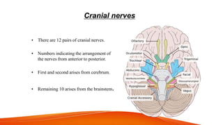
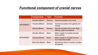
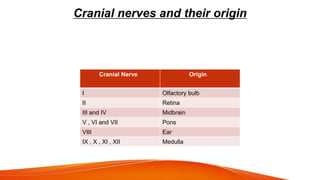
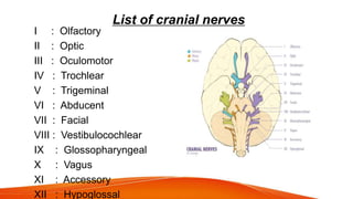
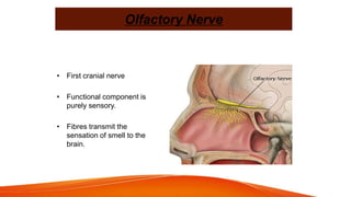
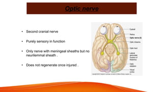
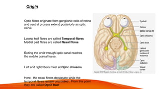
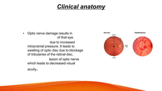
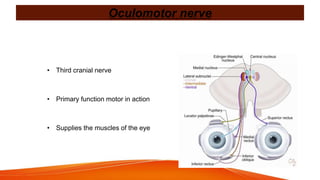
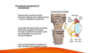
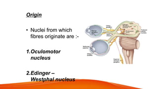
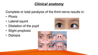
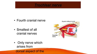
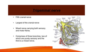
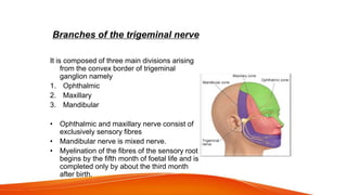
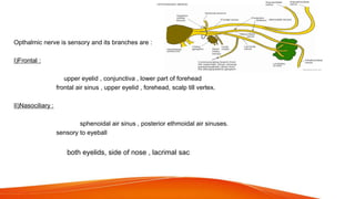
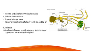
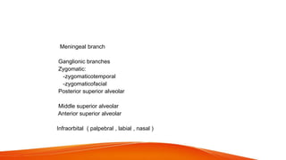
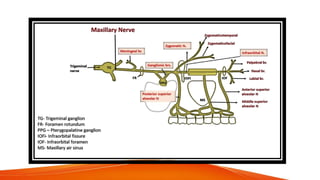
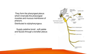
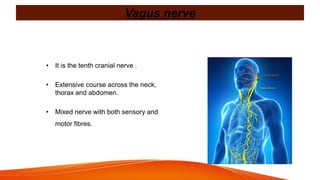
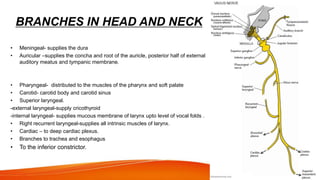
3. Introduction
• Cranial nerves originate directly from the brain.
• They reach the head and neck region by traversing
through numerous foramina present in base of skull.
• They can be motor, sensory or mixed .
4. DEFINITIONS
Nerve tract :
A bundle of myelinated nerve fibers that traverses the brain.
Ganglion :
Group of neuronal cell bodies sharing the same function, located outside
the CNS .
Afferent fibers :
Axons that carry impulses towards CNS, also called sensory fibres.
Efferent fibers :
Axons that carry impulses away from the CNS, also called motor fibres.
5. Cranial nerves
• There are 12 pairs of cranial nerves.
• Numbers indicating the arrangement of
the nerves from anterior to posterior.
• First and second arises from cerebrum.
• Remaining 10 arises from the brainstem.
6. Functional component of cranial nerves
Components Type Function
Somatic afferent Sensory General sensation from body
Visceral afferent Sensory General sensation from glands and
viscera
Special afferent Sensory Special sensation like smell, taste,
hearing, sight and balance
Somatic efferent Motor Motor supply to muscles derived
from somites
Visceral efferent Motor Motor supply to muscles derived
from branchial arches
Branchial efferent Motor Autonomic motor to smooth muscles
and glands
AFFERENT
EFFERENT
7. Cranial nerves and their origin
Cranial Nerve Origin
I Olfactory bulb
II Retina
III and IV Midbrain
V , VI and VII Pons
VIII Ear
IX , X , XI , XII Medulla
8. List of cranial nerves
I : Olfactory
II : Optic
III : Oculomotor
IV : Trochlear
V : Trigeminal
VI : Abducent
VII : Facial
VIII : Vestibulocochlear
IX : Glossopharyngeal
X : Vagus
XI : Accessory
XII : Hypoglossal
9. Olfactory Nerve
• First cranial nerve
• Functional component is
purely sensory.
• Fibres transmit the
sensation of smell to the
brain.
10. Origin
Axons originates as network
from the olfactory cells
Olfactory cells location : superior and posterior
region of lateral nasal wall and septum
Central process of olfactory cells continues as axon and
group themselves into 15 to 20 bundles .
Pierces the cribriform plate of ethmoid bone , terminating
in glomeruli of olfactory bulb in Anterior cranial fossa
Olfactory bulb leads into olfactory tract ending in sensory
cortex
11. Clinical anatomy
• ANOSMIA: loss of olfactory fibres with ageing
• Allergic rhinitis causes temporary olfactory impairment.
• Olfactory bulbs maybe torn away from olfactory nerves on
head injury as these pass through the fractured cribriform
plate of ethmoid leading to anosmia.
• Abscess of frontal lobe can also lead to anosmia.
12. Optic nerve
• Second cranial nerve
• Purely sensory in function
• Only nerve with meningeal sheaths but no
neurilemmal sheath .
• Does not regenerate once injured .
13. Origin
Optic fibres originate from ganglionic cells of retina
and central process extend posteriorly as optic
nerve
Lateral half fibres are called Temporal fibres
Medial part fibres are called Nasal fibres
Exiting the orbit through optic canal reaches
the middle cranial fossa.
Left and right fibers meet at Optic chiasma
Here , the nasal fibres decussate while the
temporal fibres remain uncrossed . From this point
they are called Optic tract
14. Clinical anatomy
• Optic nerve damage results in
complete blindness of that eye.
• Papilloedema: due to increased
intracranial pressure. It leads to
swelling of optic disc due to blockage
of tributaries of the retinal disc.
• Optic neuritis: lesion of optic nerve
which leads to decreased visual
acuity.
15. Oculomotor nerve
• Third cranial nerve
• Primary function motor in action
• Supplies the muscles of the eye
16. Functional component of
oculomotor :
General somatic efferent fibres :
Extraocular muscles except
Superior oblique and Lateral rectus
, Levator palpebrae superioris
General visceral efferent fibres :
innervate the intraocular muscles,
sphincter pupillae and ciliaris
muscle to perform functions such
as contraction of pupil and
accommodation
General somatic afferent fibres :
carry proprioceptive impulses of
eye muscles and relayed in brain
17. Origin
• Nuclei from which
fibres originate are :-
1.Oculomotor
nucleus
2.Edinger –
Westphal nucleus
18. Clinical anatomy
Complete or total paralysis of the third nerve results in:
• Ptosis
• Lateral squint
• Dilatation of the pupil
• Slight proptosis
• Diplopia
19. Trochlear nerve
• Fourth cranial nerve
• Smallest of all
cranial nerves
• Only nerve which
arises from
dorsal aspect of the
20. Functional components of trochlear
General somatic efferent :
Superior oblique muscle helps in lateral movement of
eyeball.
General somatic afferent :
Sensory fibres from the muscle that get relayed to the
mesencephalic nucleus.
Clinical anatomy
Damaged trochlear nerve : diplopia occurs on looking
downwards
21. Trigeminal nerve
• Fifth cranial nerve
• Largest of the cranial nerve
• Mixed nerve carrying both sensory
and motor fibres.
• Comprises of three branches, two of
which are purely sensory and the
third is a mixed nerve.
22. Functional component of trigeminal nerve
• Sensory component includes sensations of pain, temperature, touch
and pressure from the skin of face and mucous membrane of nose,
most of the tongue and paranasal air sinuses.
• Motor componenets for the muscles:
Supply 4 muscles of mastication:
-lateral pterygoid muscle
-medial pterygoid muscle
-Temporalis
-Masseter
And four other muscles:
Tensor veli palatini, tensor tympani, mylohyoid and anterior belly of
digastric.
23. The fibres relay in the following
three nuclei:-
• Main sensory nucleus:
light touch fibres end here
• Spinal nucleus:
pain and temperature fibres
terminate here
• Mesencephalic nucleus:
proprioceptive fibres
terminate here
• Motor nucleus:
motor fibres begin here.
24. Branches of the trigeminal nerve
It is composed of three main divisions arising
from the convex border of trigeminal
ganglion namely
1. Ophthalmic
2. Maxillary
3. Mandibular
• Ophthalmic and maxillary nerve consist of
exclusively sensory fibres
• Mandibular nerve is mixed nerve.
• Myelination of the fibres of the sensory root
begins by the fifth month of foetal life and is
completed only by about the third month
after birth.
25. Opthalmic nerve
Opthalmic nerve is sensory and its branches are :
I)Frontal :
Supratrochlear : upper eyelid , conjunctiva , lower part of forehead
Supraorbital : frontal air sinus , upper eyelid , forehead, scalp till vertex.
II)Nasociliary :
Posterior ethmoidal : sphenoidal air sinus , posterior ethmoidal air sinuses.
Long ciliary : sensory to eyeball
Branch to ciliary ganglion
Infratrochlear : both eyelids, side of nose , lacrimal sac
26. Anterior ethmoidal
• Middle and anterior ethmoidal sinuses
• Medial internal nasal
• Lateral internal nasal
• External nasal : skin of ala of vestibule and tip of nose.
III)Lacrimal
Lateral part of upper eyelid , conveys secretomotor fibres from
zygomatic nerve to lacrimal gland.
27. Maxillary nerve division
• In middle cranial fossa :
Meningeal branch
• In pterygopalatine fossa :
Ganglionic branches
Zygomatic:
-zygomaticotemporal
-zygomaticofacial
Posterior superior alveolar
• In Infraorbital canal :
Middle superior alveolar
Anterior superior alveolar
• On face :
Infraorbital ( palpebral , labial , nasal )
30. Posterior divison :
• Auriculotemporal
• Lingual nerve :
General sensation from
anterior two third of tongue.
• Inferior alveolar nerve :lower
teeth, skin of chin.
-Nerve to mylohyoid
-Anterior belly of
digastric
31. Clinical anatomy :
• Fifth cranial nerve sub serves sensation from face and
neighboring areas. It also innervates the muscles of
mastication.
• Trigeminal ganglion can harbour the herpes zoster virus
causing shingles along the distribution of the nerve.
• Nasopalatine nerves (branch of maxillary
nerve) and vessels are present near nasopalatine
foramen, hence this area needs to be relieved
while taking impressions or else it will lead
to paraesthesia.
32. TRIGEMINAL NEURALGIA
• Affects the sensory root of V
nerve characterised by
attacks of severe pain in the
area of distribution of
maxillary and mandibular
divisions. Maxillary nerve is
most frequently involved.
33. • Hypoacusis : partial deafness to low pitched sounds due to paralysis of tensor tympani
muscles
• Flaccid paralysis of muscles of mastication in the injury of mandibular nerve leading to
decreased strength for biting .
• In injury to :-
-ophthalmic nerve: loss of corneal blink reflex
-maxillary nerve: loss of sneeze reflex
-mandibular nerve: loss of jaw jerk reflex
34. Abducent nerve
• Sixth cranial nerve
• Purely motor in function
• Supply extra ocular muscles called lateral
rectus
35. Functional component of
Abducent nerve
General somatic efferent :
These are motor fibres for lateral rectus muscle
to help in lateral movement of eyeball
General somatic afferent :
Sensory fibres from the muscle which carry
proprioceptive impulses, relayed to the
mesencephalic nucleus.
36. Facial nerve
• Seventh cranial nerve
• Mixed nerve containing both sensory and
motor fibres.
37. Functional component of facial nerve
1. Special visceral/Branchial efferent fibres: muscles for facial
expressions, elevation of hyoid bone.
2. General visceral efferent fibres : For carrying secretomotor
impulses to submandibular, sublingual salivary glands, lacrimal,
nasal glands, palate and pharynx.
3. Special visceral afferent nerve : For carrying taste sensation from
the anterior two third of tongue and palate.
4. General somatic afferent fibres : For carrying proprioceptive
impulses from muscles of face to reach mesencephalic nucleus.
38. NUCLEI
• Four nuclei in the lower pons contribute to the facial nerve formation.
1. Motor nucleus
2. Nucleus of tractus solitarius
3. Superior salivatory nucleus
4. Lacrimatory nucleus
• VII carries fibres from lacrimatory nucleus to pterygopalatine ganglion
for the lacrimal gland and glands in nasal cavity, palate and pharynx
• VII also carries fibres from superior salivatory nucleus to
submandibular ganglion for supply of submandibular, sublingual and
glands in oral cavity
39.
40. Branches
WITHIN THE FACIAL CANAL
• Greater petrosal nerve
• Nerve to the stapedius
• Chorda tympani nerve
AT ITS EXIT FROM STYLOMASTOID FORAMEN
• Posterior auricular – two branches-auricular and occipital
• Digastric-posterior belly of digastric
• Stylohyoid-stylohyoid muscle
TERMINAL BRANCHES WITHIN PAROTID
• Temporal-frontalis , orbicularis oculi and corrugator muscle and joins the lacrimal
branch of ophthalmic nerve.
• Zygomatic-orbicularis oculi
• Marginal mandibular-muscles of the lower lip and chin
• Cervical –platysma muscle
COMMUNICATING BRANCHES WITH ADJACENT CRANIAL AND SPINAL
NERVES
41. • Bells palsy : sudden paralysis of facial
nerve at the stylomastoid foramen.
42. Clinical anatomy
• Lesion above the origin of chorda tympani nerve will show symptoms of bells
palsy plus loss of taste sensation from anterior 2/3rd of tongue except vallate
papillae.
• Lesion above the origin of nerve to stapedius will cause all the above
symptoms with hyperacusis.
43. Vestibulocochlear nerve
• Eighth cranial nerve
• Purely sensory nerve with two different
types of fibres:-
1. Vestibular nerve ( nerve of balance )
2. Cochlear nerve ( nerve of hearing )
• Functional component :
Special somatic afferent for hearing and
equilibrium of the head.
44. Vestibular nerve
• It is the nerve of equilibration .
• The bipolar neurons which gives rise to this nerve are located in the vestibular
ganglion.
• Ganglion is present in the outer end of the internal acoustic meatus.
• Four vestibular nuclei : superior, spinal, medial, lateral exist where the vestibular
nerve fibres are relayed.
45. Cochlear nerve
• It is the nerve of hearing
• Fibres arise from bipolar cells in spiral ganglion of cochlea present in the
osseous spiral lamina of the ear.
• Nerve runs along the internal auditory meatus along with the vestibular
nerve and across the subarachnoid space and terminates in the cochlear
nucleus.
46. Glossopharyngeal nerve
• It is the ninth cranial nerve.
• Mixed nerve containing both motor and
sensory fibres.
• Innervates the tongue and pharynx.
• Carries general sensation from mucous
membrane of pharynx, fauces and palatine
tonsil and taste sensation from posterior
part of tongue.
47. Functional component of glossopharyngeal nerve
• Special visceral efferent fibres(SVE) : supply the stylopharyngeus
muscle.
• General visceral efferent fibres (GVE): supply the parotid gland.
• Special visceral afferent , general visceral afferent and general
somatic afferent fibres : carry sensations from posteior 1/3rd of
the tongue and sensation from mucous membrane of pharynx.
48. NUCLEI
Three nuclei in medulla give rise to fibres :
1. Nucleus ambiguus
2. Inferior salivatory nucleus
3. Nucleus of tractus solitarius
IX nerve carries fibres from inferior
salivatory nucleus to the otic ganglion for
supply of parotid gland.
49. Branches
Tympanic nerve : Nerve of jacobson
• Emerges from petrous ganglion and
reaches the tympanic cavity
• Within the cavity , splits into numerous
branches to form the tympanic plexus
supplying mucous membrane of
tympanic cavity and mastoid air cells.
• Innervate the trunk of internal carotid
artery
50. Ph They form the pharyngeal plexus
which innervate the pharyngeal
muscles and mucous membrane of
pharynx.
Distributed to stylopharyngeus.
• bSupply palatine tonsil , soft palate
and fauces through a tonsillar plexus
.
51. Clinical anatomy :
Lesion of this nerve causes :
• Absence of secretions from parotid gland
• Absence of taste from posterior one third of tongue
• Loss of pain sensation from tongue, tonsil, pharynx and soft palate .
• Gag reflex is absent
• Pharyngitis may cause referred pain to ear as both are supplied by
IX nerve.
52. Vagus nerve
• It is the tenth cranial nerve .
• Extensive course across the neck,
thorax and abdomen.
• Mixed nerve with both sensory and
motor fibres.
53. Functional component of vagus
Special efferent fibres : For the movement of palate, pharynx and
larynx.
General visceral efferent fibres (GVE): distributed to thoracic and
abdominal viscera.
General visceral afferent fibres(GVA) : For sensation from pharynx,
larynx, trachea, oesophagus and from abdominal and thoracic
viscera.
Special visceral afferent fibres : For taste from the posterior most
region of tongue and epiglottis
General somatic afferent fibres : distributed to the skin of the external
ear .
54. NUCLEI
• Nucleus ambiguus
• Dorsal nucleus of vagus
• Nucleus of tractus solitarius
• Nucleus of spinal tract of trigeminal
X nerve carries fibres from dorsal nucleus of vagus for the glands
in the respiratory tract and glands in digestive tract till right two-
thirds of the transverse colon.
55. BRANCHES IN HEAD AND NECK
FROM SUPERIOR GANGLION
• Meningeal- supplies the dura
• Auricular –supplies the concha and root of the auricle, posterior half of external
auditory meatus and tympanic membrane.
FROM INFERIOR GANGLION
• Pharyngeal- distributed to the muscles of the pharynx and soft palate
• Carotid- carotid body and carotid sinus
• Superior laryngeal.
-external laryngeal-supply cricothyroid
-internal laryngeal- supplies mucous membrane of larynx upto level of vocal folds .
• Right recurrent laryngeal-supplies all intrinsic muscles of larynx.
• Cardiac – to deep cardiac plexus.
• Branches to trachea and esophagus
• To the inferior constrictor.
56. Clinical anatomy
• The vagus nerve is tested clinically by comparing the
palatal arches on the two sides. On the paralysed side,
there is no arching and uvula is pulled to the normal side.
• Paralysis of vagus nerve produces:
Nasal regurgitation of swallowed liquids
Nasal twang in voice
Hoarseness of voice
Flattening of palatal arch
Dysphagia
57. Spinal accessory nerve
• Eleventh cranial nerve
• Purely motor in function
• Two roots:
Cranial root : Adjunct to vagus nerve and
supplies the muscles of palate , pharynx ,
larynx and heart .
Spinal root : Supplies sternocleidomastoid and
trapezius muscle. More independent course .
58. Functional components
• Cranial root is special visceral efferent: distributed
through branches of vagus to palate, pharynx, larynx and
possibly the heart.
• Spinal root is also special visceral efferent: supplies
sternocledomastoid and trapezius.
NUCLEI:
Cranial root arises from nucleus ambiguus
Spinal root arises from spinal nucleus.
59. Clinical anatomy
• Lesions of spinal root cause drooping
of shoulder and inability to turn chin to
opposite side
• Tested clinically
-by asking patients to shrug shoulders
against resistance to the opposite side
and comparing the power on both sides.
Or
-by turning the chin to opposite side
against resistance and comparing both
sides
60. Hypoglossal nerve
• Twelfth cranial nerve
• Exclusively motor function
• Major nerve supplying the
muscle of the tongue.
61. Functional component of hypoglossal nerve
• General somatic efferent fibres :
Motor fibres arising from the hypoglossal nucleus and innervate all
muscles of tongue except the palatoglossus.
• General somatic afferent fibres :
the nucelus is mesencephalic nucleus of fifth cranial nerve where
proprioceptive fibres from the tongue end.
• NUCLEI: hypoglossal nuclei
62. Branches of hypoglossal nerve
• Innervating fibres of hypoglossal
nerve may be categorised into two :
1. Branches of hypoglossal nerve
proper
2. Branches containing the spinal nerve
C1
63. Branches of the hypoglossal nerve proper
• Thin delicate muscular branch innervating from the inferior
surface of tongue
• Extrinsic muscles : Styloglossus, Genioglossus, Hyoglossus
• Intrinsic muscles : Superior longitudinal, inferior longitudinal,
transverse and vertical
64.
65. Branches containing fibres of nerve C1
• Meningeal branch: contains sensory fibres. Enters
skull through hypoglossal canal and supplies bone
and meninges of posterior cranial fossa.
• Descending branch: continues as descendans
hypoglossi
• Branches are also given to thyrohyoid and
geniohyoid muscle.
66. Clinical anatomy
• Hypoglossal nerve is tested clinically by asking the patient
to protrude his/her tongue. Normally the tongue protrudes
straight. If nerve is paralysed, tongue deviates to
paralyzed side.
• Infranuclear lesion: paralysis of tongue on that side.
Gradual atrophy of paralyzed half of tongue. tongue looks
shrunken.
• Supranuclear lesion: Paralysis without wasting. Tongue
moves sluggishly producing defective speech. On
protrusion tongue deviates to opposite side.
67. Prosthodontic consideration
• Moebius/Mӧbius Syndrome (MS) is an extremely rare congenital
disorder characterized by uni- or bilateral palsy of the abducens (VI)
and the facial (VII) nerves, causing facial paralysis. Dysfunction of
cranial nerves III through XII is common, most often the
glossopharyngeal (IX) and hypoglossus (XII) are involved.
• Patients with Moebius syndrome seeking prosthodontic care present
multiple challenges, particularly in removable prosthodontics.
• This is mainly due to the paralysis of all facial muscles. Small mouth
opening, speech difficulties, and compromised neuromuscular
control present a challenge to successful restoration.
Condition 1 : Moebius
syndrome
68.
69. • Maxillary overdentures supported by two implants might be a
minimally invasive treatment alternative worth consideration in
specific patients, but the increased risk for bone loss needs to be
considered and might have a negative effect on the clinical long-term
outcome of this treatment option.
• Lack of muscle tone and extremely flaccid lips, coupled with the
need for a maxillary removable complete denture prosthesis,
presented unique challenges that required alteration of traditional
denture fabrication techniques. One particularly important deviation
from normal denture fabrication was the removal of the facial flange
of the maxillary complete denture to provide lip competence.
• Thus, monitoring over a longer period is certainly needed.
70. • The common clinical features of facial paralysis (LMN) are facial
asymmetry, drooping of the corner of mouth, inability to close/wink
the eye, loss of wrinkles of forehead, mask-like appearance, difficulty
in speech and eating .
• The denture bearing areas were captured in the impressions with
borders intentionally made thicker within the physiological limits, on
the affected side to support the flaccid musculature
• The occlusal plane was oriented parallel to a line joining the angles
of the functionally deviated mouth.
• The midline for trial dentures was marked in the center of deviated
mouth.
Condition 2 : Unilateral facial
paralysis
71. • The anterior teeth were set according to the shifted midline (almost 6
mm to the right), the posteriors were arranged accordingly and the
numbers of posterior teeth were not equal on both sides .
72. Condition-3:-Implant related nerve injury
• Nerve injuries in the maxillofacial region may happen as a result of
trauma, neoplasms, infections, or secondary to a surgical
procedure.
• Implant-related nerve injuries can be from drilling during the
osteotomy, placement of the implant, or both.
• If the site is not overprepared vertically, then it is unlikely that the
implant will enter the nerve canal and directly injure the
neurovascular bundle; this is true for normal to dense bone.
• In situations in which the bone quality is poor, implants may be
placed apical to the prepared osteotomy and directly compress or
injure the nerve .
73. • Careful preoperative planning and the availability of appropriate
radiographs are important not only for the proper placement of
implants for restorative reasons but also to avoid injuries to the
important structures in the surgical region.
• This possibility is important in the second premolar region where the
apex of the tooth socket may be close to the mental foramen.
74. CONCLUSION
Cranial nerves transmit information between the brain and
parts of the human body, especially to and from the head
and the neck area. Thus, a knowledge of cranial nerves is
important to prevent any nerve injuries related to dental
practice and to avoid and manage the same.
75. REFERENCES
• B D chaurasia’s , Human anatomy : Head and Neck (2013) vol 3,
CBS publishers , New delhi.c2018 ), ACP, LA.
• Prosthodontic management of a patient with Moebius syndrome: A
clinical report. By Ahmed Mahrous et al.
• Saumya Pandey, Kusum Datta , Prosthodontic management of a
completely edentulous patient with unilateral facial paralysis , (
2007) , vol7 , issue 4 , Amrister .
• Mark J. Steinberg , Implant-related Nerve Injuries ( 2014 ), DCNA .
Mesenceph nucleus- one of the 4 trigeminal nucelus.
Proprioception
Nerve of 1st brachial arch
Auriculotemporal: auricular, superficial temporal, articular to TMJ., secretomotor to parotid.
Reflex contraction of masseter muscle ,moving jaw briskly upwards
Nerve of 2nd brachial arch
Greater petrosal nerve : Contains sensory fibres to mucous membrane of the soft palate.
Nerve to the stapedius : Innervates the stapedius muscle.
Chorda tympani nerve :
Originates at approximately 6 mm from the stylomastoid foramen .
Carries efferent fibres receives from motor root which reached submandibular and sublingual gland.
Carries afferent fibres to innervate the mucous memebrane covering anterior two third of tongue.
LMN– paralysis of ipsilteral upper and lower half
UMN- contralateral lower quadrant
Ramsay hunt syndrome: facial palsy+ ear pain+vesicles in auditory canal . Antivirals+ STEROIDS
Abnormal sensitivity to sounds—hyperacusis
Cavity surrounding bones of middle ear-tymnpanic
In mastoid process of temporal bone—pneumatic structres . Can get infected