Downloaded 883 times


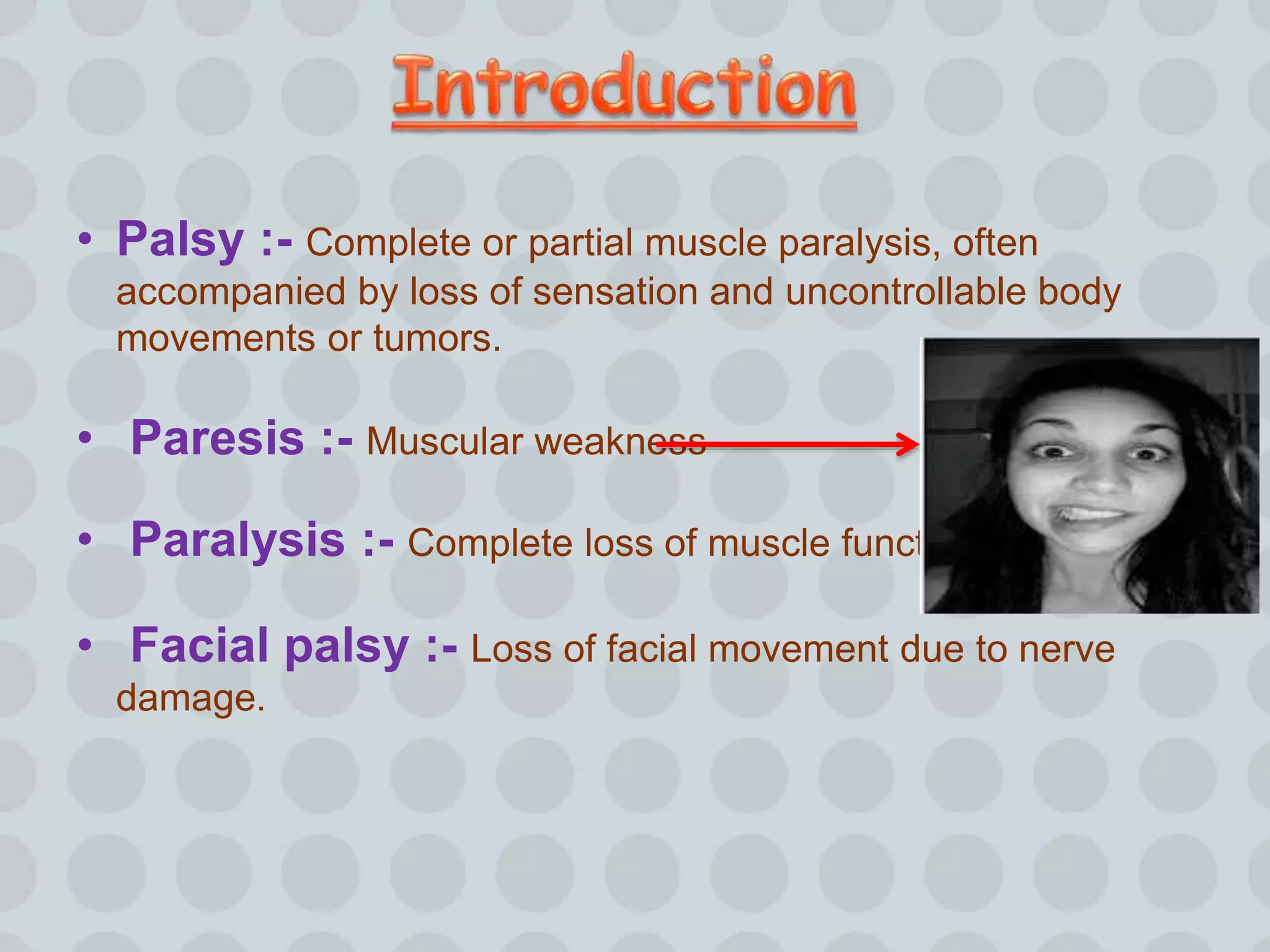


This document discusses facial palsy, also known as Bell's palsy. It is a condition that causes partial or complete paralysis of the facial nerve resulting in drooping of the facial muscles. The document outlines the types of facial palsy as central or peripheral, and the potential causes such as idiopathic (Bell's palsy), trauma, infection, tumors, or Guillain-Barre syndrome. Signs and symptoms, diagnostic tests like EMG and imaging, and treatment options including corticosteroids, antivirals, and eye care are summarized. The prognosis is generally good with 85% of idiopathic cases fully recovering within 3 weeks, but recovery can take longer or be incomplete


