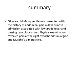
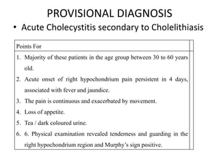
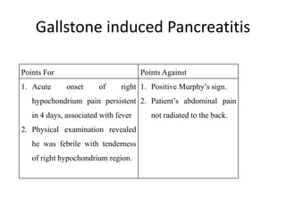
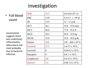
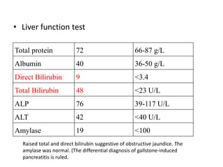
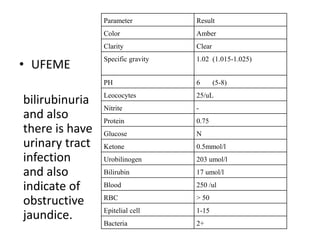
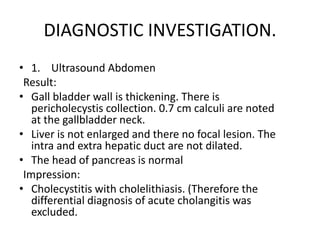
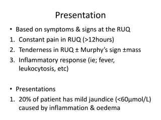
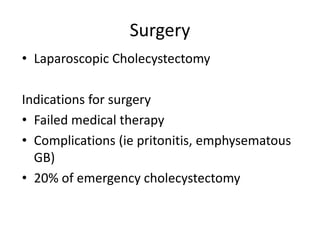
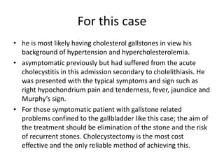
Cpc: abdomen
Siti hamidah binti mahbud
Year 4 MBBS student UniSZA
Chief complain
• right upper abdominal pain 4
days prior to admission.
history
• started 4 days ago when he suddenly developed
right upper abdominal pain. The pain was
intermittently and gripping in nature. The pain
was localized. He describes the duration of the
pain was about in half to 2 hours every attack
and relief by taking pain killer (ponstan).
According to him, the pain was aggravated by
changing his posture especially turning on his
right; during take deep breath or coughing. The
pain is not associated with meals.
• He also experienced several episode of low-
grade fever since he has the pain. The fever was
on and off. The fever is still persistent until day
of admission. There is no chill and rigors.
• Beside that, he also complaint of passing tea
colour urine 2 days after he experience the
pain. He did not complaint any itchiness and
pale stool. He also complained of loss in
appetite but did not lose any weight. He did
not complaint any nausea and vomiting.
• There is no history of hepatitis viral B and C
infection, previous blood transfusion or
history of jungle tracking recently. He claims
that he got hepatitis B vaccination before.
Physical examination
• elderly Malay gentleman who is slightly overweight,
lying flat on the bed with one pillow.
• conscious, alert, responding well to command and
well orientated. He is not in pain and not in
respiratory distress. Both his nutritional and
hydrational status is good.
His vital signs are as follows:
• Pulse Rate : 104 beats/minute, with regular
rhythm and normal volume and character.
• Blood Pressure : 140/75 mmhg
• Respiratory Rate : 16 breaths / minute
• Temperature : 38.4oC
• peripheral examination  the hands were
pale and warm. There was no clubbing or sign
of chronic liver disease such as palmar
erythema, spider naevi, Dupuytren
contacture, loss of axillary hair, and
gynecomastia. On examination of the face, the
conjunctivae were pale and there was no
jaundice detected.
Abdominal examination
• inspection the abdomen was not distended and was
moved normally with respiration. Umbilicus was
centrally located.
• On palpation, the abdomen was soft but there was
tenderness at the right hypochondrium. Murphy’s sign
was positive. There is no hepatosplenomegaly. Kidneys
were not ballotable. There was also no shifting dullness
detected.
• On auscultation, bowel sounds was audible and was in
normal character.
• Per rectal examination  no mass detected, normal
brown to yellowish stool but no maleana or fresh blood.
summary
• 50 years old Malay gentleman presented with
the history of abdominal pain 4 days prior to
admission associated with low-grade fever and
passing tea colour urine . Physical examination
revealed pain at the right hypochondrium region
and Murphy’s sign positive.
PROVISIONAL DIAGNOSIS
• Acute Cholecystitis secondary to Cholelithiasis
Points For
1. Majority of these patients in the age group between 30 to 60 years
old.
2. Acute onset of right hypochondrium pain persistent in 4 days,
associated with fever and jaundice.
3. The pain is continuous and exacerbated by movement.
4. Loss of appetite.
5. Tea / dark coloured urine.
6. 6. Physical examination revealed tenderness and guarding in the
right hypochondrium region and Murphy’s sign positive.
DIFFERENTIAL DIAGNOSIS
• Acute Cholangitis secondary to Cholelithiasis
• Gallstone induced Pancreatitis
Acute Cholangitis secondary to
Cholelithiasis
Points For Points Against
1. Acute onset of right hypochondrium pain
persistent in 4 days, associated with fever
2. Experience of tea / dark coloured urine.
3. Physical examination revealed he was
febrile and tenderness of right
hypochondrium region.
1. Positive Murphy’s
sign.
2. There is no chill and
rigor.
Gallstone induced Pancreatitis
Points For Points Against
1. Acute onset of right
hypochondrium pain persistent
in 4 days, associated with fever
2. Physical examination revealed
he was febrile with tenderness
of right hypochondrium region.
1. Positive Murphy’s sign.
2. Patient’s abdominal pain
not radiated to the back.
investigation
• Full blood
count
WBC 23.7 4.0-10.0 103/ ul
RBC 5.59 4.5-6.3 x 10⁶/ul
Hb 15.9 12 – 17 g/dL
HCT 48.1 39.0 – 52.0 ratio
MCV 86.0 77.0 – 91.0 fl
MCH 28.4 26.0 – 32.0 pg
MCHC 33.0 32.0 – 36.0 g/dL
Platelet 114 150 – 400 x 109 /L
Neutrofil 27.5 1.6-7.5 x 10⁹/L
Eosinofil 0 0.04-0.5 x 10⁹/L
Basophile 0 0-0. 1 x 10⁹/L
Lymphocyte 1.8 0.8-4.5 x 10⁹/L
Monosit 3.4 0.08-1.0 x 10⁹/L
Leucocytosis
suggests there
was underlying
inflammation
taken place and
most probably
due to bacterial
infection.
• Liver function test
Total protein 72 66-87 g/L
Albumin 40 36-50 g/L
Direct Bilirubin 9 <3.4
Total Bilirubin 48 <23 U/L
ALP 76 39-117 U/L
ALT 42 <40 U/L
Amylase 19 <100
Raised total and direct bilirubin suggestive of obstructive jaundice. The
amylase was normal. (The differential diagnosis of gallstone-induced
pancreatitis is ruled.
• UFEME
bilirubinuria
and also
there is have
urinary tract
infection
and also
indicate of
obstructive
jaundice.
Parameter Result
Color Amber
Clarity Clear
Specific gravity 1.02 (1.015-1.025)
PH 6 (5-8)
Leococytes 25/uL
Nitrite -
Protein 0.75
Glucose N
Ketone 0.5mmol/l
Urobilinogen 203 umol/l
Bilirubin 17 umol/l
Blood 250 /ul
RBC > 50
Epitelial cell 1-15
Bacteria 2+
DIAGNOSTIC INVESTIGATION.
• 1. Ultrasound Abdomen
Result:
• Gall bladder wall is thickening. There is
pericholecystis collection. 0.7 cm calculi are noted
at the gallbladder neck.
• Liver is not enlarged and there no focal lesion. The
intra and extra hepatic duct are not dilated.
• The head of pancreas is normal
Impression:
• Cholecystitis with cholelithiasis. (Therefore the
differential diagnosis of acute cholangitis was
excluded.
Acute Cholecystitis
Definition
• Inflammation of the gallbladder
• Most commonest cause
– Gallstone impaction on the Hartmann’s pouch
– Rarer causes worms, cholangiocarcinoma
• 10% of population has GB; only 20% are
symptomatic; only 1-3% of symptomatic GB
stone is due to cholecystitis
Pathogenesis
90% caused by gallstone or sludge impacted at
the neck of GB
Caused increased intraluminal pressure
+
Chemical irritation of the cholesterol
supersaturated bile
Triggers an acute inflammatory response
Pathogenesis
• Only 15% bile from GB cultures are positive
• Predominant organism
– E. coli (60%)
– Klebsiella (22%)
– Streptococcus (18%)
• Secondary / superimposed bacterial infection
 poorer prognosis
Presentation
• Based on symptoms & signs at the RUQ
1. Constant pain in RUQ (>12hours)
2. Tenderness in RUQ ± Murphy’s sign ±mass
3. Inflammatory response (ie; fever,
leukocytosis, etc)
• Presentations
1. 20% of patient has mild jaundice (<60µmol/L)
caused by inflammation & oedema
Investigations
• FBC
• LFT
• CRP / ESR
• Amylase
• CXR
• ECG
Investigations
• Ultrasonograpy
– Pericholecystic collection
– Thickened GB wall
– Distended GB
• Plain abdominal X-rays
– Gallstone in 10%
– Emphysematous cholecystitis
Management
Non surgical
Surgical
Non surgical management
• Majority (80%) response to conservative
(impacted Gallstone and falls back into GB)
• Rest the GB (ie : NBM)
• Rehydrate with IV fluids
• Adequate analgesia ( NSAID’s)
• Start iv antibiotics empirically
gallstone dissolution therapy, extra corporeal shock
wave lithotripsy, and mechanical litholysis
Surgery
• Laparoscopic Cholecystectomy
Indications for surgery
• Failed medical therapy
• Complications (ie pritonitis, emphysematous
GB)
• 20% of emergency cholecystectomy
Sequelae of Acute Cholecystitis
• Empyema gallbladder
• Gangrenous GB
• Perforated GB
• Pericholecystic abscess
• Cholecystoenteric fistulas
• Gallstone ileus
• Emphysematous cholecystitis
For this case
• he is most likely having cholesterol gallstones in view his
background of hypertension and hypercholesterolemia.
• asymptomatic previously but had suffered from the acute
cholecystitis in this admission secondary to cholelithiasis. He
was presented with the typical symptoms and sign such as
right hypochondrium pain and tenderness, fever, jaundice and
Murphy’s sign.
• For those symptomatic patient with gallstone related
problems confined to the gallbladder like this case; the aim of
the treatment should be elimination of the stone and the risk
of recurrent stones. Cholecystectomy is the most cost
effective and the only reliable method of achieving this.

Cpc.ABDOMEN

  • 1.
    Cpc: abdomen Siti hamidahbinti mahbud Year 4 MBBS student UniSZA
  • 2.
    Chief complain • rightupper abdominal pain 4 days prior to admission.
  • 3.
    history • started 4days ago when he suddenly developed right upper abdominal pain. The pain was intermittently and gripping in nature. The pain was localized. He describes the duration of the pain was about in half to 2 hours every attack and relief by taking pain killer (ponstan). According to him, the pain was aggravated by changing his posture especially turning on his right; during take deep breath or coughing. The pain is not associated with meals. • He also experienced several episode of low- grade fever since he has the pain. The fever was on and off. The fever is still persistent until day of admission. There is no chill and rigors.
  • 4.
    • Beside that,he also complaint of passing tea colour urine 2 days after he experience the pain. He did not complaint any itchiness and pale stool. He also complained of loss in appetite but did not lose any weight. He did not complaint any nausea and vomiting. • There is no history of hepatitis viral B and C infection, previous blood transfusion or history of jungle tracking recently. He claims that he got hepatitis B vaccination before.
  • 5.
    Physical examination • elderlyMalay gentleman who is slightly overweight, lying flat on the bed with one pillow. • conscious, alert, responding well to command and well orientated. He is not in pain and not in respiratory distress. Both his nutritional and hydrational status is good. His vital signs are as follows: • Pulse Rate : 104 beats/minute, with regular rhythm and normal volume and character. • Blood Pressure : 140/75 mmhg • Respiratory Rate : 16 breaths / minute • Temperature : 38.4oC
  • 6.
    • peripheral examination the hands were pale and warm. There was no clubbing or sign of chronic liver disease such as palmar erythema, spider naevi, Dupuytren contacture, loss of axillary hair, and gynecomastia. On examination of the face, the conjunctivae were pale and there was no jaundice detected.
  • 7.
    Abdominal examination • inspectionthe abdomen was not distended and was moved normally with respiration. Umbilicus was centrally located. • On palpation, the abdomen was soft but there was tenderness at the right hypochondrium. Murphy’s sign was positive. There is no hepatosplenomegaly. Kidneys were not ballotable. There was also no shifting dullness detected. • On auscultation, bowel sounds was audible and was in normal character. • Per rectal examination  no mass detected, normal brown to yellowish stool but no maleana or fresh blood.
  • 8.
    summary • 50 yearsold Malay gentleman presented with the history of abdominal pain 4 days prior to admission associated with low-grade fever and passing tea colour urine . Physical examination revealed pain at the right hypochondrium region and Murphy’s sign positive.
  • 9.
    PROVISIONAL DIAGNOSIS • AcuteCholecystitis secondary to Cholelithiasis Points For 1. Majority of these patients in the age group between 30 to 60 years old. 2. Acute onset of right hypochondrium pain persistent in 4 days, associated with fever and jaundice. 3. The pain is continuous and exacerbated by movement. 4. Loss of appetite. 5. Tea / dark coloured urine. 6. 6. Physical examination revealed tenderness and guarding in the right hypochondrium region and Murphy’s sign positive.
  • 10.
    DIFFERENTIAL DIAGNOSIS • AcuteCholangitis secondary to Cholelithiasis • Gallstone induced Pancreatitis
  • 11.
    Acute Cholangitis secondaryto Cholelithiasis Points For Points Against 1. Acute onset of right hypochondrium pain persistent in 4 days, associated with fever 2. Experience of tea / dark coloured urine. 3. Physical examination revealed he was febrile and tenderness of right hypochondrium region. 1. Positive Murphy’s sign. 2. There is no chill and rigor.
  • 12.
    Gallstone induced Pancreatitis PointsFor Points Against 1. Acute onset of right hypochondrium pain persistent in 4 days, associated with fever 2. Physical examination revealed he was febrile with tenderness of right hypochondrium region. 1. Positive Murphy’s sign. 2. Patient’s abdominal pain not radiated to the back.
  • 13.
    investigation • Full blood count WBC23.7 4.0-10.0 103/ ul RBC 5.59 4.5-6.3 x 10⁶/ul Hb 15.9 12 – 17 g/dL HCT 48.1 39.0 – 52.0 ratio MCV 86.0 77.0 – 91.0 fl MCH 28.4 26.0 – 32.0 pg MCHC 33.0 32.0 – 36.0 g/dL Platelet 114 150 – 400 x 109 /L Neutrofil 27.5 1.6-7.5 x 10⁹/L Eosinofil 0 0.04-0.5 x 10⁹/L Basophile 0 0-0. 1 x 10⁹/L Lymphocyte 1.8 0.8-4.5 x 10⁹/L Monosit 3.4 0.08-1.0 x 10⁹/L Leucocytosis suggests there was underlying inflammation taken place and most probably due to bacterial infection.
  • 14.
    • Liver functiontest Total protein 72 66-87 g/L Albumin 40 36-50 g/L Direct Bilirubin 9 <3.4 Total Bilirubin 48 <23 U/L ALP 76 39-117 U/L ALT 42 <40 U/L Amylase 19 <100 Raised total and direct bilirubin suggestive of obstructive jaundice. The amylase was normal. (The differential diagnosis of gallstone-induced pancreatitis is ruled.
  • 15.
    • UFEME bilirubinuria and also thereis have urinary tract infection and also indicate of obstructive jaundice. Parameter Result Color Amber Clarity Clear Specific gravity 1.02 (1.015-1.025) PH 6 (5-8) Leococytes 25/uL Nitrite - Protein 0.75 Glucose N Ketone 0.5mmol/l Urobilinogen 203 umol/l Bilirubin 17 umol/l Blood 250 /ul RBC > 50 Epitelial cell 1-15 Bacteria 2+
  • 16.
    DIAGNOSTIC INVESTIGATION. • 1.Ultrasound Abdomen Result: • Gall bladder wall is thickening. There is pericholecystis collection. 0.7 cm calculi are noted at the gallbladder neck. • Liver is not enlarged and there no focal lesion. The intra and extra hepatic duct are not dilated. • The head of pancreas is normal Impression: • Cholecystitis with cholelithiasis. (Therefore the differential diagnosis of acute cholangitis was excluded.
  • 17.
  • 18.
    Definition • Inflammation ofthe gallbladder • Most commonest cause – Gallstone impaction on the Hartmann’s pouch – Rarer causes worms, cholangiocarcinoma • 10% of population has GB; only 20% are symptomatic; only 1-3% of symptomatic GB stone is due to cholecystitis
  • 19.
    Pathogenesis 90% caused bygallstone or sludge impacted at the neck of GB Caused increased intraluminal pressure + Chemical irritation of the cholesterol supersaturated bile Triggers an acute inflammatory response
  • 20.
    Pathogenesis • Only 15%bile from GB cultures are positive • Predominant organism – E. coli (60%) – Klebsiella (22%) – Streptococcus (18%) • Secondary / superimposed bacterial infection  poorer prognosis
  • 21.
    Presentation • Based onsymptoms & signs at the RUQ 1. Constant pain in RUQ (>12hours) 2. Tenderness in RUQ ± Murphy’s sign ±mass 3. Inflammatory response (ie; fever, leukocytosis, etc) • Presentations 1. 20% of patient has mild jaundice (<60µmol/L) caused by inflammation & oedema
  • 22.
    Investigations • FBC • LFT •CRP / ESR • Amylase • CXR • ECG
  • 23.
    Investigations • Ultrasonograpy – Pericholecysticcollection – Thickened GB wall – Distended GB • Plain abdominal X-rays – Gallstone in 10% – Emphysematous cholecystitis
  • 24.
  • 25.
    Non surgical management •Majority (80%) response to conservative (impacted Gallstone and falls back into GB) • Rest the GB (ie : NBM) • Rehydrate with IV fluids • Adequate analgesia ( NSAID’s) • Start iv antibiotics empirically gallstone dissolution therapy, extra corporeal shock wave lithotripsy, and mechanical litholysis
  • 26.
    Surgery • Laparoscopic Cholecystectomy Indicationsfor surgery • Failed medical therapy • Complications (ie pritonitis, emphysematous GB) • 20% of emergency cholecystectomy
  • 27.
    Sequelae of AcuteCholecystitis • Empyema gallbladder • Gangrenous GB • Perforated GB • Pericholecystic abscess • Cholecystoenteric fistulas • Gallstone ileus • Emphysematous cholecystitis
  • 28.
    For this case •he is most likely having cholesterol gallstones in view his background of hypertension and hypercholesterolemia. • asymptomatic previously but had suffered from the acute cholecystitis in this admission secondary to cholelithiasis. He was presented with the typical symptoms and sign such as right hypochondrium pain and tenderness, fever, jaundice and Murphy’s sign. • For those symptomatic patient with gallstone related problems confined to the gallbladder like this case; the aim of the treatment should be elimination of the stone and the risk of recurrent stones. Cholecystectomy is the most cost effective and the only reliable method of achieving this.