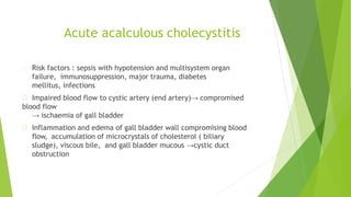
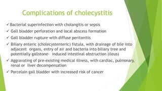
Gall bladder-Benign
conditions
Dr Joel Danie Mathew
Assistant Professor
Dept. of General Surgery
BCMCH
An Overview of this class….
Cholelithiasis
Cholecystitis
Empyema Gallbladder
Gallbladder polyps
Choledochal cyst
Miscellaneous conditions
CHOLELITHIASIS
Cholelithiasis
• Types of gall stones :
– Cholesterol stones
– Pigment stones
– Mixed stones
• In the West, about 80% are cholesterol stones.
• Pigment gall stone is predominant in non- Western population –
associated with bacterial infection of biliary tree and parasitic
infestations.
Risk factors:
Demography [Europe, N & S America, Mexico]
Advancing age
Female sex
Obesity
Rapid weight reduction
Gallbladder stasis
Hyerlipidaemia
Chronic haemolytic syndromes
Biliary infection
Gastrointestinal disorders: [CD, CF, pancreatic insufficiency]
Acquired disorders. Gallbladder stasis, either neurogenic or hormonal.
Hereditary factors. Genes encoding hepatocyte proteins that transport biliary
lipids, known as ATP-binding cassette (ABC) transporters have associations
with gallstone formation
Factors associated with gallstone formation
Pathogenesis of Cholesterol Stones:
Cholesterol is rendered soluble in bile by aggregation with water-soluble bile salts
and water-insoluble lecithins, both of which act as detergents.
When cholesterol concentrations exceed the solubilizing capacity of bile
(supersaturation), cholesterol can no longer remain dispersed and nucleates into
solid cholesterol monohydrate crystals.
Pathogenesis of Pigment Stones
Complex mixtures of insoluble calcium salts of unconjugated bilirubin
along with inorganic calcium salts.
Hemolytic anaemias and infections of the biliary tract
→ increased unconjugated bilirubin in the biliary tree
→ form precipitates : insoluble calcium bilirubinate salts.
Brown pigment stones contain calcium bilirubinate, calcium palmitate and calcium
stearate, as well as cholesterol.
Brown stones are rare in the gall bladder. They form in the bile duct and are related to
bile stasis and infected bile.
 Brown pigment stones are also associated with the presence of foreign bodies within
the bile ducts, such as endoprosthesis(stents), or parasites, such as Clonorchis sinensis
and Ascaris lumbricoides.
Black stones are largely composed of an insoluble bilirubin pigment polymer mixed
with calcium phosphate and calcium bicarbonate.
Black stones are associated with haemolysis, usually hereditary spherocytosis or sickle
cell disease.
Effects and
complications
of GB stones
MIRIZZI’S SYNDROME
• The prerequisites for this syndrome,
characterized by gallbladder disease
causing obstructive jaundice, include a
cystic duct that courses parallel to the
common hepatic duct, an impacted
stone in the gallbladder neck or cystic
duct, and an obstruction of the
common hepatic duct caused by the
stone orinflammatory response.
• The resultant inflammation can cause a
cholecystocholedochal fistula.
• The treatment of Mirizzi syndrome is
cholecystectomy, which may require
repair of the common duct; when a
large fistula exists, a
choledochojejunostomy may be
necessary.
Cholecystitis
Def: Inflammation of the gall bladder
Can be divided into
–Acute cholecystitis
–Chronic cholecystitis
–Acute on chronic cholecystitis
Acute cholecystitis
Can be divided into :
– Acute Calculous CS: 85-90% of the cases. Most common complication of gall
stones and emergency cholecystectomy
– Acute Acalculous CS (10-15% of cases)
Acute acalculous cholecystitis is particularly seen in critically ill patients and those
recovering from major surgery, trauma and burns
Acute acalculous cholecystitis
Risk factors : sepsis with hypotension and multisystem organ
failure, immunosuppression, major trauma, diabetes
mellitus, infections
Impaired blood flow to cystic artery (end artery)→ compromised
blood flow
→ ischaemia of gall bladder
Inflammation and edema of gall bladder wall compromising blood
flow, accumulation of microcrystals of cholesterol ( biliary
sludge), viscous bile, and gall bladder mucous →cystic duct
obstruction
Pathology
Gross :
Enlarged, tense, edematous, red or violaceous colour (subserosal
haemorrhage)
Fibrinous /fibrinopurulent exudate covering the serosa
+- stones obstructing the neck or
cystic duct– Lumen contains blood and pus(empyema)
Green black necrotic- gangrenous
Microscopic :
acute inflammation in the wall
mucosal ulceration.
May be associated with abscess
formation or gangrenous necrosis
Murphy’s sign
Murphy's sign is elicited in patients with acute
cholecystitis by asking the patient to take in and
hold a deep breath while palpating the right
subcostal area. If pain occurs when the inflamed
gallbladder comes into contact with the
examiner's hand, patient will have a catch in
inspiration.
This is positive Murphy's sign.
Chronic cholecystitis
Chronic cholecystitis may be a sequel to repeated bouts of mild to severe acute
cholecystitis, but in many instances it develops in the apparent absence of
antecedent attacks.
Associated with cholelithiasis > 90% of cases
• Pathogenesis : supersaturation of bile predisposes to both chronic inflammation and
stone formation.
• 1/3 of cases : E.coli and enterococci can be isolated from the bile
Microscopic appearance
Reactive proliferation of mucosa
Inflammation (lymphocytes, plasma cells, and macrophages in the
mucosa and
in the subserosal fibrous tissue). May be minimal.
Prominent outpouching of the mucosal epithelium through the wall
(Rokitansky Aschoff sinuses)
Marked subepithelial and subserosal fibrosis
+-Superimposed acute inflammation
+-Extensive calcification within the wall →porcelain gall bladder
→increase risk of cancer
Variants of chronic cholecystitis
Cholecystitis glandularis, when the mucosal folds fuse together
due to inflammation and result in formation of crypts of
epithelium buried in the gallbladder wall.
Porcelain gallbladder is the pattern when the gallbladder wall is
calcified and cracks like an egg-shell.
Acute on chronic cholecystitis is the term used for the morphologic
changes of acute cholecystitis superimposed on changes of chronic
cholecystitis.
Complications of cholecystitis
 Bacterial superinfection with cholangitis or sepsis
 Gall bladder perforation and local abscess formation
 Gall bladder rupture with diffuse peritonitis
 Biliary enteric (cholecystenteric) fistula, with drainage of bile into
adjacent organs, entry of air and bacteria into biliary tree and
potentially gallstone- induced intestinal obstruction (ileus)
 Aggravating of pre-existing medical illness, with cardiac, pulmonary,
renal or liver decompensation
 Porcelain gall bladder with increased risk of cancer
TREATMENT OF CHOLECYSTITIS
Acute cholecystitis can be managed conservatively in more than 90%
of cases ,with th epillars of treatment being NPO,IV antibiotics and
analgesics.
An interval elective cholecystectomy after 6 weeks is advocated if
patient is responding to conservative management.
Laparoscopic cholecystectomy is the gold standard of treatment
Gangrenous
cholecystitis
a/c cholecystitis c/c cholecystitis
EMPYEMA GB
 Empyema may be a sequel of acute cholecystitis or the result of a mucocoele
becoming infected.
 The gall bladder is distended with pus.
 The optimal treatment is drainage (cholecystostomy) and later, cholecystectomy.
GB POLYPS
• A gallbladder polyp (GBP) is an elevation of the GB mucosa that
protrudes into the gallbladder lumen
• 5% prevalence in the global adult population, only 5% of these are
true polyps.
• Increased detection due to frequent use of abdominal imaging.
• EUS(Endoscopic ultrasound) is more sensitive and specific than
transabdominal Ultrasound to detect GB polyps.
Risk factors for malignancy in polyps
POLYPS
1. Size: > 10 mm, Increase in size
2. Morphology : sessile, focal GB
wall thickening > 4mm.
3. Presence of symptoms or gall
stones : Relationship with
malignancy not well
established.
PATIENT
1. Age >50
2. History of primary sclerosing
cholangitis (PSC)
3. Indian ethnicity
Management
• Cholecystectomy is recommended for polyps >10 mm
• Management of polyps <10 mm depends on patient, symptoms
and polyp characteristics
CHOLEDOCHAL CYST
• Choledochal cysts are congenital dilations of the intra- and/or
extrahepatic biliary system.
• Anomalous junctions of the biliary pancreatic junction are
frequently observed, but whether or not these play a role in the
pathogenesis of the condition is unclear.
• Patients may present at any age with jaundice, fever, abdominal
pain and a right upper quadrant mass on examination,however, 60
per cent of cases are diagnosed before the age of ten years.
• Pancreatitis is not an infrequent presentation in adults.
• Patients with choledochal cysts have an increased risk of developing
cholangiocarcinoma
TODANI CLASSIFICATION
PATHOGENESIS may be congenital or acquired
Congenital cysts may result from an unequal proliferation of
embryologic biliary epithelial cells before bile duct cannulation is
complete.
• Fetal viral infection may also have a role
• cyst formation may be the result of ductal obstruction or
distension during the prenatal or neonatal period.
Acquired cysts may be the result of an abnormal
pancreaticobiliary junction (APBJ).
• APBJ is a rare congenital anomaly, with a prevalence
of 0.03%
• It is present in about 70 percent of patients with biliary cysts
and may be a significant risk factor for malignancy with the
cyst.
• In addition, patients with APBJ without biliary cysts appear
to be at a markedly increased risk for gallbladder cancer
Abnormal pancreaticobiliary junction
INVESTIGATIONS
Ultrasonography will confirm the presence of an abnormal cyst.
Magnetic resonance imaging (MRI/MRCP) will reveal the
anatomy, in particular the relationship between the lower end of
the bile duct and the pancreatic duct.
CT is also useful for delineating the extent of the intra- or
extrahepatic dilation
Surgical Treatment
For types I, II, and IV –
• Excision of the extrahepatic biliary tree – including
cholecystectomy,with a Roux-en-Y hepaticojejunostomy are
ideal.
• In some difficult case, some surgeons advocate leaving posterior
cyst wall intact with mucosectomy
• In type IV, additional segmental resection of the liver
may be appropriate
For type III
• can often be managed with endoscopic
sphincterotomy or endoscopic resection
For type V — As with type IVA cysts,
• some patients with type V cysts will eventually require
liver transplantation.
Lily’s operation
• Occasionally, the cyst adheres densely to the portal vein secondary to long-
standing inflammatory reaction. In this situation, a complete, full-thickness
excision of the cyst may not be possible.
• In the Lilly technique, the serosal surface of the duct is left adhering to the
portal vein, while the mucosa of the cyst wall is obliterated by curettage or
cautery.
• Theoretically, this removes the risk of malignant transformation in that
segment of the duct
MISCELLANEOUS CONDITIONS
CHOLESTEROSIS
 “Strawberry GB”
 In the fresh state, the interior of the gall bladder looks something like a strawberry;
the yellow specks (submucous aggregations of cholesterol crystals and cholesterol
esters) correspond to the seeds.
 It may be associated with cholesterol stones.
Diverticulosis of GB
 Diverticulosis of the gall bladder is usually manifest as black pigment
stones impacted in the outpouchings of the lacunae of Luschka.
 Diverticulosis of the gall bladder may be demonstrated by
cholecystography, especially when the gall bladder contracts after a
fatty meal.
 Rx- Cholecystectomy
Torsion of GB
• This is very rare and requires a long mesentery, which often
occurs in an older patient with a large mucocoele of the gall
bladder.
• The patient presents with an acute abdomen.
• Immediate exploration is indicated, with cholecystectomy as
the only treatment
Based on Bailey & Love’s Short Practice of
Surgery, 26th edition
THANK YOU!

Gallbladder benign conditions

  • 1.
    Gall bladder-Benign conditions Dr JoelDanie Mathew Assistant Professor Dept. of General Surgery BCMCH
  • 2.
    An Overview ofthis class…. Cholelithiasis Cholecystitis Empyema Gallbladder Gallbladder polyps Choledochal cyst Miscellaneous conditions
  • 3.
  • 4.
    Cholelithiasis • Types ofgall stones : – Cholesterol stones – Pigment stones – Mixed stones • In the West, about 80% are cholesterol stones. • Pigment gall stone is predominant in non- Western population – associated with bacterial infection of biliary tree and parasitic infestations.
  • 5.
    Risk factors: Demography [Europe,N & S America, Mexico] Advancing age Female sex Obesity Rapid weight reduction Gallbladder stasis Hyerlipidaemia Chronic haemolytic syndromes Biliary infection Gastrointestinal disorders: [CD, CF, pancreatic insufficiency] Acquired disorders. Gallbladder stasis, either neurogenic or hormonal. Hereditary factors. Genes encoding hepatocyte proteins that transport biliary lipids, known as ATP-binding cassette (ABC) transporters have associations with gallstone formation
  • 6.
    Factors associated withgallstone formation
  • 7.
    Pathogenesis of CholesterolStones: Cholesterol is rendered soluble in bile by aggregation with water-soluble bile salts and water-insoluble lecithins, both of which act as detergents. When cholesterol concentrations exceed the solubilizing capacity of bile (supersaturation), cholesterol can no longer remain dispersed and nucleates into solid cholesterol monohydrate crystals.
  • 8.
    Pathogenesis of PigmentStones Complex mixtures of insoluble calcium salts of unconjugated bilirubin along with inorganic calcium salts. Hemolytic anaemias and infections of the biliary tract → increased unconjugated bilirubin in the biliary tree → form precipitates : insoluble calcium bilirubinate salts.
  • 10.
    Brown pigment stonescontain calcium bilirubinate, calcium palmitate and calcium stearate, as well as cholesterol. Brown stones are rare in the gall bladder. They form in the bile duct and are related to bile stasis and infected bile.  Brown pigment stones are also associated with the presence of foreign bodies within the bile ducts, such as endoprosthesis(stents), or parasites, such as Clonorchis sinensis and Ascaris lumbricoides. Black stones are largely composed of an insoluble bilirubin pigment polymer mixed with calcium phosphate and calcium bicarbonate. Black stones are associated with haemolysis, usually hereditary spherocytosis or sickle cell disease.
  • 13.
  • 15.
    MIRIZZI’S SYNDROME • Theprerequisites for this syndrome, characterized by gallbladder disease causing obstructive jaundice, include a cystic duct that courses parallel to the common hepatic duct, an impacted stone in the gallbladder neck or cystic duct, and an obstruction of the common hepatic duct caused by the stone orinflammatory response. • The resultant inflammation can cause a cholecystocholedochal fistula. • The treatment of Mirizzi syndrome is cholecystectomy, which may require repair of the common duct; when a large fistula exists, a choledochojejunostomy may be necessary.
  • 16.
    Cholecystitis Def: Inflammation ofthe gall bladder Can be divided into –Acute cholecystitis –Chronic cholecystitis –Acute on chronic cholecystitis
  • 17.
    Acute cholecystitis Can bedivided into : – Acute Calculous CS: 85-90% of the cases. Most common complication of gall stones and emergency cholecystectomy – Acute Acalculous CS (10-15% of cases) Acute acalculous cholecystitis is particularly seen in critically ill patients and those recovering from major surgery, trauma and burns
  • 19.
    Acute acalculous cholecystitis Riskfactors : sepsis with hypotension and multisystem organ failure, immunosuppression, major trauma, diabetes mellitus, infections Impaired blood flow to cystic artery (end artery)→ compromised blood flow → ischaemia of gall bladder Inflammation and edema of gall bladder wall compromising blood flow, accumulation of microcrystals of cholesterol ( biliary sludge), viscous bile, and gall bladder mucous →cystic duct obstruction
  • 20.
    Pathology Gross : Enlarged, tense,edematous, red or violaceous colour (subserosal haemorrhage) Fibrinous /fibrinopurulent exudate covering the serosa +- stones obstructing the neck or cystic duct– Lumen contains blood and pus(empyema) Green black necrotic- gangrenous Microscopic : acute inflammation in the wall mucosal ulceration. May be associated with abscess formation or gangrenous necrosis
  • 22.
    Murphy’s sign Murphy's signis elicited in patients with acute cholecystitis by asking the patient to take in and hold a deep breath while palpating the right subcostal area. If pain occurs when the inflamed gallbladder comes into contact with the examiner's hand, patient will have a catch in inspiration. This is positive Murphy's sign.
  • 24.
    Chronic cholecystitis Chronic cholecystitismay be a sequel to repeated bouts of mild to severe acute cholecystitis, but in many instances it develops in the apparent absence of antecedent attacks. Associated with cholelithiasis > 90% of cases • Pathogenesis : supersaturation of bile predisposes to both chronic inflammation and stone formation. • 1/3 of cases : E.coli and enterococci can be isolated from the bile
  • 25.
    Microscopic appearance Reactive proliferationof mucosa Inflammation (lymphocytes, plasma cells, and macrophages in the mucosa and in the subserosal fibrous tissue). May be minimal. Prominent outpouching of the mucosal epithelium through the wall (Rokitansky Aschoff sinuses) Marked subepithelial and subserosal fibrosis +-Superimposed acute inflammation +-Extensive calcification within the wall →porcelain gall bladder →increase risk of cancer
  • 26.
    Variants of chroniccholecystitis Cholecystitis glandularis, when the mucosal folds fuse together due to inflammation and result in formation of crypts of epithelium buried in the gallbladder wall. Porcelain gallbladder is the pattern when the gallbladder wall is calcified and cracks like an egg-shell. Acute on chronic cholecystitis is the term used for the morphologic changes of acute cholecystitis superimposed on changes of chronic cholecystitis.
  • 27.
    Complications of cholecystitis Bacterial superinfection with cholangitis or sepsis  Gall bladder perforation and local abscess formation  Gall bladder rupture with diffuse peritonitis  Biliary enteric (cholecystenteric) fistula, with drainage of bile into adjacent organs, entry of air and bacteria into biliary tree and potentially gallstone- induced intestinal obstruction (ileus)  Aggravating of pre-existing medical illness, with cardiac, pulmonary, renal or liver decompensation  Porcelain gall bladder with increased risk of cancer
  • 28.
    TREATMENT OF CHOLECYSTITIS Acutecholecystitis can be managed conservatively in more than 90% of cases ,with th epillars of treatment being NPO,IV antibiotics and analgesics. An interval elective cholecystectomy after 6 weeks is advocated if patient is responding to conservative management. Laparoscopic cholecystectomy is the gold standard of treatment
  • 29.
  • 30.
    a/c cholecystitis c/ccholecystitis
  • 31.
  • 32.
     Empyema maybe a sequel of acute cholecystitis or the result of a mucocoele becoming infected.  The gall bladder is distended with pus.  The optimal treatment is drainage (cholecystostomy) and later, cholecystectomy.
  • 33.
    GB POLYPS • Agallbladder polyp (GBP) is an elevation of the GB mucosa that protrudes into the gallbladder lumen • 5% prevalence in the global adult population, only 5% of these are true polyps. • Increased detection due to frequent use of abdominal imaging. • EUS(Endoscopic ultrasound) is more sensitive and specific than transabdominal Ultrasound to detect GB polyps.
  • 34.
    Risk factors formalignancy in polyps POLYPS 1. Size: > 10 mm, Increase in size 2. Morphology : sessile, focal GB wall thickening > 4mm. 3. Presence of symptoms or gall stones : Relationship with malignancy not well established. PATIENT 1. Age >50 2. History of primary sclerosing cholangitis (PSC) 3. Indian ethnicity
  • 35.
    Management • Cholecystectomy isrecommended for polyps >10 mm • Management of polyps <10 mm depends on patient, symptoms and polyp characteristics
  • 36.
  • 37.
    • Choledochal cystsare congenital dilations of the intra- and/or extrahepatic biliary system. • Anomalous junctions of the biliary pancreatic junction are frequently observed, but whether or not these play a role in the pathogenesis of the condition is unclear. • Patients may present at any age with jaundice, fever, abdominal pain and a right upper quadrant mass on examination,however, 60 per cent of cases are diagnosed before the age of ten years. • Pancreatitis is not an infrequent presentation in adults. • Patients with choledochal cysts have an increased risk of developing cholangiocarcinoma
  • 38.
  • 40.
    PATHOGENESIS may becongenital or acquired Congenital cysts may result from an unequal proliferation of embryologic biliary epithelial cells before bile duct cannulation is complete. • Fetal viral infection may also have a role • cyst formation may be the result of ductal obstruction or distension during the prenatal or neonatal period.
  • 41.
    Acquired cysts maybe the result of an abnormal pancreaticobiliary junction (APBJ). • APBJ is a rare congenital anomaly, with a prevalence of 0.03% • It is present in about 70 percent of patients with biliary cysts and may be a significant risk factor for malignancy with the cyst. • In addition, patients with APBJ without biliary cysts appear to be at a markedly increased risk for gallbladder cancer
  • 42.
  • 43.
    INVESTIGATIONS Ultrasonography will confirmthe presence of an abnormal cyst. Magnetic resonance imaging (MRI/MRCP) will reveal the anatomy, in particular the relationship between the lower end of the bile duct and the pancreatic duct. CT is also useful for delineating the extent of the intra- or extrahepatic dilation
  • 44.
    Surgical Treatment For typesI, II, and IV – • Excision of the extrahepatic biliary tree – including cholecystectomy,with a Roux-en-Y hepaticojejunostomy are ideal. • In some difficult case, some surgeons advocate leaving posterior cyst wall intact with mucosectomy • In type IV, additional segmental resection of the liver may be appropriate
  • 45.
    For type III •can often be managed with endoscopic sphincterotomy or endoscopic resection For type V — As with type IVA cysts, • some patients with type V cysts will eventually require liver transplantation.
  • 47.
    Lily’s operation • Occasionally,the cyst adheres densely to the portal vein secondary to long- standing inflammatory reaction. In this situation, a complete, full-thickness excision of the cyst may not be possible. • In the Lilly technique, the serosal surface of the duct is left adhering to the portal vein, while the mucosa of the cyst wall is obliterated by curettage or cautery. • Theoretically, this removes the risk of malignant transformation in that segment of the duct
  • 48.
  • 49.
    CHOLESTEROSIS  “Strawberry GB” In the fresh state, the interior of the gall bladder looks something like a strawberry; the yellow specks (submucous aggregations of cholesterol crystals and cholesterol esters) correspond to the seeds.  It may be associated with cholesterol stones.
  • 51.
    Diverticulosis of GB Diverticulosis of the gall bladder is usually manifest as black pigment stones impacted in the outpouchings of the lacunae of Luschka.  Diverticulosis of the gall bladder may be demonstrated by cholecystography, especially when the gall bladder contracts after a fatty meal.  Rx- Cholecystectomy
  • 52.
    Torsion of GB •This is very rare and requires a long mesentery, which often occurs in an older patient with a large mucocoele of the gall bladder. • The patient presents with an acute abdomen. • Immediate exploration is indicated, with cholecystectomy as the only treatment
  • 53.
    Based on Bailey& Love’s Short Practice of Surgery, 26th edition
  • 54.