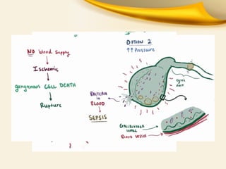
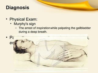
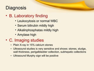
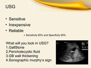
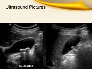
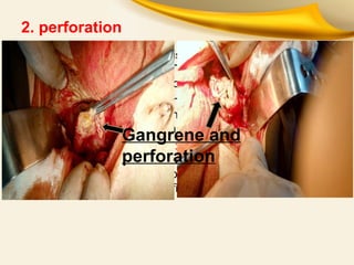
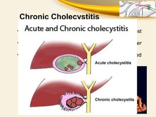
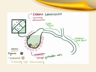
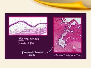
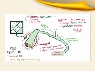
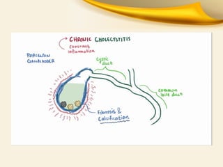
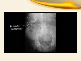
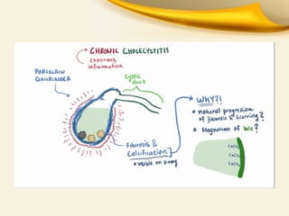
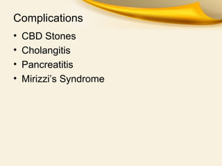
Acute & Chronic Lithiatic
And Non Lithiatic
Cholecystitis
By:Moh.Mujib Munirzai
Amiri Medical Complex
Date:19/11/2016
ACUTE CHOLECYSTITIS
• Acute cholecystitis is inflammation of the
gallbladder that develops over hours, usually
because a gallstone obstructs the cystic duct.
• Most patients have had prior attacks of biliary
colic or acute cholecystitis.
• Pain lasts longer (i.e. >6hr) than in biliary
colic and more severe.
• Acute cholecystitis begins to subside in 2 to 3
days and resolves within 1 week in 85% of
patients.
Pathogenesis
• Obstruction of the cysticduct
• Brief impaction may cause pain only
• Inflammationthe gallbladder
– Enlarged
– Tense
– Reddened
– Wallthickening
– Exudate of peri-cholecystic fluid
• BACTERIA
– E.coli
– Enterococci
– Anerobes(bacteriods)
– klebsilla
• The wall of the gallbladder may undergo
necrosis and gangrene (gangrenous
cholecystitis).
• Bacterial super-infection with gas-forming
organisms may lead to gas in the wall or
lumen of the gallbladder(emphysematous
cholecystitis).
Diagnosis
• Clinical findings
• The main symptom of uncomplicated
– biliarycolic
– caused by the obstruction of the gallbladder neck bya stone.
• The pain is characteristically
• Episodic
• Severe
• Located in the epigastrium or RUQ.
• Radiates into the back
– It frequentlyfollows after
• food intake or comes on at night.
• Accompanied by nauseaand vomiting.
Diagnosis
• Physical Exam:
• Murphy's sign
– The arrest of inspirationwhile palpating the gallbladder
during a deep breath.
• Palpable gallbladder because of fibroses,
empyema and hydrops GB
Diagnosis
• B. Laboratory finding
• Leukocytosis or normal WBC
• Serum bilirubin mildly high
• Alkalinphosphatas mildly high
• Amylase high
• C. Imaging studies
– Plain X-ray in 15% calcium stones
– Ultrasound studies is very sensitive and shows: stones, sludge,
wall thickness, perigalbladder collection, subhepatic collections
– Ultrasound Murphy sign will be positive
USG
• Sensitive
• Inexpensive
• Reliable
» Sensitivity 85% and Specificity 95%
What will you look in USG?
1.GallStone
2.Pericholecystic fluid
3.GB wall thickening
4.Sonographic murphy’s sign
Ultrasound Pictures
Differential diagnosis
• Acute peptic ulcer with or without
perforation, by radiography abdomen with
pneumopritoneum
• Acute appendicitis specially in subhepatic
location by scan, ultrasound
• Acute pancreatitis by lipase, CT Scan
Complications
1. empyema( supporative cholecystitis)
Thick wall GB, fever, toxication,
chills, WBC
Treatment: urgent
cholecystectomy or
cholecystostomy
2. perforation
a. Pericholecystic abscess: is common, palpable mass,
toxication, fever, WBC . Treats by cholecystectomy in
poor condition subcutaneous cholecystostomy.
b. Free perforation: occurs in1-2% in early gangrene
before adhesion formation and in rupture of localized
abscess with sudden pain.
3. Cholecystoenteric fistula: with stomach, duodenum,
colon adherent and necrosis and then fistula formation,
gallstone ileus, malabsorption and steatorhea. In most
cases fistula has no significant symptoms and clinic.
Gangrene and
perforation
3 main things to take therapeutic
decision:
I. Diagnosis is established
II. Susceptible general condition by
coexistent diseases
III. Sigs of local complications of acute
cholecystitis
Emergency cholecystectomy perform
in:
 Empyema gallbladder: high fever, leukocytosis, chills
 Nonlithiatic cholecystitis
 Signs of local complications of acute cholecystitis:
subhepatic local collection, perigalbladder collection,
sludge bile
 Free perforation: sudden abdominal pain on the period of
acute cholecystitis
 All patients needs urgent cholecystectomy, but in poor
condition patients percutaneous cholecystostomy
advised
Management of Acute Cholecystitis
1. NPO
2. RYLES TUBE ASPIRATE
3. IV Fluids
4.BROAD SPECTRUM ANTIBIOTICS
5.IV ANALGESICS
6. OBSERVATION
Surgery in a/c Cholecystitis
When presents within 2 to 3 days LAP CHOLECYSTECTOMY
When presents more than 3 days INTERVAL CHOLECYSTECTOMY
after 6 weeks
Empyema, Persisting and Progressing Symptoms EMERGENCY
CHOLECYSTECTOMY
Acute acalculous cholecystitis
• 5-10% of cases of acute cholecystitis
• Seen in critically ill pts or prolonged TPN
• More likely to progress to gangrene, empyema & perforation due
to ischemia
• Caused by gallbladder stasis from lack of enteral stimulation by
cholecystokinin
• Emergent operation is needed
Chronic Cholecystitis
• Long-standing gallbladder inflammation almost
always due to gallstones.
• Chronically Inflammed Thickened Gallbladder
which is NONFunctioning NONdistending
• Extensive calcification due to fibrosis is called
porcelain gallbladder.
Complications
• CBD Stones
• Cholangitis
• Pancreatitis
• Mirizzi’s Syndrome
Treatment of Chronic Cholecystitis is
CHOLECYSTECTOMY
summary
Cholecystectomy is preferable method for
treatment of acute cholecystitis
In poor condition patients percutaneous
cholecystostomy advised
Just evacuation of bile is enough not
stones
Post improving of general condition
cholecystectomy should be done
THANKS FROM YOUR PATIANCE

Acute and chronic cholicystitis

  • 1.
    Acute & ChronicLithiatic And Non Lithiatic Cholecystitis By:Moh.Mujib Munirzai Amiri Medical Complex Date:19/11/2016
  • 2.
    ACUTE CHOLECYSTITIS • Acutecholecystitis is inflammation of the gallbladder that develops over hours, usually because a gallstone obstructs the cystic duct. • Most patients have had prior attacks of biliary colic or acute cholecystitis. • Pain lasts longer (i.e. >6hr) than in biliary colic and more severe. • Acute cholecystitis begins to subside in 2 to 3 days and resolves within 1 week in 85% of patients.
  • 3.
    Pathogenesis • Obstruction ofthe cysticduct • Brief impaction may cause pain only • Inflammationthe gallbladder – Enlarged – Tense – Reddened – Wallthickening – Exudate of peri-cholecystic fluid
  • 4.
    • BACTERIA – E.coli –Enterococci – Anerobes(bacteriods) – klebsilla • The wall of the gallbladder may undergo necrosis and gangrene (gangrenous cholecystitis). • Bacterial super-infection with gas-forming organisms may lead to gas in the wall or lumen of the gallbladder(emphysematous cholecystitis).
  • 8.
    Diagnosis • Clinical findings •The main symptom of uncomplicated – biliarycolic – caused by the obstruction of the gallbladder neck bya stone. • The pain is characteristically • Episodic • Severe • Located in the epigastrium or RUQ. • Radiates into the back – It frequentlyfollows after • food intake or comes on at night. • Accompanied by nauseaand vomiting.
  • 9.
    Diagnosis • Physical Exam: •Murphy's sign – The arrest of inspirationwhile palpating the gallbladder during a deep breath. • Palpable gallbladder because of fibroses, empyema and hydrops GB
  • 10.
    Diagnosis • B. Laboratoryfinding • Leukocytosis or normal WBC • Serum bilirubin mildly high • Alkalinphosphatas mildly high • Amylase high • C. Imaging studies – Plain X-ray in 15% calcium stones – Ultrasound studies is very sensitive and shows: stones, sludge, wall thickness, perigalbladder collection, subhepatic collections – Ultrasound Murphy sign will be positive
  • 11.
    USG • Sensitive • Inexpensive •Reliable » Sensitivity 85% and Specificity 95% What will you look in USG? 1.GallStone 2.Pericholecystic fluid 3.GB wall thickening 4.Sonographic murphy’s sign
  • 12.
  • 13.
    Differential diagnosis • Acutepeptic ulcer with or without perforation, by radiography abdomen with pneumopritoneum • Acute appendicitis specially in subhepatic location by scan, ultrasound • Acute pancreatitis by lipase, CT Scan
  • 14.
    Complications 1. empyema( supporativecholecystitis) Thick wall GB, fever, toxication, chills, WBC Treatment: urgent cholecystectomy or cholecystostomy
  • 15.
    2. perforation a. Pericholecysticabscess: is common, palpable mass, toxication, fever, WBC . Treats by cholecystectomy in poor condition subcutaneous cholecystostomy. b. Free perforation: occurs in1-2% in early gangrene before adhesion formation and in rupture of localized abscess with sudden pain. 3. Cholecystoenteric fistula: with stomach, duodenum, colon adherent and necrosis and then fistula formation, gallstone ileus, malabsorption and steatorhea. In most cases fistula has no significant symptoms and clinic. Gangrene and perforation
  • 16.
    3 main thingsto take therapeutic decision: I. Diagnosis is established II. Susceptible general condition by coexistent diseases III. Sigs of local complications of acute cholecystitis
  • 17.
    Emergency cholecystectomy perform in: Empyema gallbladder: high fever, leukocytosis, chills  Nonlithiatic cholecystitis  Signs of local complications of acute cholecystitis: subhepatic local collection, perigalbladder collection, sludge bile  Free perforation: sudden abdominal pain on the period of acute cholecystitis  All patients needs urgent cholecystectomy, but in poor condition patients percutaneous cholecystostomy advised
  • 18.
    Management of AcuteCholecystitis 1. NPO 2. RYLES TUBE ASPIRATE 3. IV Fluids 4.BROAD SPECTRUM ANTIBIOTICS 5.IV ANALGESICS 6. OBSERVATION
  • 19.
    Surgery in a/cCholecystitis When presents within 2 to 3 days LAP CHOLECYSTECTOMY When presents more than 3 days INTERVAL CHOLECYSTECTOMY after 6 weeks Empyema, Persisting and Progressing Symptoms EMERGENCY CHOLECYSTECTOMY
  • 20.
    Acute acalculous cholecystitis •5-10% of cases of acute cholecystitis • Seen in critically ill pts or prolonged TPN • More likely to progress to gangrene, empyema & perforation due to ischemia • Caused by gallbladder stasis from lack of enteral stimulation by cholecystokinin • Emergent operation is needed
  • 21.
    Chronic Cholecystitis • Long-standinggallbladder inflammation almost always due to gallstones. • Chronically Inflammed Thickened Gallbladder which is NONFunctioning NONdistending • Extensive calcification due to fibrosis is called porcelain gallbladder.
  • 28.
    Complications • CBD Stones •Cholangitis • Pancreatitis • Mirizzi’s Syndrome
  • 29.
    Treatment of ChronicCholecystitis is CHOLECYSTECTOMY
  • 30.
    summary Cholecystectomy is preferablemethod for treatment of acute cholecystitis In poor condition patients percutaneous cholecystostomy advised Just evacuation of bile is enough not stones Post improving of general condition cholecystectomy should be done
  • 31.