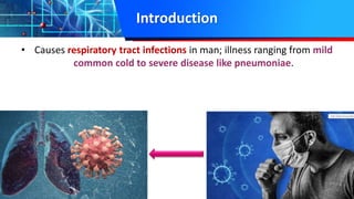
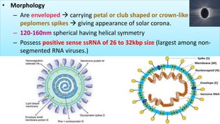
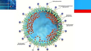
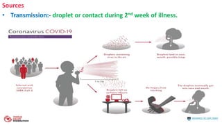
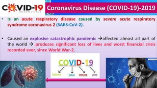
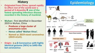
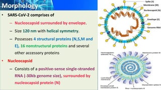
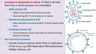
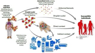
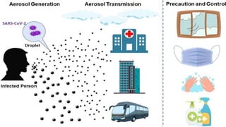
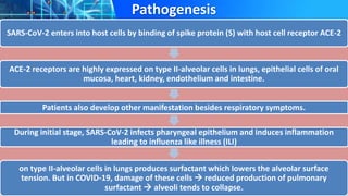
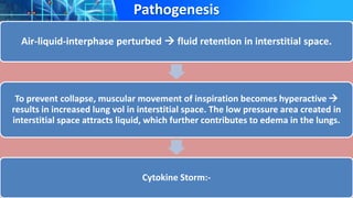
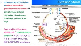
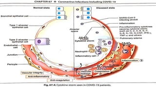
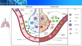
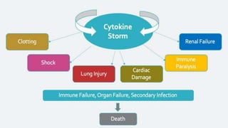
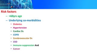
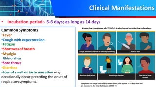
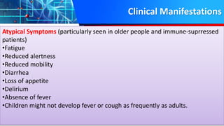
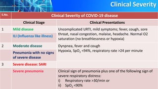
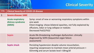
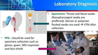
Coronavirus Infections including COVID-19
Dr. Rakesh Prasad Sah
Associate Professor, Microbiology
Introduction
• Causes respiratory tract infections in man; illness ranging from mild
common cold to severe disease like pneumoniae.
• Morphology
– Are enveloped  carrying petal or club shaped or crown-like
peplomers spikes  giving appearance of solar corona.
– 120-160nm spherical having helical symmetry
– Possess positive sense ssRNA of 26 to 32kbp size (largest among non-
segmented RNA viruses.)
Classification
• Belongs to Coronaviridae.
• Infect mammals and birds  causing ds of respiratory tract, GIT, liver, kidney and
nervous system.
• Are six genera
– Alphacoronavirus
– Betacoronavirus
– Gammacoronavirus
– Deltacoronavirus
– Bafinivirus
– Torovirus
• Alpha, beta & toro  contains viruses infect humans.
Human Coronaviruses
Human Coronaviruses
Coronaviruses that produces Milder diseases
1 Human Coronavirus 229E
2 Human Coronavirus NL63
3 Human Coronavirus OC43
4 Human Coronavirus HKU1
Coronaviruses that produces severe diseases
5 SARS-CoV :- explosive epidemic in China in 2003
6 MERS-CoV (Middle East respiratory syndrome coronavirus-2):- explosive epidemic in
Middle East in 2012.
7 SARS-CoV-2 (Severe acute respiratory syndrome coronavirus-2):- ongoing explosive
pandemics affecting whole world in 2019-21; called COVID-19
Severe Acute Respiratory Syndrome (SARS)
• History:-
– first recognized in China in 2003 by WHO physician Dr. Carlo Urbani.
– Diagnosed in a businessman  travelled from China through Hong
Kong, to Hanoi, Vietnam.
– Both died from the illness.
• Epidemiology
– Spread from Asia to various other 29 countries of the world
– Nearly 8098 cases & 774 deaths
– India remained free
Sources
• Transmission:- droplet or contact during 2nd week of illness.
Clinical manifestations
– Severe LRTI
– Muscle pain
– Headache
– Sore throat
– Fever
– Followed by onset of respiratory symptoms cough, dyspnea and pneumonia;
in some cases it may progress to acute respiratory distress syndrome.
• Treatment:- no effective drug or vaccine available. Cases managed by symptomatic
treatment.
• Infection Control: Standard Infection Control  main reason behind global
outbreak to an end.
Middle East Respiratory Syndrome (MERS)
• Causes a severe form of lower respiratory illness with a mortality rate of 35%.
• Epidemiology
– First reported in Saudi Arabia in 2012.
– 2519 confirmed cases; 858 deaths (mortality rate 34.3%).
– 27 countries reported but india was not affected.
– Origin  Bat  Camel  Human
• Transmission:- Zoonotic (Camel to human) and Human to Human
• High risk for severe disease:- Elderly people with diabetes, renal failure, Chronic
lung disease and immunocompromised persons from MERS-CoV infection.
Clinical Manifestations
– Fever
– Cough
– Shortness of breath
– Pneumonia
– GIT symptoms
– Acute respiratory distress syndrome and
– Kidney failure
• Lab Diagnosis:- Abs, Ags, RT-PCR
• Treatment:- no effective drug or vaccine available. Cases managed by
symptomatic treatment.
Coronavirus Disease (COVID-19)-2019
• Is an acute respiratory disease caused by severe acute respiratory
syndrome coronavirus 2 (SARS-CoV-2).
• Caused an explosive catastrophic pandemic affected almost all part of
the world  produces significant loss of lives and worst financial crisis
recorded ever, since World War-2.
Epidemiology
History
• Originated from China; spread rapidly
to affect rest of the world over a
period of 3-4months one of the
fastest spreading infectious disease
recorded in the history of mankind.
• Wuhan:- first identified in December
2019 in Wuhan, China
– Produces a large cluster of
pneumonia cases
– Hence called ‘Wuhan Virus’.
– Named as 2019-novel coronavirus
(2019-nCoV).
• Origin:- is a β-Coronavirus with highly
identical genome (96%) to SARS-like
bat coronavirus
• PHEIC:- cases contineud to rise  otubreak was declared as “Public Health
Emergency of International Concern (PHIEC)” on 30th Jan 2020.
• Nomenclature:- On 11th Feb 2020, WHO announced official name ‘COVID-
19’ for new coronavirus disease  renamed as SARS-CoV-2  because it’s
genome related to SARS-CoV.
Pandemic
• Disease spread rapidly in an explosive manner
• On 11th March 2020, WHO declared it as a global pandemic; by that time
almost 114 countries were affected with >118,000 cases and 4291 deaths.
• Explosive spread continued, affecting over 200 countries in next couple of
months.
Situation in India
• First report  Kerala in Jan 2020 where 3 imported cases were there 
since after a month  slow rise of cases boosted by certain clusters of
outbreaks.
• Italian visitors  was the first cluster reported in India  15 Italian visitors
were detected to be positive at Jaipur in March 2020.
• TJ cluster a religious congregation Delhi’s Nizamuddin Markaz Mosque
early in March 2020  where explosive outbreak of COVID-19 occurred
with >4000 cases.
• Koyambedu cluster  Another large local cluster of COVID-19 cases was
identified in Koyambedu market of Chennai with >3000 cases.
• Maharashtra cluster 
• As of now 
Morphology
• SARS-CoV-2 comprises of
– Nucleocapsid surrounded by envelope.
– Size 120 nm with helical symmetry.
– Possesses 4 structural proteins (N,S,M and
E), 16 nonstructural proteins and several
other accessory proteins
• Nucleocapsid
– Consists of a positive-sense single-stranded
RNA (-30kb genome size), surrounded by
nucleocapsid protein (N)
• Envelope is lipoprotein in nature  lipid is derived
from host in which proteins are embedded.
– Spike protein (S)
• Helps in the attachment to the host cells.
• Neutralizing Abs  are protective in nature.
– Membrane glycoprotein (M)
• Most abundant structural protein  gives shape to the
virus.
– Envelope protein (E)
• Transmembrane protein and with ion channel activity;
found in small qty
• Non-structural proteins
– Include several enzymes which help in replication
of the virus, e.g. RNA-dependent RNA polymerase
(RdRp), helicase, etc.
Pathogenesis
• MOT  respiratory droplets and contact routes.
• Droplet Transmission
– Person in close contact (within 1 meter) with infected person.
– Through coughing, sneezing, contact resulting in inoculation of entry portals such
as mouth, nose or conjunctiva.
– Use mask to prevent droplet transmission.
• Contact Transmission
– Direct contact with infected people
– Objects used on or by the infected person e.g. Stethoscope or Thermometer
– Through fomites
• Aerosol Transmission
– Spread of the infected droplet nuclei beyond 1 meter
– Aerosol generating procedures e.g. Endotracheal intubation
• Presymptomatic Transmission
Sources
• Transmission:- droplet or contact during 2nd week of illness.
Pathogenesis
SARS-CoV-2 enters into host cells by binding of spike protein (S) with host cell receptor ACE-2
ACE-2 receptors are highly expressed on type II-alveolar cells in lungs, epithelial cells of oral
mucosa, heart, kidney, endothelium and intestine.
Patients also develop other manifestation besides respiratory symptoms.
During initial stage, SARS-CoV-2 infects pharyngeal epithelium and induces inflammation
leading to influenza like illness (ILI)
on type II-alveolar cells in lungs produces surfactant which lowers the alveolar surface
tension. But in COVID-19, damage of these cells  reduced production of pulmonary
surfactant  alveoli tends to collapse.
Pathogenesis
Air-liquid-interphase perturbed  fluid retention in interstitial space.
To prevent collapse, muscular movement of inspiration becomes hyperactive 
results in increased lung vol in interstitial space. The low pressure area created in
interstitial space attracts liquid, which further contributes to edema in the lungs.
Cytokine Storm:-
Cytokine Storm
• Presence of SARS-CoV-2 in lung
 induces uncontrolled
generalized immune response 
Several immune cells like
neutrophils, T-lymphocytes,
macrphages recruited to the
lungs.
• Acute cytokine influx:- these
immune cells  proinflammatory
cytokines IL-2, IL-2R, IL-6, IL-7,
IL-8, IL-10, G-CSF, IFN-ϒ, IP-10,
MCP-1, MCP-3, TNF-α and others
Host Injury:- elevated cytokines leads to various consequences 
• Tissue damage and necrosis
• Further recruitment of leukocytes
• Impaired gas exchange:- reduced blood oxygenation and tissue hypoxia.
• Endothelial damage:- pulmonary vasculature, leading to vasodilation, microvascular
thrombosis and hemorrhage and hypercoagulability.
• Dilatation of blood vessels underlying alveoli:- allows passage of fluid from blood
vessels to lungs  pulmonary edema  infiltrates appear as ‘ground-glass’ in chest
imaging.
• Fibrosis:- in later stage, recruitment of fibroblast  lung fibrosis  respiratory failure
 stage is called as acute respiratory distress syndrome (ARDS) respiratory failure.
• Multiorgan failure:- induce damage to other organs e.g. heart, kidney, liver etc.
Risk factors
– >60yrs age
– Underlying co-morbidities
• Diabetes
• Hypertension
• Cardiac Ds
• COPD
• Cerebrovascular Ds
• CKD
• Immune-suppression And
• Cancer
Clinical Manifestations
• Incubation period:- 5-6 days; as long as 14 days
Common Symptoms
•Fever
•Cough with expectoration
•Fatigue
•Shortness of breath
•Myalgia
•Rhinorrhea
•Sore throat
•Diarrhea
•Loss of smell or taste sensation may
occasionally occur preceding the onset of
respiratory symptoms.
Clinical Manifestations
Atypical Symptoms (particularly seen in older people and immune-suprressed
patients)
•Fatigue
•Reduced alertness
•Reduced mobility
•Diarrhea
•Loss of appetite
•Delirium
•Absence of fever
•Children might not develop fever or cough as frequently as adults.
Clinical Severity
S.No. Clinical Severity of COVID-19 disease
Clinical Stage Clinical Presentations
1 Mild disease Uncomplicated URTI, mild symptoms; fever, cough, sore
throat, nasal congestion, malaise, headache. Normal O2
saturation (no breathlessness or hypoxia)
ILI (Influenza like illness)
2 Moderate disease Dynpnea, fever and cough
Hypoxia, SpO2 <94%, respiratory rate >24 per minute
Pneumonia with no signs
of severe disease
3 Severe disease: SARI
Severe pneumonia Clinical sign of pneumonia plus one of the following sign of
severe respiratory distress:
i) Respiratory rate >30/min or
ii) SpO2 <90%
Clinical Severity
S.No. Clinical Severity of COVID-19 disease
3 Severe disease: SARI
Acute respiratory
distress syndrome
(ARDS)
Symp: onset of new or worsening respiratory symptoms within
one week
Chest imaging: shows bilateral opacities, not fully explained by
effusions, lobar or lung collapse or nodules
Decreased PaO2/FiO2
Sepsis Acute life-threatening multiorgan dysfunction: clinically
diagnosed by SOFA (Sequential organ failure
assessment)
Septic shock Persisting hypotension despite volume resuscitation,
requiring vasopressors to maintain mean arterial pressure >
65mmHg and serum lactate level >18mg/dl
Laboratory Diagnosis
• PPE:- should be used for
specimen collection such as
gloves, gown, N95 respirator
and face shield
• Specimens: Throat and Nasal swabs
(Nasopharyngeal swabs are
preffered). Dacron or polyester
flocked swabs are used  VTM after
collection.
Laboratory Diagnosis
• Specimen transport and packing:-
– properly labeled
– Packed in three layers
– Transported in adequate cold chain
• Storage:- <40C for <5 days and –770C for >5
days
• Biosafety precaution:- Initial processing of the specimen should take place
in a biological safety cabinet. The lab should have following biosafety facility
– For NAAT:- require BSL-2 facility
– For Culture:- require BSL-3 facility
– For point-of-care test (Ag detection): can be employed without BSC.
Nucleic Acid Amplification Testing (NAAT)
• Real-Time RT-PCR
– Gold std for COVID-19
– 4-5 hrs
– >90 sample at a time
Nucleic Acid Amplification Testing (NAAT)
• Gene Targets
– Screening and Confirmatory
– Gene targets for screening are
genus specific; i.e. specific for
Betacoronavirus
• Spike protein (S)
• Envelope protein (E)
• Membrane protein (M)
• Nucleocapsid protein (N)
• Gene targets for confirmation are
species specific i.e. specific for
SARS-CoV-2
• RNA-dependent RNA-
dependent polymerase (RdRp)
• Open reading frames
(ORF1a/b)
• N2 nucleocapsid
Antigen Detection
• Nasopharyngeal swab
• Squeezed in extraction buffer
• Buffer inactivates virus releasing Ag
• Point-of-care test
• Conducted within 1hr
• Highly specific 99-100%
• Sensitivity 50-84%
• Symptomatic but negative patients should be essentially referred for a RT-PCR test.
Non-specific Tests
• Prognostic markers
– Elevated IL-6 level (cytokine storm)
– Elevated D-dimer (high level of fibrin degradation  suggesting underlying
coagulopathy)
– Elevated serum ferritin (inflammation)
– Severe lymphopenia
– Elevated CRP (Acute Inflammation)
• CT scan of lungs shows ground glass appearance and/or consolidation.
Prophylaxis
Infection Prevention and
Control
• Most effective way to
prevent COVID-19 infection.
• IPC measures at Healthcare Facility
• Hand Hygiene
– Hand Rub
– Hand wash
Infection Prevention and Control
• PPE
– Gloves
– N95 mask
– Face shield
– Head cap
– Gown
– Shoe cover
Environmental Cleaning
• Floor and Surfaces:- cleaned with detergent followed by Sodium hypochlorite 0.5%.
• Cleaning of equipments:-
– Stethoscope
– BP apparatus
• High touch surfaces
– lift button
– rail of the staircase
– patient trolley
– bed rails
– bed frames
– bedside table
– door handles
• Terminal disinfection in patient care room after discharge/transfer of patients.
(1000ppm chlorine releasing agent or a chlorine dioxide solution)
Alcohol (70%)
Alcohol (70%)
Other IPC measures
• Respiratory hygiene and cough etiquette
– Wearing a medical mask by all individuals
who are symptomatic
– Hand was immediately after sneezing
and coughing
• Biomedical waste management
• Laundry:- All linens used for COVID-19
patients washed at 60-900C with laundry
detergent  followed by soaking 0.1%
sodium hypochlorite for 30 mins.
IPC for General Public
• Hand wash:- frequently after contact with
– individuals
– High-touch area
– Public places
– After recipients of any items
– After blowing nose
– Coughing
– Sneezing
– Touching eyes, nose and mouth with unwashed hands
• Social distancing : 1 meter (prevent droplet transmission)
• Environmental cleaning: frequently touched surfaces
• Cloth Mask (Non-medical Mask)
Measures taken by Government
• Quarantine
• Lockdown
• Cluster Containment Strategy
COVID-19

COVID-19

  • 1.
    Coronavirus Infections includingCOVID-19 Dr. Rakesh Prasad Sah Associate Professor, Microbiology
  • 2.
    Introduction • Causes respiratorytract infections in man; illness ranging from mild common cold to severe disease like pneumoniae.
  • 3.
    • Morphology – Areenveloped  carrying petal or club shaped or crown-like peplomers spikes  giving appearance of solar corona. – 120-160nm spherical having helical symmetry – Possess positive sense ssRNA of 26 to 32kbp size (largest among non- segmented RNA viruses.)
  • 5.
    Classification • Belongs toCoronaviridae. • Infect mammals and birds  causing ds of respiratory tract, GIT, liver, kidney and nervous system. • Are six genera – Alphacoronavirus – Betacoronavirus – Gammacoronavirus – Deltacoronavirus – Bafinivirus – Torovirus • Alpha, beta & toro  contains viruses infect humans.
  • 7.
    Human Coronaviruses Human Coronaviruses Coronavirusesthat produces Milder diseases 1 Human Coronavirus 229E 2 Human Coronavirus NL63 3 Human Coronavirus OC43 4 Human Coronavirus HKU1 Coronaviruses that produces severe diseases 5 SARS-CoV :- explosive epidemic in China in 2003 6 MERS-CoV (Middle East respiratory syndrome coronavirus-2):- explosive epidemic in Middle East in 2012. 7 SARS-CoV-2 (Severe acute respiratory syndrome coronavirus-2):- ongoing explosive pandemics affecting whole world in 2019-21; called COVID-19
  • 8.
    Severe Acute RespiratorySyndrome (SARS) • History:- – first recognized in China in 2003 by WHO physician Dr. Carlo Urbani. – Diagnosed in a businessman  travelled from China through Hong Kong, to Hanoi, Vietnam. – Both died from the illness. • Epidemiology – Spread from Asia to various other 29 countries of the world – Nearly 8098 cases & 774 deaths – India remained free
  • 9.
    Sources • Transmission:- dropletor contact during 2nd week of illness.
  • 11.
    Clinical manifestations – SevereLRTI – Muscle pain – Headache – Sore throat – Fever – Followed by onset of respiratory symptoms cough, dyspnea and pneumonia; in some cases it may progress to acute respiratory distress syndrome. • Treatment:- no effective drug or vaccine available. Cases managed by symptomatic treatment. • Infection Control: Standard Infection Control  main reason behind global outbreak to an end.
  • 12.
    Middle East RespiratorySyndrome (MERS) • Causes a severe form of lower respiratory illness with a mortality rate of 35%. • Epidemiology – First reported in Saudi Arabia in 2012. – 2519 confirmed cases; 858 deaths (mortality rate 34.3%). – 27 countries reported but india was not affected. – Origin  Bat  Camel  Human • Transmission:- Zoonotic (Camel to human) and Human to Human • High risk for severe disease:- Elderly people with diabetes, renal failure, Chronic lung disease and immunocompromised persons from MERS-CoV infection.
  • 14.
    Clinical Manifestations – Fever –Cough – Shortness of breath – Pneumonia – GIT symptoms – Acute respiratory distress syndrome and – Kidney failure • Lab Diagnosis:- Abs, Ags, RT-PCR • Treatment:- no effective drug or vaccine available. Cases managed by symptomatic treatment.
  • 15.
    Coronavirus Disease (COVID-19)-2019 •Is an acute respiratory disease caused by severe acute respiratory syndrome coronavirus 2 (SARS-CoV-2). • Caused an explosive catastrophic pandemic affected almost all part of the world  produces significant loss of lives and worst financial crisis recorded ever, since World War-2.
  • 16.
    Epidemiology History • Originated fromChina; spread rapidly to affect rest of the world over a period of 3-4months one of the fastest spreading infectious disease recorded in the history of mankind. • Wuhan:- first identified in December 2019 in Wuhan, China – Produces a large cluster of pneumonia cases – Hence called ‘Wuhan Virus’. – Named as 2019-novel coronavirus (2019-nCoV). • Origin:- is a β-Coronavirus with highly identical genome (96%) to SARS-like bat coronavirus
  • 18.
    • PHEIC:- casescontineud to rise  otubreak was declared as “Public Health Emergency of International Concern (PHIEC)” on 30th Jan 2020. • Nomenclature:- On 11th Feb 2020, WHO announced official name ‘COVID- 19’ for new coronavirus disease  renamed as SARS-CoV-2  because it’s genome related to SARS-CoV.
  • 19.
    Pandemic • Disease spreadrapidly in an explosive manner • On 11th March 2020, WHO declared it as a global pandemic; by that time almost 114 countries were affected with >118,000 cases and 4291 deaths. • Explosive spread continued, affecting over 200 countries in next couple of months.
  • 20.
    Situation in India •First report  Kerala in Jan 2020 where 3 imported cases were there  since after a month  slow rise of cases boosted by certain clusters of outbreaks. • Italian visitors  was the first cluster reported in India  15 Italian visitors were detected to be positive at Jaipur in March 2020. • TJ cluster a religious congregation Delhi’s Nizamuddin Markaz Mosque early in March 2020  where explosive outbreak of COVID-19 occurred with >4000 cases. • Koyambedu cluster  Another large local cluster of COVID-19 cases was identified in Koyambedu market of Chennai with >3000 cases. • Maharashtra cluster 
  • 21.
    • As ofnow 
  • 22.
    Morphology • SARS-CoV-2 comprisesof – Nucleocapsid surrounded by envelope. – Size 120 nm with helical symmetry. – Possesses 4 structural proteins (N,S,M and E), 16 nonstructural proteins and several other accessory proteins • Nucleocapsid – Consists of a positive-sense single-stranded RNA (-30kb genome size), surrounded by nucleocapsid protein (N)
  • 23.
    • Envelope islipoprotein in nature  lipid is derived from host in which proteins are embedded. – Spike protein (S) • Helps in the attachment to the host cells. • Neutralizing Abs  are protective in nature. – Membrane glycoprotein (M) • Most abundant structural protein  gives shape to the virus. – Envelope protein (E) • Transmembrane protein and with ion channel activity; found in small qty • Non-structural proteins – Include several enzymes which help in replication of the virus, e.g. RNA-dependent RNA polymerase (RdRp), helicase, etc.
  • 24.
    Pathogenesis • MOT respiratory droplets and contact routes. • Droplet Transmission – Person in close contact (within 1 meter) with infected person. – Through coughing, sneezing, contact resulting in inoculation of entry portals such as mouth, nose or conjunctiva. – Use mask to prevent droplet transmission. • Contact Transmission – Direct contact with infected people – Objects used on or by the infected person e.g. Stethoscope or Thermometer – Through fomites
  • 25.
    • Aerosol Transmission –Spread of the infected droplet nuclei beyond 1 meter – Aerosol generating procedures e.g. Endotracheal intubation • Presymptomatic Transmission
  • 26.
    Sources • Transmission:- dropletor contact during 2nd week of illness.
  • 30.
    Pathogenesis SARS-CoV-2 enters intohost cells by binding of spike protein (S) with host cell receptor ACE-2 ACE-2 receptors are highly expressed on type II-alveolar cells in lungs, epithelial cells of oral mucosa, heart, kidney, endothelium and intestine. Patients also develop other manifestation besides respiratory symptoms. During initial stage, SARS-CoV-2 infects pharyngeal epithelium and induces inflammation leading to influenza like illness (ILI) on type II-alveolar cells in lungs produces surfactant which lowers the alveolar surface tension. But in COVID-19, damage of these cells  reduced production of pulmonary surfactant  alveoli tends to collapse.
  • 31.
    Pathogenesis Air-liquid-interphase perturbed fluid retention in interstitial space. To prevent collapse, muscular movement of inspiration becomes hyperactive  results in increased lung vol in interstitial space. The low pressure area created in interstitial space attracts liquid, which further contributes to edema in the lungs. Cytokine Storm:-
  • 32.
    Cytokine Storm • Presenceof SARS-CoV-2 in lung  induces uncontrolled generalized immune response  Several immune cells like neutrophils, T-lymphocytes, macrphages recruited to the lungs. • Acute cytokine influx:- these immune cells  proinflammatory cytokines IL-2, IL-2R, IL-6, IL-7, IL-8, IL-10, G-CSF, IFN-ϒ, IP-10, MCP-1, MCP-3, TNF-α and others
  • 34.
    Host Injury:- elevatedcytokines leads to various consequences  • Tissue damage and necrosis • Further recruitment of leukocytes • Impaired gas exchange:- reduced blood oxygenation and tissue hypoxia. • Endothelial damage:- pulmonary vasculature, leading to vasodilation, microvascular thrombosis and hemorrhage and hypercoagulability. • Dilatation of blood vessels underlying alveoli:- allows passage of fluid from blood vessels to lungs  pulmonary edema  infiltrates appear as ‘ground-glass’ in chest imaging. • Fibrosis:- in later stage, recruitment of fibroblast  lung fibrosis  respiratory failure  stage is called as acute respiratory distress syndrome (ARDS) respiratory failure. • Multiorgan failure:- induce damage to other organs e.g. heart, kidney, liver etc.
  • 38.
    Risk factors – >60yrsage – Underlying co-morbidities • Diabetes • Hypertension • Cardiac Ds • COPD • Cerebrovascular Ds • CKD • Immune-suppression And • Cancer
  • 39.
    Clinical Manifestations • Incubationperiod:- 5-6 days; as long as 14 days Common Symptoms •Fever •Cough with expectoration •Fatigue •Shortness of breath •Myalgia •Rhinorrhea •Sore throat •Diarrhea •Loss of smell or taste sensation may occasionally occur preceding the onset of respiratory symptoms.
  • 40.
    Clinical Manifestations Atypical Symptoms(particularly seen in older people and immune-suprressed patients) •Fatigue •Reduced alertness •Reduced mobility •Diarrhea •Loss of appetite •Delirium •Absence of fever •Children might not develop fever or cough as frequently as adults.
  • 41.
    Clinical Severity S.No. ClinicalSeverity of COVID-19 disease Clinical Stage Clinical Presentations 1 Mild disease Uncomplicated URTI, mild symptoms; fever, cough, sore throat, nasal congestion, malaise, headache. Normal O2 saturation (no breathlessness or hypoxia) ILI (Influenza like illness) 2 Moderate disease Dynpnea, fever and cough Hypoxia, SpO2 <94%, respiratory rate >24 per minute Pneumonia with no signs of severe disease 3 Severe disease: SARI Severe pneumonia Clinical sign of pneumonia plus one of the following sign of severe respiratory distress: i) Respiratory rate >30/min or ii) SpO2 <90%
  • 42.
    Clinical Severity S.No. ClinicalSeverity of COVID-19 disease 3 Severe disease: SARI Acute respiratory distress syndrome (ARDS) Symp: onset of new or worsening respiratory symptoms within one week Chest imaging: shows bilateral opacities, not fully explained by effusions, lobar or lung collapse or nodules Decreased PaO2/FiO2 Sepsis Acute life-threatening multiorgan dysfunction: clinically diagnosed by SOFA (Sequential organ failure assessment) Septic shock Persisting hypotension despite volume resuscitation, requiring vasopressors to maintain mean arterial pressure > 65mmHg and serum lactate level >18mg/dl
  • 43.
    Laboratory Diagnosis • PPE:-should be used for specimen collection such as gloves, gown, N95 respirator and face shield • Specimens: Throat and Nasal swabs (Nasopharyngeal swabs are preffered). Dacron or polyester flocked swabs are used  VTM after collection.
  • 44.
    Laboratory Diagnosis • Specimentransport and packing:- – properly labeled – Packed in three layers – Transported in adequate cold chain • Storage:- <40C for <5 days and –770C for >5 days
  • 45.
    • Biosafety precaution:-Initial processing of the specimen should take place in a biological safety cabinet. The lab should have following biosafety facility – For NAAT:- require BSL-2 facility – For Culture:- require BSL-3 facility – For point-of-care test (Ag detection): can be employed without BSC.
  • 46.
    Nucleic Acid AmplificationTesting (NAAT) • Real-Time RT-PCR – Gold std for COVID-19 – 4-5 hrs – >90 sample at a time
  • 47.
    Nucleic Acid AmplificationTesting (NAAT) • Gene Targets – Screening and Confirmatory – Gene targets for screening are genus specific; i.e. specific for Betacoronavirus • Spike protein (S) • Envelope protein (E) • Membrane protein (M) • Nucleocapsid protein (N) • Gene targets for confirmation are species specific i.e. specific for SARS-CoV-2 • RNA-dependent RNA- dependent polymerase (RdRp) • Open reading frames (ORF1a/b) • N2 nucleocapsid
  • 48.
    Antigen Detection • Nasopharyngealswab • Squeezed in extraction buffer • Buffer inactivates virus releasing Ag • Point-of-care test • Conducted within 1hr • Highly specific 99-100% • Sensitivity 50-84% • Symptomatic but negative patients should be essentially referred for a RT-PCR test.
  • 49.
    Non-specific Tests • Prognosticmarkers – Elevated IL-6 level (cytokine storm) – Elevated D-dimer (high level of fibrin degradation  suggesting underlying coagulopathy) – Elevated serum ferritin (inflammation) – Severe lymphopenia – Elevated CRP (Acute Inflammation) • CT scan of lungs shows ground glass appearance and/or consolidation.
  • 50.
  • 51.
    Infection Prevention and Control •Most effective way to prevent COVID-19 infection. • IPC measures at Healthcare Facility • Hand Hygiene – Hand Rub – Hand wash
  • 53.
    Infection Prevention andControl • PPE – Gloves – N95 mask – Face shield – Head cap – Gown – Shoe cover
  • 54.
    Environmental Cleaning • Floorand Surfaces:- cleaned with detergent followed by Sodium hypochlorite 0.5%. • Cleaning of equipments:- – Stethoscope – BP apparatus • High touch surfaces – lift button – rail of the staircase – patient trolley – bed rails – bed frames – bedside table – door handles • Terminal disinfection in patient care room after discharge/transfer of patients. (1000ppm chlorine releasing agent or a chlorine dioxide solution) Alcohol (70%) Alcohol (70%)
  • 55.
    Other IPC measures •Respiratory hygiene and cough etiquette – Wearing a medical mask by all individuals who are symptomatic – Hand was immediately after sneezing and coughing • Biomedical waste management • Laundry:- All linens used for COVID-19 patients washed at 60-900C with laundry detergent  followed by soaking 0.1% sodium hypochlorite for 30 mins.
  • 56.
    IPC for GeneralPublic • Hand wash:- frequently after contact with – individuals – High-touch area – Public places – After recipients of any items – After blowing nose – Coughing – Sneezing – Touching eyes, nose and mouth with unwashed hands • Social distancing : 1 meter (prevent droplet transmission) • Environmental cleaning: frequently touched surfaces • Cloth Mask (Non-medical Mask)
  • 57.
    Measures taken byGovernment • Quarantine • Lockdown • Cluster Containment Strategy